HAF017). A specific band was detected for Angiogenin at approximately 14 kDa (as indicated). This experiment was conducted under reducing conditions and using
Immunoblot Buffer Group 1." class="big_lightbox">
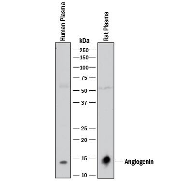
HAF017). A specific band was detected for Angiogenin at approximately 14 kDa (as indicated). This experiment was conducted under reducing conditions and using
Immunoblot Buffer Group 1." alt="Western blot shows human plasma and rat plasma. PVDF membrane was probed with 0.5 µg/mL of Goat Anti-Human/Rat Angiogenin Antigen Affinity-purified Polyclonal Antibody (Catalog # AF265) followed by HRP-conjugated Anti-Goat IgG Secondary Antibody (Catalog #
HAF017). A specific band was detected for Angiogenin at approximately 14 kDa (as indicated). This experiment was conducted under reducing conditions and using
Immunoblot Buffer Group 1." class="big_thumb" />
VC004). Before incubation with the primary antibody, tissue was subjected to heat-induced epitope retrieval using Antigen Retrieval Reagent-Basic (
CTS013). Tissue was stained using DAB (brown) and counterstained with hematoxylin (blue). Specific staining was localized to convoluted tubules. Staining was performed using our protocol for IHC Staining with VisUCyte HRP Polymer Detection Reagents." class="big_lightbox">
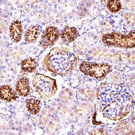
VC004). Before incubation with the primary antibody, tissue was subjected to heat-induced epitope retrieval using Antigen Retrieval Reagent-Basic (
CTS013). Tissue was stained using DAB (brown) and counterstained with hematoxylin (blue). Specific staining was localized to convoluted tubules. Staining was performed using our protocol for IHC Staining with VisUCyte HRP Polymer Detection Reagents." class="big_thumb" />
VC004). Before incubation with the primary antibody, tissue was subjected to heat-induced epitope retrieval using Antigen Retrieval Reagent-Basic (
CTS013). Tissue was stained using DAB (brown) and counterstained with hematoxylin (blue). Specific staining was localized to hepatocytes. Staining was performed using our protocol for IHC Staining with VisUCyte HRP Polymer Detection Reagents." class="big_lightbox">
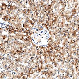
VC004). Before incubation with the primary antibody, tissue was subjected to heat-induced epitope retrieval using Antigen Retrieval Reagent-Basic (
CTS013). Tissue was stained using DAB (brown) and counterstained with hematoxylin (blue). Specific staining was localized to hepatocytes. Staining was performed using our protocol for IHC Staining with VisUCyte HRP Polymer Detection Reagents." class="big_thumb" />
HAF109). This experiment was conducted under reducing conditions and using the 12-230 kDa separation system." class="big_lightbox">
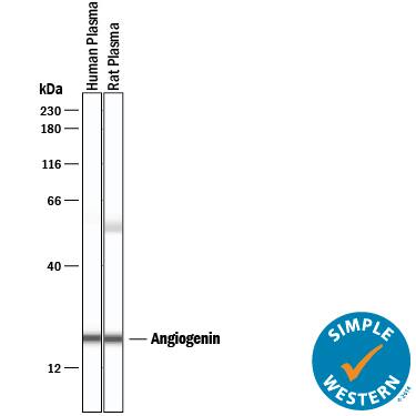
HAF109). This experiment was conducted under reducing conditions and using the 12-230 kDa separation system." class="big_thumb" />
265-AN) was serially diluted and captured by Mouse Anti-Human Angiogenin Monoclonal Antibody (Catalog #
MAB265) coated on a Clear Polystyrene Microplate (Catalog #
DY990). Goat Anti-Human/Rat Angiogenin Antigen Affinity-purified Polyclonal Antibody (Catalog # AF265) was biotinylated and incubated with the protein captured on the plate. Detection of the standard curve was achieved by incubating Streptavidin-HRP (Catalog #
DY998)" class="big_lightbox">

265-AN) was serially diluted and captured by Mouse Anti-Human Angiogenin Monoclonal Antibody (Catalog #
MAB265) coated on a Clear Polystyrene Microplate (Catalog #
DY990). Goat Anti-Human/Rat Angiogenin Antigen Affinity-purified Polyclonal Antibody (Catalog # AF265) was biotinylated and incubated with the protein captured on the plate. Detection of the standard curve was achieved by incubating Streptavidin-HRP (Catalog #
DY998)" class="big_thumb" />
 HAF017). A specific band was detected for Angiogenin at approximately 14 kDa (as indicated). This experiment was conducted under reducing conditions and using Immunoblot Buffer Group 1." alt="Western blot shows human plasma and rat plasma. PVDF membrane was probed with 0.5 µg/mL of Goat Anti-Human/Rat Angiogenin Antigen Affinity-purified Polyclonal Antibody (Catalog # AF265) followed by HRP-conjugated Anti-Goat IgG Secondary Antibody (Catalog # HAF017). A specific band was detected for Angiogenin at approximately 14 kDa (as indicated). This experiment was conducted under reducing conditions and using Immunoblot Buffer Group 1." class="big_thumb" />
 265-AN) was serially diluted and captured by Mouse Anti-Human Angiogenin Monoclonal Antibody (Catalog # MAB265) coated on a Clear Polystyrene Microplate (Catalog # DY990). Goat Anti-Human/Rat Angiogenin Antigen Affinity-purified Polyclonal Antibody (Catalog # AF265) was biotinylated and incubated with the protein captured on the plate. Detection of the standard curve was achieved by incubating Streptavidin-HRP (Catalog # DY998)" alt="Recombinant Human Angiogenin (Catalog # 265-AN) was serially diluted and captured by Mouse Anti-Human Angiogenin Monoclonal Antibody (Catalog # MAB265) coated on a Clear Polystyrene Microplate (Catalog # DY990). Goat Anti-Human/Rat Angiogenin Antigen Affinity-purified Polyclonal Antibody (Catalog # AF265) was biotinylated and incubated with the protein captured on the plate. Detection of the standard curve was achieved by incubating Streptavidin-HRP (Catalog # DY998)" class="big_thumb" />


 VC004). Before incubation with the primary antibody, tissue was subjected to heat-induced epitope retrieval using Antigen Retrieval Reagent-Basic (
 VC004). Before incubation with the primary antibody, tissue was subjected to heat-induced epitope retrieval using Antigen Retrieval Reagent-Basic (
 HAF109). This experiment was conducted under reducing conditions and using the 12-230 kDa separation system." class="big_thumb" />
 265-AN) was serially diluted and captured by Mouse Anti-Human Angiogenin Monoclonal Antibody (Catalog #
![Western Blot: Angiogenin Antibody [NBP2-41185] - Analysis of ANG in rat liver tissue lysate with Ang antibody at (A) 1 and (B) 2 ug/mL.](http://images.novusbio.com/fullsize/Angiogenin-Antibody-Western-Blot-NBP2-41185-img0001.jpg)
![Western Blot: Angiogenin Antibody - BSA Free [NBP2-41185] - Angiogenin & EPCs modulation after mouse cerebral ischemia & rehabilitation. (A) Infarct & contralateral mouse brain cortical homogenates were analyzed by western blot to quantify angiogenin protein (n = 3). The densitometry results (arbitrary units) were corrected by the actin load & are represented as fold-change of the ipsilateral vs. contralateral signal. (B) Bar graphs representing the angiogenin RNA expression in the ischemic & contralateral cortex in the short-term (n = 6–9) & long-term rehabilitation groups (n = 5), **p < 0.01, *p < 0.05 as indicated by horizontal lines. Non-RHB ipsilateral short-term vs. long-term; &p < 0.05. Pasta Matrix & Treadmill contralateral short-term vs. long-term; †p < 0.05. (C) Bar graphs showing the percentage of the Angiogenin+ area in the pasta matrix long-term rehabilitation group (n = 6) together with images of representative brains with inserts showing co-localization of angiogenin in neurons: scale bar represents 100 μm. *p < 0.05. (D) Graph showing the plasma angiogenin temporal profile of No-RHB, Pasta matrix, & Treadmill groups (n = 3–4/group); *p < 0.05. (E) Box plots representing the cell density of EPCs from the three different RHB groups (n = 3–5 short-term; n = 6–7 long-term) & representative images of the primary cultures; scale bar represents 250 μm. Data are represented as the mean ± SEM or as box plots indicating the median (IQR). IP, ipsilateral; CL, contralateral; ST-R, short-term rehabilitation; LT-R, long-term rehabilitation; No-RHB, No-Rehabilitation; EPC, endothelial progenitor cells. Image collected & cropped by CiteAb from the following publication (//www.frontiersin.org/article/10.3389/fneur.2018.00508/full), licensed under a CC-BY license. Not internally tested by Novus Biologicals.](http://images.novusbio.com/fullsize/nbp2-41185_rabbit-polyclonal-angiogenin-antibody-31020241612238.jpg)
![Immunohistochemistry: Angiogenin Antibody - BSA Free [NBP2-41185] - Immunofluorescence of Angiogenin in human liver tissue with Angiogenin antibody at 20 ug/mL.](http://images.novusbio.com/fullsize/nbp2-41185_rabbit-polyclonal-angiogenin-antibody-2711202420171783.jpg)
![Western Blot: Angiogenin Antibody (HL2215) - Azide and BSA Free [NBP3-25361] - HepG2 whole cell extract and conditioned medium (30 ug) were separated by 15% SDS-PAGE, and the membrane was blotted with Angiogenin antibody [HL2215] (NBP3-25361) diluted at 1:1000. The HRP-conjugated anti-rabbit IgG antibody was used to detect the primary antibody.](http://images.novusbio.com/fullsize/nbp3-25361_rabbit-angiogenin-mab-hl2215-azide-and-bsa-free-2120249383160.jpg "Western Blot: Angiogenin Antibody (HL2215) - Azide and BSA Free [NBP3-25361] - HepG2 whole cell extract and conditioned medium (30 ug) were separated by 15% SDS-PAGE, and the membrane was blotted with Angiogenin antibody [HL2215] (NBP3-25361) diluted at 1:1000. The HRP-conjugated anti-rabbit IgG antibody was used to detect the primary antibody.")
![Immunohistochemistry-Paraffin: Angiogenin Antibody (HL2215) - Azide and BSA Free [NBP3-25361] - Angiogenin antibody [HL2215] detects Angiogenin protein at cytoplasm by immunohistochemical analysis. Sample: Paraffin-embedded rat tissue. Angiogenin stained by Angiogenin antibody [HL2215] (NBP3-25361) diluted at 1:200. Antigen Retrieval: Citrate buffer, pH 6.0, 15 min](http://images.novusbio.com/fullsize/nbp3-25361_rabbit-angiogenin-mab-hl2215-azide-and-bsa-free-2120249371537.jpg)
![Immunocytochemistry/Immunofluorescence: Angiogenin Antibody (HL2215) - Azide and BSA Free [NBP3-25361] - Angiogenin antibody [HL2215] detects Angiogenin protein at cytoplasm by immunofluorescent analysis. Sample: HepG2 cells were fixed in 4% paraformaldehyde at RT for 15 min. Green: Angiogenin stained by Angiogenin antibody [HL2215] (NBP3-25361) diluted at 1:200. Blue: Fluoroshield with DAPI .](http://images.novusbio.com/fullsize/nbp3-25361_rabbit-angiogenin-mab-hl2215-azide-and-bsa-free-2120249393520.jpg)
![Western Blot: Angiogenin Antibody (HL2214) - Azide and BSA Free [NBP3-25360] - HepG2 whole cell extract and conditioned medium (30 ug) were separated by 15% SDS-PAGE, and the membrane was blotted with Angiogenin antibody [HL2214] (NBP3-25360) diluted at 1:1000. The HRP-conjugated anti-rabbit IgG antibody was used to detect the primary antibody.](http://images.novusbio.com/fullsize/nbp3-25360_rabbit-angiogenin-mab-hl2214-azide-and-bsa-free-212024939157.jpg "Western Blot: Angiogenin Antibody (HL2214) - Azide and BSA Free [NBP3-25360] - HepG2 whole cell extract and conditioned medium (30 ug) were separated by 15% SDS-PAGE, and the membrane was blotted with Angiogenin antibody [HL2214] (NBP3-25360) diluted at 1:1000. The HRP-conjugated anti-rabbit IgG antibody was used to detect the primary antibody.")
![Immunohistochemistry-Paraffin: Angiogenin Antibody (HL2214) - Azide and BSA Free [NBP3-25360] - Angiogenin antibody [HL2214] detects Angiogenin protein by immunohistochemical analysis. Sample: Paraffin-embedded mouse tissues. Angiogenin stained by Angiogenin antibody [HL2214] (NBP3-25360) diluted at 1:100. Antigen Retrieval: Citrate buffer, pH 6.0, 15 min](http://images.novusbio.com/fullsize/nbp3-25360_rabbit-angiogenin-mab-hl2214-azide-and-bsa-free-2120249362764.jpg)
![Immunocytochemistry/Immunofluorescence: Angiogenin Antibody (HL2214) - Azide and BSA Free [NBP3-25360] - Angiogenin antibody [HL2214] detects Angiogenin protein at cytoplasm by immunofluorescent analysis. Sample: HepG2 cells were fixed in 4% paraformaldehyde at RT for 15 min. Green: Angiogenin stained by Angiogenin antibody [HL2214] (NBP3-25360) diluted at 1:500. Blue: Fluoroshield with DAPI .](http://images.novusbio.com/fullsize/nbp3-25360_rabbit-angiogenin-mab-hl2214-azide-and-bsa-free-2120249351353.jpg)
![Immunohistochemistry-Paraffin: Angiogenin Antibody (C1) [NBP3-41634] - DAB staining on IHC-P; Sample: Human Kidney Tissue; Primary Ab: 40ug/ml NBP3-41634; Second Ab: 2ug/mL HRP-Linked Caprine Anti-Mouse IgG Polyclonal Antibody](http://images.novusbio.com/fullsize/nbp3-41634_mouse-angiogenin-mab-c1-61220242151853.jpg "Immunohistochemistry-Paraffin: Angiogenin Antibody (C1) [NBP3-41634] - DAB staining on IHC-P; Sample: Human Kidney Tissue; Primary Ab: 40ug/ml NBP3-41634; Second Ab: 2ug/mL HRP-Linked Caprine Anti-Mouse IgG Polyclonal Antibody")
![Immunohistochemistry-Paraffin: Angiogenin Antibody (C1) [NBP3-41634] - DAB staining on IHC-P; Sample: Human Liver Tissue; Primary Ab: 40ug/ml NBP3-41634; Second Ab: 2ug/mL HRP-Linked Caprine Anti-Mouse IgG Polyclonal Antibody](http://images.novusbio.com/fullsize/nbp3-41634_mouse-angiogenin-mab-c1-6122024211627.jpg)
![Immunohistochemistry-Paraffin: Angiogenin Antibody [NBP2-98773] - Immunochemical staining of human Angiogenin in human liver with rabbit polyclonal antibody (1:1000, formalin-fixed paraffin embedded sections).](http://images.novusbio.com/fullsize/Angiogenin-Antibody-Immunohistochemistry-Paraffin-NBP2-98773-img0002.jpg "Immunohistochemistry-Paraffin: Angiogenin Antibody [NBP2-98773] - Immunochemical staining of human Angiogenin in human liver with rabbit polyclonal antibody (1:1000, formalin-fixed paraffin embedded sections).")
![Immunohistochemistry-Paraffin: Angiogenin Antibody [NBP2-98773] - Immunochemical staining of human Angiogenin in human kidney with rabbit polyclonal antibody (1:1000, formalin-fixed paraffin embedded sections).](http://images.novusbio.com/fullsize/Angiogenin-Antibody-Immunohistochemistry-Paraffin-NBP2-98773-img0001.jpg)
![Western Blot: Angiogenin Antibody (C5) [NBP3-41553] - Sample: Hela cell lysate; Primary Ab: 3ug/ml NBP3-41553; Second Ab: 0.2ug/mL HRP-Linked Caprine Anti-Mouse IgG Polyclonal Antibody](http://images.novusbio.com/fullsize/nbp3-41553_mouse-angiogenin-mab-c5-612202421175911.jpg "Western Blot: Angiogenin Antibody (C5) [NBP3-41553] - Sample: Hela cell lysate; Primary Ab: 3ug/ml NBP3-41553; Second Ab: 0.2ug/mL HRP-Linked Caprine Anti-Mouse IgG Polyclonal Antibody")
![Western Blot: Angiogenin Antibody (C5) [NBP3-41553] - Sample: Hela cell lysate; Primary Ab: 3ug/ml NBP3-41553; Second Ab: 0.2ug/mL HRP-Linked Caprine Anti-Mouse IgG Polyclonal Antibody](http://images.novusbio.com/fullsize/nbp3-41553_mouse-angiogenin-mab-c5-612202421283889.jpg)
![Western Blot: Angiogenin Antibody (2A7) [H00000283-M05] - Analysis of ANG expression in transfected 293T cell line by ANG monoclonal antibody (M05), clone 2A7. Lane 1: ANG transfected lysatE (16.6 KDa). Lane 2: Non-transfected lysate.](http://images.novusbio.com/fullsize/Angiogenin-Antibody-2A7-Western-Blot-H00000283-M05-img0002.jpg "Western Blot: Angiogenin Antibody (2A7) [H00000283-M05] - Analysis of ANG expression in transfected 293T cell line by ANG monoclonal antibody (M05), clone 2A7. Lane 1: ANG transfected lysatE (16.6 KDa). Lane 2: Non-transfected lysate.")
![ELISA: Angiogenin Antibody (2A7) [H00000283-M05] - Detection limit for recombinant GST tagged ANG is 1 ng/ml as a capture antibody.](http://images.novusbio.com/fullsize/Angiogenin-Antibody-2A7-ELISA-H00000283-M05-img0001.jpg)

![Immunohistochemistry-Paraffin: Angiogenin Antibody (C6) [NBP3-41635] - DAB staining on IHC-P; Sample: Human Liver Tissue; Primary Ab: 20ug/ml NBP3-41635; Second Ab: 2ug/mL HRP-Linked Caprine Anti-Mouse IgG Polyclonal Antibody](http://images.novusbio.com/fullsize/nbp3-41635_mouse-angiogenin-mab-c6-61220242126296.jpg "Immunohistochemistry-Paraffin: Angiogenin Antibody (C6) [NBP3-41635] - DAB staining on IHC-P; Sample: Human Liver Tissue; Primary Ab: 20ug/ml NBP3-41635; Second Ab: 2ug/mL HRP-Linked Caprine Anti-Mouse IgG Polyclonal Antibody")


