CTS008) and counterstained with hematoxylin (blue). Specific labeling was localized to the cytoplasm of lymphocytes. View our protocol for
Chromogenic IHC Staining of Frozen Tissue Sections." class="big_lightbox">
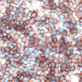
CTS008) and counterstained with hematoxylin (blue). Specific labeling was localized to the cytoplasm of lymphocytes. View our protocol for
Chromogenic IHC Staining of Frozen Tissue Sections." alt="Galectin‑1 was detected in perfusion fixed frozen sections of mouse thymus using Goat Anti-Mouse Galectin‑1 Antigen Affinity-purified Polyclonal Antibody (Catalog # AF1245) at 5 µg/mL overnight at 4 °C. Tissue was stained using the Anti-Goat HRP-DAB Cell & Tissue Staining Kit (brown; Catalog #
CTS008) and counterstained with hematoxylin (blue). Specific labeling was localized to the cytoplasm of lymphocytes. View our protocol for
Chromogenic IHC Staining of Frozen Tissue Sections." class="big_thumb" />
FAB4841A) and either (A) Normal Goat IgG Control (Catalog #
AB-108-C) or (B) Goat Anti-Mouse Galectin-1 Antigen Affinity-purified Polyclonal Antibody (Catalog # AF1245) followed by Phycoerythrin-conjugated Anti-Goat IgG Secondary Antibody (Catalog #
F0107). To facilitate intracellular staining, cells were fixed with paraformaldehyde and permeabilized with saponin." class="big_lightbox">
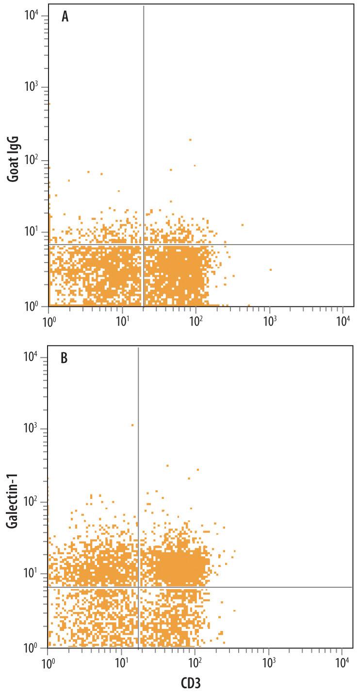
FAB4841A) and either (A) Normal Goat IgG Control (Catalog #
AB-108-C) or (B) Goat Anti-Mouse Galectin-1 Antigen Affinity-purified Polyclonal Antibody (Catalog # AF1245) followed by Phycoerythrin-conjugated Anti-Goat IgG Secondary Antibody (Catalog #
F0107). To facilitate intracellular staining, cells were fixed with paraformaldehyde and permeabilized with saponin." class="big_thumb" />

HAF017). A specific band was detected for Galectin‑1 at approximately 14 kDa (as indicated). This experiment was conducted under reducing conditions and using Western Blot Buffer Group 1." class="big_lightbox">

HAF017). A specific band was detected for Galectin‑1 at approximately 14 kDa (as indicated). This experiment was conducted under reducing conditions and using Western Blot Buffer Group 1." class="big_thumb" />
1245-GA) was serially diluted and captured by Goat Anti-Mouse Galectin‑1 Antigen Affinity-purified Polyclonal Antibody (Catalog # AF1245) coated on a Clear Polystyrene Microplate (Catalog #
DY990). Goat Anti-Mouse Galectin‑1 Antigen Affinity-purified Polyclonal Antibody (Catalog #
AF1245) was biotinylated and incubated with the protein captured on the plate. Detection of the standard curve was achieved by incubating Streptavidin-HRP (Catalog #
DY998)" class="big_lightbox">

1245-GA) was serially diluted and captured by Goat Anti-Mouse Galectin‑1 Antigen Affinity-purified Polyclonal Antibody (Catalog # AF1245) coated on a Clear Polystyrene Microplate (Catalog #
DY990). Goat Anti-Mouse Galectin‑1 Antigen Affinity-purified Polyclonal Antibody (Catalog #
AF1245) was biotinylated and incubated with the protein captured on the plate. Detection of the standard curve was achieved by incubating Streptavidin-HRP (Catalog #
DY998)" class="big_thumb" />
1245-GA) was serially diluted and captured by Goat Anti-Mouse Galectin‑1 Antigen Affinity-purified Polyclonal Antibody (Catalog #
AF1245) coated on a Clear Polystyrene Microplate (Catalog #
DY990). Goat Anti-Mouse Galectin‑1 Antigen Affinity-purified Polyclonal Antibody (Catalog # AF1245) was biotinylated and incubated with the protein captured on the plate. Detection of the standard curve was achieved by incubating Streptavidin-HRP (Catalog #
DY998)" class="big_lightbox">

1245-GA) was serially diluted and captured by Goat Anti-Mouse Galectin‑1 Antigen Affinity-purified Polyclonal Antibody (Catalog #
AF1245) coated on a Clear Polystyrene Microplate (Catalog #
DY990). Goat Anti-Mouse Galectin‑1 Antigen Affinity-purified Polyclonal Antibody (Catalog # AF1245) was biotinylated and incubated with the protein captured on the plate. Detection of the standard curve was achieved by incubating Streptavidin-HRP (Catalog #
DY998)" class="big_thumb" />
 CTS008) and counterstained with hematoxylin (blue). Specific labeling was localized to the cytoplasm of lymphocytes. View our protocol for Chromogenic IHC Staining of Frozen Tissue Sections." alt="Galectin‑1 was detected in perfusion fixed frozen sections of mouse thymus using Goat Anti-Mouse Galectin‑1 Antigen Affinity-purified Polyclonal Antibody (Catalog # AF1245) at 5 µg/mL overnight at 4 °C. Tissue was stained using the Anti-Goat HRP-DAB Cell & Tissue Staining Kit (brown; Catalog # CTS008) and counterstained with hematoxylin (blue). Specific labeling was localized to the cytoplasm of lymphocytes. View our protocol for Chromogenic IHC Staining of Frozen Tissue Sections." class="big_thumb" />
 HAF109). A specific band was detected for Galectin-1 at approximately 14 kDa (as indicated). This experiment was conducted under reducing conditions and using Immunoblot Buffer Group 1." alt="Western blot shows lysates of HeLa human cervical epithelial carcinoma cell line and A549 human lung carcinoma cell line. PVDF membrane was probed with 0.1 µg/mL of Goat Anti-Human Galectin-1 Antigen Affinity-purified Polyclonal Antibody (Catalog # AF1152) followed by HRP-conjugated Anti-Goat IgG Secondary Antibody (HAF109). A specific band was detected for Galectin-1 at approximately 14 kDa (as indicated). This experiment was conducted under reducing conditions and using Immunoblot Buffer Group 1." class="big_thumb" />
 HAF018). A specific band was detected for Galectin-1 at approximately 14 kDa (as indicated). This experiment was conducted under reducing conditions and using Immunoblot Buffer Group 1." alt="Western blot shows lysates of HeLa human cervical epithelial carcinoma cell line and A549 human lung carcinoma cell line. PVDF membrane was probed with 2 µg/mL of Mouse Anti-Human Galectin-1 Monoclonal Antibody (Catalog # MAB1152) followed by HRP-conjugated Anti-Mouse IgG Secondary Antibody (Catalog # HAF018). A specific band was detected for Galectin-1 at approximately 14 kDa (as indicated). This experiment was conducted under reducing conditions and using Immunoblot Buffer Group 1." class="big_thumb" />


 FAB4841A) and either (A) Normal Goat IgG Control (Catalog #
 HAF017). A specific band was detected for Galectin‑1 at approximately 14 kDa (as indicated). This experiment was conducted under reducing conditions and using Western Blot Buffer Group 1." class="big_thumb" />
 1245-GA) was serially diluted and captured by Goat Anti-Mouse Galectin‑1 Antigen Affinity-purified Polyclonal Antibody (Catalog # AF1245) coated on a Clear Polystyrene Microplate (Catalog #
  1245-GA) was serially diluted and captured by Goat Anti-Mouse Galectin‑1 Antigen Affinity-purified Polyclonal Antibody (Catalog #
  042-206). This experiment was conducted under reducing conditions and using the 2-40kDa separation system." class="big_thumb" />
 CTS008) and counterstained with hematoxylin (blue). View our protocol for
  CTS008) and counterstained with hematoxylin (blue). Specific labeling was localized to the cytoplasm of stromal cells and nuclei of epithelial cells. View our protocol for
  FAB3832P) and either (A) Normal Goat IgG Control (
 HAF109). This experiment was conducted under reducing conditions and using the 12-230 kDa separation system." class="big_thumb" />

![N/A Galectin-1 [HRP]](http://images.novusbio.com/fullsize/elisa/DATA_Galectin1_DGAL10_ELISA_687.jpg "N/A Galectin-1 [HRP]")
![N/A Galectin-1 [HRP]](http://images.novusbio.com/fullsize/elisa/Galectin1_DGAL10_ELISA_447.jpg)
![N/A Galectin-1 [Biotin]](http://images.novusbio.com/fullsize/elisa/DATA_Galectin1_DY1245_ELISA_1612.jpg "N/A Galectin-1 [Biotin]")
 CTS002) and counterstained with hematoxylin (blue). Specific staining was localized to stromal cells. View our protocol for

![Immunohistochemistry-Paraffin: Galectin-1 Antibody [NBP1-89791] - Staining of human liver shows moderate cytoplasmic positivity in a subset of hepatocytes.](http://images.novusbio.com/fullsize/Galectin-1-Antibody-Immunohistochemistry-Paraffin-NBP1-89791-img0016.jpg "Immunohistochemistry-Paraffin: Galectin-1 Antibody [NBP1-89791] - Staining of human liver shows moderate cytoplasmic positivity in a subset of hepatocytes.")
![Immunohistochemistry-Paraffin: Galectin-1 Antibody [NBP1-89791] - Staining of human endometrium shows strong cytoplasmic positivity in cells in endometrial stroma.](http://images.novusbio.com/fullsize/Galectin-1-Antibody-Immunohistochemistry-Paraffin-NBP1-89791-img0012.jpg)
![Immunohistochemistry-Paraffin: Galectin-1 Antibody [NBP1-89791] - Staining of human placenta shows moderate cytoplasmic positivity in decidual cells.](http://images.novusbio.com/fullsize/Galectin-1-Antibody-Immunohistochemistry-Paraffin-NBP1-89791-img0014.jpg)
![Immunohistochemistry-Paraffin: Galectin-1 Antibody [NBP1-89791] - Staining of human skin shows moderate cytoplasmic positivity in dermal cells.](http://images.novusbio.com/fullsize/Galectin-1-Antibody-Immunohistochemistry-Paraffin-NBP1-89791-img0015.jpg)
![<b>Orthogonal Strategies Validation. </b>Western Blot: Galectin-1 Antibody [NBP1-89791] - Analysis in human cell lines PC-3 and Caco-2 using anti-LGALS1 antibody. Corresponding LGALS1 RNA-seq data are presented for the same cell lines. Loading control: anti-HSP90B1.](http://images.novusbio.com/fullsize/Galectin-1-Antibody-Western-Blot-NBP1-89791-img0011.jpg)


![Immunocytochemistry/Immunofluorescence: Galectin-1 Antibody [NBP2-16580] - Galectin 1 antibody detects Galectin 1 protein at cytoplasm by immunofluorescent analysis. Sample: SK-N-SH cells were fixed in 4% paraformaldehyde at RT for 15 min.Green: Galectin 1 stained by Galectin 1 antibody diluted at 1:1000.Blue: Fluoroshield with DAPI. Scale bar= 10 um.](http://images.novusbio.com/fullsize/Galectin-1-Antibody-Immunocytochemistry-Immunofluorescence-NBP2-16580-img0016.jpg "Immunocytochemistry/Immunofluorescence: Galectin-1 Antibody [NBP2-16580] - Galectin 1 antibody detects Galectin 1 protein at cytoplasm by immunofluorescent analysis. Sample: SK-N-SH cells were fixed in 4% paraformaldehyde at RT for 15 min.Green: Galectin 1 stained by Galectin 1 antibody diluted at 1:1000.Blue: Fluoroshield with DAPI. Scale bar= 10 um.")
![Western Blot: Galectin 1 Antibody [NBP2-16580] - Sample (30 ug of whole cell lysate) A: BCL-1 15% SDS PAGE gel, diluted at 1:10000.](http://images.novusbio.com/fullsize/Galectin-1-Antibody-Western-Blot-NBP2-16580-img0001.jpg)
![Western Blot: Galectin 1 Antibody [NBP2-16580] - Sample (30 ug of whole cell lysate) A: NCI-H929 15% SDS PAGE gel, diluted at 1:10000.](http://images.novusbio.com/fullsize/Galectin-1-Antibody-Western-Blot-NBP2-16580-img0002.jpg)
![Western Blot: Galectin-1 Antibody [NBP2-16580] - Non-transfected (-) and transfected (+) HeLa whole cell extracts (30 ug) were separated by 15% SDS-PAGE, and the membrane was blotted with Galectin-1 antibody diluted at 1:10000. HRP-conjugated anti-rabbit IgG antibody was used to detect the primary antibody.](http://images.novusbio.com/fullsize/Galectin-1-Antibody-Western-Blot-NBP2-16580-img0014.jpg)
![Immunohistochemistry-Paraffin: Galectin-1 Antibody [NBP2-16580] - Human colon carcinoma, using Galectin 1antibody at 1:500 dilution. Antigen Retrieval: Trilogy™ (EDTA based, pH 8.0) buffer, 15min.](http://images.novusbio.com/fullsize/Galectin-1-Antibody-Immunohistochemistry-Paraffin-NBP2-16580-img0003.jpg)
![Galectin 1 antibody detects Galectin 1 protein expression by immunohistochemical analysis.<br>Sample: Frozen sectioned E13.5 Rat brain. <br>Green: Galectin 1 protein stained by Galectin 1 antibody (NBP2-16580) diluted at 1:250.<br>Red: beta Tubulin 3/ TUJ1, a mature neuron marker, stained by beta Tubulin 3/ TUJ1 antibody [GT11710] diluted at 1:500.<br>Blue: Fluoroshield with DAPI .<br>](http://images.novusbio.com/fullsize/nbp2-16580_rabbit-polyclonal-galectin-1-antibody-immunohistochemistry-frozen-2611202518354321.jpg)


![Western Blot: Galectin-1 Antibody (GAL1/1831) [NBP2-53212] - Western Blot Analysis of JEG-3 and K562 cell lysate using Galectin-1 antibody (GAL1/1831).](http://images.novusbio.com/fullsize/Galectin-1-Antibody-GAL1-1831-Western-Blot-NBP2-53212-img0006.jpg "Western Blot: Galectin-1 Antibody (GAL1/1831) [NBP2-53212] - Western Blot Analysis of JEG-3 and K562 cell lysate using Galectin-1 antibody (GAL1/1831).")
![Immunocytochemistry/Immunofluorescence: Galectin-1 Antibody (GAL1/1831) [NBP2-53212] - Confocal immunofluorescence image of HeLa cells using Galectin-1 Antibody (GAL1/1831). Green (CF488) and Red Dot is used to label the nuclei Red.](http://images.novusbio.com/fullsize/Galectin-1-Antibody-GAL1-1831-Immunocytochemistry-Immunofluorescence-NBP2-53212-img0003.jpg)
![Immunohistochemistry-Paraffin: Galectin-1 Antibody (GAL1/1831) [NBP2-53212] - Formalin-fixed, paraffin-embedded Human Prostate Carcinoma stained with Galectin-1 Monoclonal Antibody (GAL1/1831).](http://images.novusbio.com/fullsize/Galectin-1-Antibody-GAL1-1831-Immunohistochemistry-Paraffin-NBP2-53212-img0001.jpg)
![Western Blot: Galectin-1 Antibody (GAL1/1831) [NBP2-53212] - Western Blot of HeLa, A431, K562 and 293 Cell Lysates using Galectin-1 Monoclonal Antibody (GAL1/1831).](http://images.novusbio.com/fullsize/Galectin-1-Antibody-GAL1-1831-Western-Blot-NBP2-53212-img0002.jpg)
![Western Blot: Galectin-1 Antibody (GAL1/1831) [NBP2-53212] - Western Blot Analysis of human HeLa cell lysate using Galectin-1 Antibody (GAL1/1831).](http://images.novusbio.com/fullsize/Galectin-1-Antibody-GAL1-1831-Western-Blot-NBP2-53212-img0005.jpg)
![Protein Array: Galectin-1 Antibody (GAL1/1831) [NBP2-53212] - Analysis of Protein Array containing more than 19,000 full-length human proteins using Galectin-1 Antibody (GAL1/1831). Z- and S- Score: The Z-score represents the strength of a signal that a monoclonal antibody (MAb) (in combination with a fluorescently-tagged anti-IgG secondary antibody) produces when binding to a particular protein on the HuProt(TM) array. Z-scores are described in units of standard deviations (SD's) above the mean value of all signals generated on that array. If targets on HuProt(TM) are arranged in descending order of the Z-score, the S-score is the difference (also in units of SD's) between the Z-score. S-score therefore represents the relative target specificity of a MAb to its intended target. A MAb is considered to specific to its intended target, if the MAb has an S-score of at least 2.5.](http://images.novusbio.com/fullsize/Galectin-1-Antibody-GAL1-1831-Protein-Array-NBP2-53212-img0004.jpg)
![Western Blot: Galectin-1 Antibody (1E8-1B2) [H00003956-M01] - Analysis of LGALS1 expression in transfected 293T cell line by LGALS1 monoclonal antibody (M01), clone 1E8-1B2.Lane 1: LGALS1 transfected lysate(14.7 KDa).Lane 2: Non-transfected lysate.](http://images.novusbio.com/fullsize/Galectin-1-Antibody-1E8-1B2-Western-Blot-H00003956-M01-img0012.jpg "Western Blot: Galectin-1 Antibody (1E8-1B2) [H00003956-M01] - Analysis of LGALS1 expression in transfected 293T cell line by LGALS1 monoclonal antibody (M01), clone 1E8-1B2.Lane 1: LGALS1 transfected lysate(14.7 KDa).Lane 2: Non-transfected lysate.")
![Immunocytochemistry/Immunofluorescence: Galectin-1 Antibody (1E8-1B2) [H00003956-M01] - Analysis of monoclonal antibody to LGALS1 on HeLa cell. Antibody concentration 10 ug/ml.](http://images.novusbio.com/fullsize/Galectin-1-Antibody-1E8-1B2-Immunocytochemistry-Immunofluorescence-H00003956-M01-img0007.jpg)
![Western Blot: Galectin-1 Antibody (1E8-1B2) [H00003956-M01] - LGALS1 monoclonal antibody (M01), clone 1E8-1B2 Analysis of LGALS1 expression in HepG2.](http://images.novusbio.com/fullsize/Galectin-1-Antibody-1E8-1B2-Western-Blot-H00003956-M01-img0011.jpg)
![Immunoprecipitation: Galectin-1 Antibody (1E8-1B2) [H00003956-M01] - Analysis of LGALS1 transfected lysate using anti-LGALS1 monoclonal antibody and Protein A Magnetic Bead, and immunoblotted with LGALS1 MaxPab rabbit polyclonal antibody.](http://images.novusbio.com/fullsize/Galectin-1-Antibody-1E8-1B2-Immunoprecipitation-H00003956-M01-img0008.jpg)
![Sandwich ELISA: Galectin-1 Antibody (1E8-1B2) [H00003956-M01] - Detection limit for recombinant GST tagged LGALS1 is 1 ng/ml as a capture antibody.](http://images.novusbio.com/fullsize/Galectin-1-Antibody-1E8-1B2-Sandwich-ELISA-H00003956-M01-img0009.jpg)
![Sandwich ELISA: Galectin-1 Antibody (1E8-1B2) [H00003956-M01] - Detection limit for recombinant GST tagged LGALS1 is 0.03 ng/ml as a capture antibody.](http://images.novusbio.com/fullsize/Galectin-1-Antibody-1E8-1B2-Sandwich-ELISA-H00003956-M01-img0010.jpg)
![Immunocytochemistry/Immunofluorescence: Galectin-1 Antibody [NBP2-16581] - A431.](http://images.novusbio.com/fullsize/Galectin-1-Antibody-Immunocytochemistry-Immunofluorescence-NBP2-16581-img0005.jpg "Immunocytochemistry/Immunofluorescence: Galectin-1 Antibody [NBP2-16581] - A431.")
![<b>Orthogonal Strategies Validation. </b>Western Blot: Galectin-1 Antibody [NBP2-16581] - Various whole cell extracts (30 ug) were separated by 15% SDS-PAGE, and the membrane was blotted with Galectin 1 antibody diluted at 1:2000. The HRP-conjugated anti-rabbit IgG antibody was used to detect the primary antibody. Corresponding RNA expression data for the same cell lines are based on Human Protein Atlas program.](http://images.novusbio.com/fullsize/nbp2-16581_rabbit-polyclonal-galectin-1-antibody-1610202419414557.jpg)


