F0105B) and Rat Anti-Mouse CD4 APC-conjugated Monoclonal Antibody (
FAB554A). Quadrant markers were set based on control antibody staining (
MAB006). To facilitate intracellular staining, cells were fixed with Flow Cytometry Fixation Buffer (
FC004) and permeabilized with Flow Cytometry Permeabilization/Wash Buffer I (
FC005). View our protocol for
Staining Intracellular Molecules." class="big_lightbox">
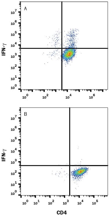
F0105B) and Rat Anti-Mouse CD4 APC-conjugated Monoclonal Antibody (
FAB554A). Quadrant markers were set based on control antibody staining (
MAB006). To facilitate intracellular staining, cells were fixed with Flow Cytometry Fixation Buffer (
FC004) and permeabilized with Flow Cytometry Permeabilization/Wash Buffer I (
FC005). View our protocol for
Staining Intracellular Molecules." alt="Mouse splenocytes either (A) stimulated to induce Th1 cells or (B) unstimulated were stained with Rat Anti-Mouse IFN-gamma Monoclonal Antibody (Catalog # MAB485) followed by Phycoerythrin-conjugated Anti-Rat IgG Secondary Antibody (
F0105B) and Rat Anti-Mouse CD4 APC-conjugated Monoclonal Antibody (
FAB554A). Quadrant markers were set based on control antibody staining (
MAB006). To facilitate intracellular staining, cells were fixed with Flow Cytometry Fixation Buffer (
FC004) and permeabilized with Flow Cytometry Permeabilization/Wash Buffer I (
FC005). View our protocol for
Staining Intracellular Molecules." class="big_thumb" />
5476) or (B) unstimulated were stained with Rat Anti-Mouse IFN-gamma Monoclonal Antibody (Catalog # MAB485) followed by Phycoerythrin-conjugated Anti-Rat IgG Secondary Antibody (
F0105B) and Rat Anti-Mouse CD4 APC-conjugated Monoclonal Antibody (
FAB554A). Quadrant markers were set based on control antibody staining (
MAB006). To facilitate intracellular staining, cells were fixed with Flow Cytometry Fixation Buffer (
FC004) and permeabilized with Flow Cytometry Permeabilization/Wash Buffer I (
FC005). Staining was performed using our Staining Intracellular Molecules protocol." class="big_lightbox">
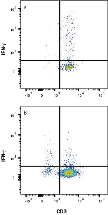
5476) or (B) unstimulated were stained with Rat Anti-Mouse IFN-gamma Monoclonal Antibody (Catalog # MAB485) followed by Phycoerythrin-conjugated Anti-Rat IgG Secondary Antibody (
F0105B) and Rat Anti-Mouse CD4 APC-conjugated Monoclonal Antibody (
FAB554A). Quadrant markers were set based on control antibody staining (
MAB006). To facilitate intracellular staining, cells were fixed with Flow Cytometry Fixation Buffer (
FC004) and permeabilized with Flow Cytometry Permeabilization/Wash Buffer I (
FC005). Staining was performed using our Staining Intracellular Molecules protocol." class="big_thumb" />
485-MI) reduces the Encephalomyocarditis Virus (EMCV)-induced cytopathy in the L-929 mouse fibroblast cell line in a dose-dependent manner (orange line), as measured by crystal violet staining. Inhibition of EMCV activity elicited by Recombinant Mouse IFN-gamma (2.5 ng/mL) is neutralized (green line) by increasing concentrations of Rat Anti-Mouse IFN-gamma Monoclonal Antibody (Catalog # MAB485). The ND
50 is typically 0.075-0.3 µg/mL." class="big_lightbox">
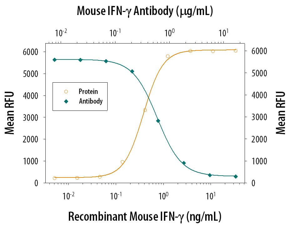
485-MI) reduces the Encephalomyocarditis Virus (EMCV)-induced cytopathy in the L-929 mouse fibroblast cell line in a dose-dependent manner (orange line), as measured by crystal violet staining. Inhibition of EMCV activity elicited by Recombinant Mouse IFN-gamma (2.5 ng/mL) is neutralized (green line) by increasing concentrations of Rat Anti-Mouse IFN-gamma Monoclonal Antibody (Catalog # MAB485). The ND
50 is typically 0.075-0.3 µg/mL." class="big_thumb" />
VC005). Before incubation with the primary antibody, tissue was subjected to heat-induced epitope retrieval using VisUCyte Antigen Retrieval Reagent-Basic (Catalog #
VCTS021). Tissue was stained using DAB (brown) and counterstained with hematoxylin (blue). Specific staining was localized to cytoplasm in lymphocytes. View our protocol for
IHC Staining with VisUCyte HRP Polymer Detection Reagents." class="big_lightbox">

VC005). Before incubation with the primary antibody, tissue was subjected to heat-induced epitope retrieval using VisUCyte Antigen Retrieval Reagent-Basic (Catalog #
VCTS021). Tissue was stained using DAB (brown) and counterstained with hematoxylin (blue). Specific staining was localized to cytoplasm in lymphocytes. View our protocol for
IHC Staining with VisUCyte HRP Polymer Detection Reagents." class="big_thumb" />
 NL007) and counterstained with DAPI (blue). View our protocol for Fluorescent ICC Staining of Non-adherent Cells." alt="IFN-gamma was detected in immersion fixed human peripheral blood mononuclear cells (PBMCs) stimulated with PMA and ionomycin using 10 µg/mL Human IFN-gamma Monoclonal Antibody (Catalog # MAB285) for 3 hours at room temperature. Cells were stained with the NorthernLights™ 557-conjugated Anti-Mouse IgG Secondary Antibody (red; Catalog # NL007) and counterstained with DAPI (blue). View our protocol for Fluorescent ICC Staining of Non-adherent Cells." class="big_thumb" />
 F0105B) and Rat Anti-Mouse CD4 APC-conjugated Monoclonal Antibody (FAB554A). Quadrant markers were set based on control antibody staining (MAB006). To facilitate intracellular staining, cells were fixed with Flow Cytometry Fixation Buffer (FC004) and permeabilized with Flow Cytometry Permeabilization/Wash Buffer I (FC005). View our protocol for Staining Intracellular Molecules." alt="Mouse splenocytes either (A) stimulated to induce Th1 cells or (B) unstimulated were stained with Rat Anti-Mouse IFN-gamma Monoclonal Antibody (Catalog # MAB485) followed by Phycoerythrin-conjugated Anti-Rat IgG Secondary Antibody (F0105B) and Rat Anti-Mouse CD4 APC-conjugated Monoclonal Antibody (FAB554A). Quadrant markers were set based on control antibody staining (MAB006). To facilitate intracellular staining, cells were fixed with Flow Cytometry Fixation Buffer (FC004) and permeabilized with Flow Cytometry Permeabilization/Wash Buffer I (FC005). View our protocol for Staining Intracellular Molecules." class="big_thumb" />
 MAB003) followed by anti-Mouse IgG PE-conjugated secondary antibody (Catalog # F0102B) and Mouse Anti-Human CD3 epsilon APC-conjugated Monoclonal Antibody (Catalog # FAB100A). To facilitate intracellular staining, cells were fixed and permeabilized with FlowX FoxP3 Fixation & Permeabilization Buffer Kit (Catalog # FC012).View our protocol for Staining Intracellular Molecules." alt="Human peripheral blood mononuclear cells (PBMCs) treated with 50 ng/mL PMA, 1 ug/mL Ionomycin, and 3 uM Monensin overnight were stained with either (A) Mouse Anti-Human IFN- gamma Monoclonal Antibody (Catalog # MAB2852) or (B) Mouse IgG2A Isotype Control (Catalog # MAB003) followed by anti-Mouse IgG PE-conjugated secondary antibody (Catalog # F0102B) and Mouse Anti-Human CD3 epsilon APC-conjugated Monoclonal Antibody (Catalog # FAB100A). To facilitate intracellular staining, cells were fixed and permeabilized with FlowX FoxP3 Fixation & Permeabilization Buffer Kit (Catalog # FC012).View our protocol for Staining Intracellular Molecules." class="big_thumb" />
 285-IF) reduces the Encephalomyocarditis Virus (EMCV)-induced cytopathy in the HeLa human cervical epithelial carcinoma cell line in a dose-dependent manner (orange line), as measured by crystal violet staining. Inhibition of EMCV activity elicited by Recombinant Human IFN-gamma (5 ng/mL) is neutralized (green line) by increasing concentrations of Goat Anti-Human IFN-gamma Antigen Affinity-purified Polyclonal Antibody (Catalog # AF-285-NA). The ND50 is typically 0.0300 ‑ 0.300 µg/mL." alt="Recombinant Human IFN-gamma (285-IF) reduces the Encephalomyocarditis Virus (EMCV)-induced cytopathy in the HeLa human cervical epithelial carcinoma cell line in a dose-dependent manner (orange line), as measured by crystal violet staining. Inhibition of EMCV activity elicited by Recombinant Human IFN-gamma (5 ng/mL) is neutralized (green line) by increasing concentrations of Goat Anti-Human IFN-gamma Antigen Affinity-purified Polyclonal Antibody (Catalog # AF-285-NA). The ND50 is typically 0.0300 ‑ 0.300 µg/mL." class="big_thumb" />


![N/A IFN-gamma [Biotin]](http://images.novusbio.com/fullsize/elisa/DATA_IFNgamma_DY485_ELISA_2061.jpg "N/A IFN-gamma [Biotin]")
![N/A IFN-gamma [HRP]](http://images.novusbio.com/fullsize/elisa/DATA_IFNgamma_MIF00_ELISA_987.jpg "N/A IFN-gamma [HRP]")
![N/A IFN-gamma [HRP]](http://images.novusbio.com/fullsize/elisa/DATA_IFNgamma_MIF00_ELISA_988.jpg)
![N/A IFN-gamma [HRP]](http://images.novusbio.com/fullsize/elisa/IFN-gamma_MIF00_ELISA_280.jpg)
 MAB003) followed by anti-Mouse IgG PE-conjugated secondary antibody (Catalog #
  285-IF) reduces the Encephalomyocarditis Virus (EMCV)-induced cytopathy in the HeLa human cervical epithelial carcinoma cell line in a dose-dependent manner (orange line), as measured by crystal violet staining. Inhibition of EMCV activity elicited by Recombinant Human IFN-gamma (1 ng/mL) is neutralized (green line) by increasing concentrations of Human IFN-gamma Monoclonal Antibody (Catalog # MAB285). The ND50 is typically 0.02-0.06 µg/mL." class="big_thumb" />
 5476) or (B) unstimulated were stained with Rat Anti-Mouse IFN-gamma Monoclonal Antibody (Catalog # MAB485) followed by Phycoerythrin-conjugated Anti-Rat IgG Secondary Antibody (
 485-MI) reduces the Encephalomyocarditis Virus (EMCV)-induced cytopathy in the L-929 mouse fibroblast cell line in a dose-dependent manner (orange line), as measured by crystal violet staining. Inhibition of EMCV activity elicited by Recombinant Mouse IFN-gamma (2.5 ng/mL) is neutralized (green line) by increasing concentrations of Rat Anti-Mouse IFN-gamma Monoclonal Antibody (Catalog # MAB485). The ND50 is typically 0.075-0.3 µg/mL." class="big_thumb" />
 VC005). Before incubation with the primary antibody, tissue was subjected to heat-induced epitope retrieval using VisUCyte Antigen Retrieval Reagent-Basic (Catalog #


![N/A IFN-gamma [HRP]](http://images.novusbio.com/fullsize/elisa/DATA_IFNgamma_RIF00_ELISA_1091.jpg "N/A IFN-gamma [HRP]")
![N/A IFN-gamma [HRP]](http://images.novusbio.com/fullsize/elisa/IFN-gamma_PRIF00_ELISA_358.jpg)
 285-IF) reduces the Encephalomyocarditis Virus (EMCV)-induced cytopathy in the HeLa human cervical epithelial carcinoma cell line in a dose-dependent manner (orange line), as measured by crystal violet staining. Inhibition of EMCV activity elicited by Recombinant Human IFN-gammaamma (1 ng/mL) is neutralized (green line) by increasing concentrations of Mouse Anti-Human IFN-gammaamma Monoclonal Antibody (Catalog # MAB2852). The ND50 is typically ≤ 2 µg/mL." class="big_thumb" />
 NL001) and counterstained with DAPI (blue). View our protocol for
 ![Immunohistochemistry-Paraffin: IFN-gamma Antibody (JM10-10) [NBP2-66900] - Human liver tissue using anti-Interferon gamma antibody. The section was pre-treated using heat mediated antigen retrieval with Tris-EDTA buffer (pH 8.0-8.4) for 20 minutes.The tissues were blocked in 5% BSA for 30 minutes at room temperature, washed with ddH2O and PBS, and then probed with the primary antibody ( 1/50) for 30 minutes at room temperature. The detection was performed using an HRP conjugated compact polymer system. DAB was used as the chromogen. Tissues were counterstained with hematoxylin and mounted with DPX.](http://images.novusbio.com/fullsize/IFN-gamma-Antibody-JM10-10-Immunohistochemistry-Paraffin-NBP2-66900-img0004.jpg "<b>Biological Strategies Validation. </b>Immunohistochemistry-Paraffin: IFN-gamma Antibody (JM10-10) [NBP2-66900] - Human liver tissue using anti-Interferon gamma antibody. The section was pre-treated using heat mediated antigen retrieval with Tris-EDTA buffer (pH 8.0-8.4) for 20 minutes.The tissues were blocked in 5% BSA for 30 minutes at room temperature, washed with ddH2O and PBS, and then probed with the primary antibody ( 1/50) for 30 minutes at room temperature. The detection was performed using an HRP conjugated compact polymer system. DAB was used as the chromogen. Tissues were counterstained with hematoxylin and mounted with DPX.")
![<b>Biological Strategies Validation. </b>Flow Cytometry: IFN-gamma Antibody (JM10-10) [NBP2-66900] - Analysis of Hela cells with Interferon gamma antibody at 1/50 dilution (red) compared with an unlabelled control (cells without incubation with primary antibody; black). Alexa Fluor 488-conjugated goat anti rabbit IgG was used as the secondary antibody.](http://images.novusbio.com/fullsize/IFN-gamma-Antibody-JM10-10-Flow-Cytometry-NBP2-66900-img0001.jpg)
![<b>Biological Strategies Validation. </b>Immunohistochemistry-Paraffin: IFN-gamma Antibody (JM10-10) [NBP2-66900] - Mouse liver tissue using anti-Interferon gamma antibody. The section was pre-treated using heat mediated antigen retrieval with Tris-EDTA buffer (pH 8.0-8.4) for 20 minutes.The tissues were blocked in 5% BSA for 30 minutes at room temperature, washed with ddH2O and PBS, and then probed with the primary antibody (1/50) for 30 minutes at room temperature. The detection was performed using an HRP conjugated compact polymer system. DAB was used as the chromogen. Tissues were counterstained with hematoxylin and mounted with DPX.](http://images.novusbio.com/fullsize/IFN-gamma-Antibody-JM10-10-Immunohistochemistry-Paraffin-NBP2-66900-img0002.jpg)
![<b>Biological Strategies Validation. </b>Immunohistochemistry-Paraffin: IFN-gamma Antibody (JM10-10) [NBP2-66900] - Human liver carcinoma tissue using anti-Interferon gamma antibody. The section was pre-treated using heat mediated antigen retrieval with Tris-EDTA buffer (pH 8.0-8.4) for 20 minutes.The tissues were blocked in 5% BSA for 30 minutes at room temperature, washed with ddH2O and PBS, and then probed with the primary antibody (1/50) for 30 minutes at room temperature. The detection was performed using an HRP conjugated compact polymer system. DAB was used as the chromogen. Tissues were counterstained with hematoxylin and mounted with DPX.](http://images.novusbio.com/fullsize/IFN-gamma-Antibody-JM10-10-Immunohistochemistry-Paraffin-NBP2-66900-img0003.jpg)
![Western blot analysis on different lysates with NBP2-66900 at 1/1,000 dilution. Lane 1: Human IFN-gamma recombinant protein, 50ng/Lane Lane 2: Mouse IFN-gamma recombinant protein, 50ng/Lane Lane 3: Rat IFN-gamma recombinant protein, 50ng/Lane Exposure time: 3 minutes; 4-20% SDS-PAGE gel. Proteins were transferred to a PVDF membrane and blocked with 5% NFDM/TBST for 1 hour at room temperature. NBP2-66900 at 1/1,000 dilution was used in 5% NFDM/TBST at room temperature for 2 hours. Goat Anti-Rabbit IgG [HRP] Secondary Antibody at 1:50,000 dilution was used for 1 hour at room temperature.](http://images.novusbio.com/fullsize/nbp2-66900_-western-blot-639204586674703050.jpg)

 485-MI) was serially diluted and captured by Rat Anti-Mouse IFN‑ gamma Monoclonal Antibody (Catalog # MAB785) coated on a Clear Polystyrene Microplate (Catalog #


