DR647RB) and counterstained with DAPI (blue; Lunaphore Catalog #
DR100). Specific staining was localized to the membrane and cytoplasm. Protocol available in
COMET™ Panel Builder." class="big_lightbox">
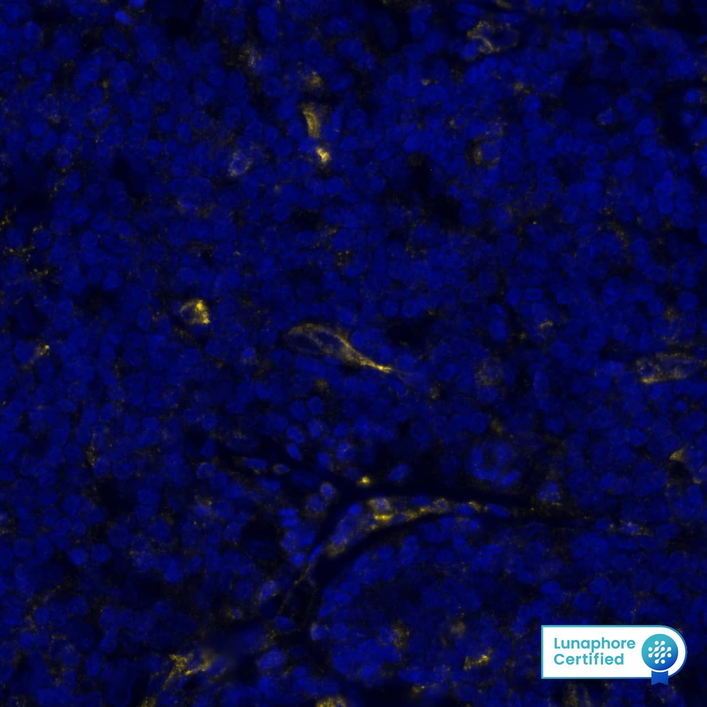
DR647RB) and counterstained with DAPI (blue; Lunaphore Catalog #
DR100). Specific staining was localized to the membrane and cytoplasm. Protocol available in
COMET™ Panel Builder." alt="VISTA was detected in immersion fixed paraffin-embedded sections of human Hodgkin's Lymphoma using Rabbit Anti-Human VISTA, Monoclonal Antibody (Catalog # MAB11712) at 5ug/mL at 27° Celsius for 2 minutes. Before incubation with the primary antibody, tissue underwent an all-in-one dewaxing and antigen retrieval preprocessing using PreTreatment Module (PT Module) and Dewax and HIER Buffer H (pH 9; Epredia Catalog # TA-999-DHBH). Tissue was stained using the Alexa Fluor™ Plus 647 Goat anti-Rabbit IgG Secondary Antibody at 1:200 at 37 ° Celsius for 2 minutes. (Yellow; Lunaphore Catalog #
DR647RB) and counterstained with DAPI (blue; Lunaphore Catalog #
DR100). Specific staining was localized to the membrane and cytoplasm. Protocol available in
COMET™ Panel Builder." class="big_thumb" />
DR647RB) and counterstained with DAPI (blue; Lunaphore Catalog #
DR100). Specific staining was localized to the membrane and cytoplasm. Protocol available in
COMET™ Panel Builder." class="big_lightbox">
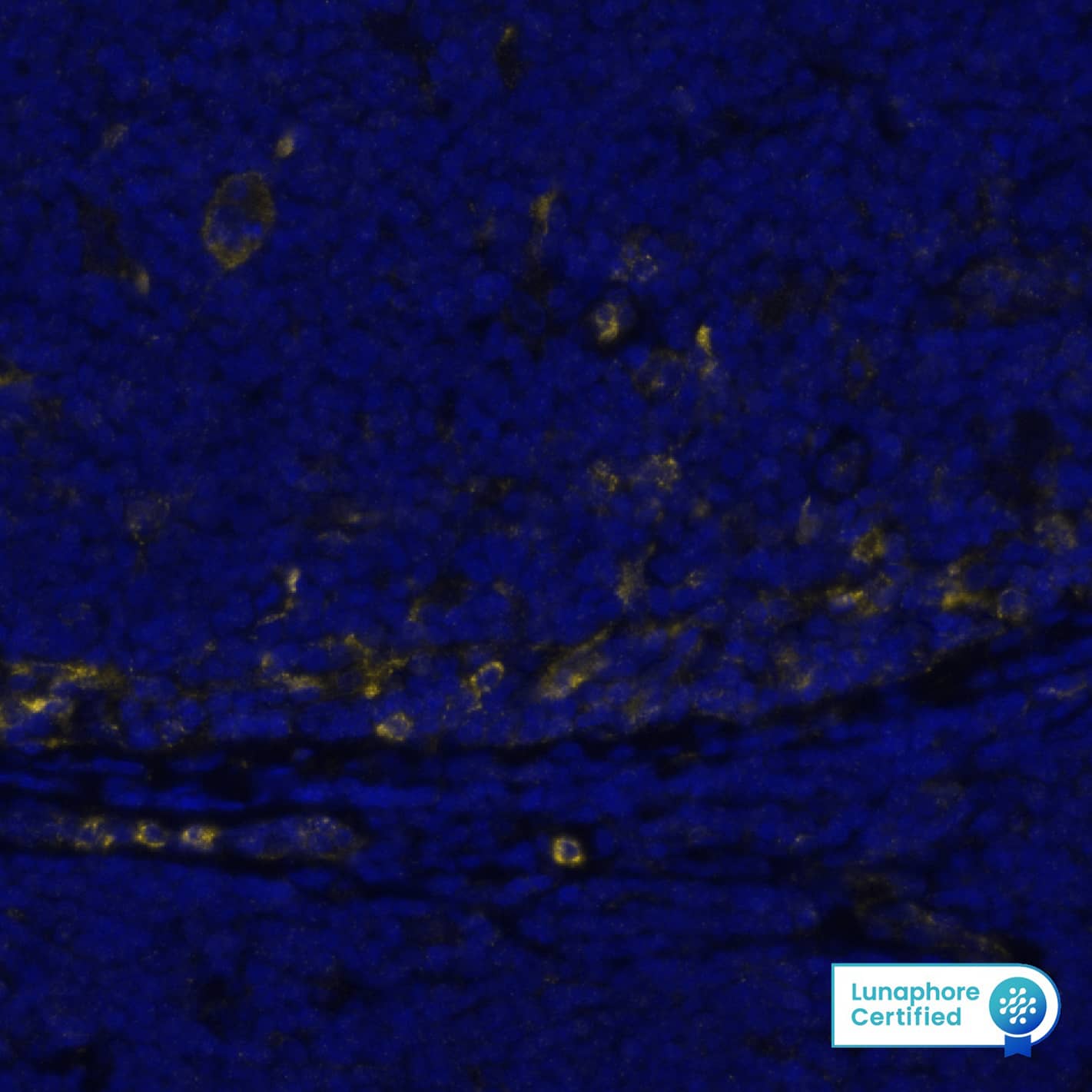
DR647RB) and counterstained with DAPI (blue; Lunaphore Catalog #
DR100). Specific staining was localized to the membrane and cytoplasm. Protocol available in
COMET™ Panel Builder." class="big_thumb" />
VC003) or the HRP-conjugated Anti-Rabbit IgG Secondary Antibody (Catalog #
HAF008). Before incubation with the primary antibody, tissue was subjected to heat-induced epitope retrieval using VisUCyte Antigen Retrieval Reagent-Basic (Catalog #
VCTS021). Tissue was stained using DAB (brown) and counterstained with hematoxylin (blue). Specific staining was localized to the cell surface. View our protocol for
IHC Staining with VisUCyte HRP Polymer Detection Reagents." class="big_lightbox">
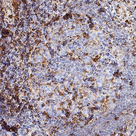
VC003) or the HRP-conjugated Anti-Rabbit IgG Secondary Antibody (Catalog #
HAF008). Before incubation with the primary antibody, tissue was subjected to heat-induced epitope retrieval using VisUCyte Antigen Retrieval Reagent-Basic (Catalog #
VCTS021). Tissue was stained using DAB (brown) and counterstained with hematoxylin (blue). Specific staining was localized to the cell surface. View our protocol for
IHC Staining with VisUCyte HRP Polymer Detection Reagents." class="big_thumb" />
VC003) or the HRP-conjugated Anti-Rabbit IgG Secondary Antibody (Catalog #
HAF008). Before incubation with the primary antibody, tissue was subjected to heat-induced epitope retrieval using VisUCyte Antigen Retrieval Reagent-Basic (Catalog #
VCTS021). Tissue was stained using DAB (brown) and counterstained with hematoxylin (blue). Specific staining was localized to the cell surface. View our protocol for
IHC Staining with VisUCyte HRP Polymer Detection Reagents." class="big_lightbox">
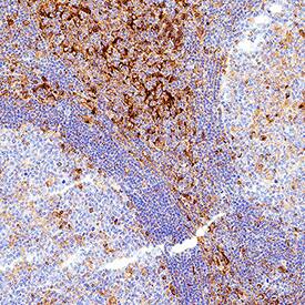
VC003) or the HRP-conjugated Anti-Rabbit IgG Secondary Antibody (Catalog #
HAF008). Before incubation with the primary antibody, tissue was subjected to heat-induced epitope retrieval using VisUCyte Antigen Retrieval Reagent-Basic (Catalog #
VCTS021). Tissue was stained using DAB (brown) and counterstained with hematoxylin (blue). Specific staining was localized to the cell surface. View our protocol for
IHC Staining with VisUCyte HRP Polymer Detection Reagents." class="big_thumb" />
HAF008). A specific band was detected for VISTA/B7-H5/PD-1H at approximately 55 kDa (as indicated). This experiment was conducted under reducing conditions and using
Western Blot Buffer Group 1." class="big_lightbox">
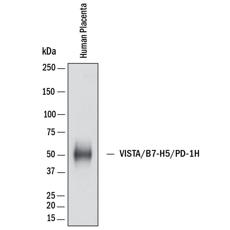
HAF008). A specific band was detected for VISTA/B7-H5/PD-1H at approximately 55 kDa (as indicated). This experiment was conducted under reducing conditions and using
Western Blot Buffer Group 1." class="big_thumb" />
 DR647RB) and counterstained with DAPI (blue; Lunaphore Catalog # DR100). Specific staining was localized to the membrane. Protocol available in COMET™ Panel Builder." alt="VISTA was detected in immersion fixed paraffin-embedded sections of human Spleen using Rabbit Anti-Human VISTA Recombinant Monoclonal Antibody (Catalog # NBP2-76406) at 1:250 at 37 ° Celsius for 4 minutes. Before incubation with the primary antibody, tissue underwent an all-in-one dewaxing and antigen retrieval preprocessing using PreTreatment Module (PT Module) and Dewax and HIER Buffer H (pH 9; Epredia Catalog # TA-999-DHBH). Tissue was stained using the Alexa Fluor™ Plus 647 Goat anti-Rabbit IgG Secondary Antibody at 1:200 at 37 ° Celsius for 2 minutes. (Yellow; Lunaphore Catalog # DR647RB) and counterstained with DAPI (blue; Lunaphore Catalog # DR100). Specific staining was localized to the membrane. Protocol available in COMET™ Panel Builder." class="big_thumb" />
 MAB0041) followed by Allophycocyanin-conjugated Anti-Mouse IgG Secondary Antibody (Catalog # F0101B) and Mouse Anti-Human Integrin aM/CD11b PE-conjugated Monoclonal Antibody (Catalog # FAB16991P). View our protocol for Staining Membrane-associated Proteins." alt="Human peripheral blood mononuclear cells (PBMCs) were stained with (A) Mouse Anti-Human VISTA/B7-H5/PD-1H Monoclonal Antibody (Catalog # MAB71261) or (B) Mouse IgG2BIsotype Control (Catalog # MAB0041) followed by Allophycocyanin-conjugated Anti-Mouse IgG Secondary Antibody (Catalog # F0101B) and Mouse Anti-Human Integrin aM/CD11b PE-conjugated Monoclonal Antibody (Catalog # FAB16991P). View our protocol for Staining Membrane-associated Proteins." class="big_thumb" />
 HAF016). A specific band was detected for VISTA/B7-H5/PD-1H at approximately 45-65 kDa (as indicated). This experiment was conducted under reducing conditions and using Immunoblot Buffer Group 1." alt="Western blot shows lysates of human placenta tissue. PVDF membrane was probed with 1 µg/mL of Sheep Anti-Human VISTA/B7-H5/PD-1H Antigen Affinity-purified Polyclonal Antibody (Catalog # AF7126) followed by HRP-conjugated Anti-Sheep IgG Secondary Antibody (Catalog # HAF016). A specific band was detected for VISTA/B7-H5/PD-1H at approximately 45-65 kDa (as indicated). This experiment was conducted under reducing conditions and using Immunoblot Buffer Group 1." class="big_thumb" />
 DR647RB) and counterstained with DAPI (blue; Lunaphore Catalog # DR100). Specific staining was localized to the membrane and cytoplasm. Protocol available in COMET™ Panel Builder." alt="VISTA was detected in immersion fixed paraffin-embedded sections of human Hodgkin's Lymphoma using Rabbit Anti-Human VISTA, Monoclonal Antibody (Catalog # MAB11712) at 5ug/mL at 27° Celsius for 2 minutes. Before incubation with the primary antibody, tissue underwent an all-in-one dewaxing and antigen retrieval preprocessing using PreTreatment Module (PT Module) and Dewax and HIER Buffer H (pH 9; Epredia Catalog # TA-999-DHBH). Tissue was stained using the Alexa Fluor™ Plus 647 Goat anti-Rabbit IgG Secondary Antibody at 1:200 at 37 ° Celsius for 2 minutes. (Yellow; Lunaphore Catalog # DR647RB) and counterstained with DAPI (blue; Lunaphore Catalog # DR100). Specific staining was localized to the membrane and cytoplasm. Protocol available in COMET™ Panel Builder." class="big_thumb" />
 HAF016). A specific band was detected for VISTA/B7-H5/PD-1H at approximately 30 kDa (as indicated). This experiment was conducted under reducing conditions and using Immunoblot Buffer Group 8." alt="Western blot shows lysates of mouse platelets. PVDF membrane was probed with 1 µg/mL of Sheep Anti-Mouse VISTA/B7-H5/PD-1H Antigen Affinity-purified Polyclonal Antibody (Catalog # AF7005) followed by HRP-conjugated Anti-Sheep IgG Secondary Antibody (Catalog # HAF016). A specific band was detected for VISTA/B7-H5/PD-1H at approximately 30 kDa (as indicated). This experiment was conducted under reducing conditions and using Immunoblot Buffer Group 8." class="big_thumb" />
 HAF005). A specific band was detected for VISTA/B7-H5/PD-1H at approximately 30 kDa (as indicated). This experiment was conducted under reducing conditions and using Immunoblot Buffer Group 1." alt="Western blot shows lysates of mouse placenta tissue, mouse spleen tissue, mouse lung tissue, and mouse platelets. PVDF membrane was probed with 0.5 µg/mL of Rat Anti-Mouse VISTA/B7-H5/ PD-1H Monoclonal Antibody (Catalog # MAB70051) followed by HRP-conjugated Anti-Rat IgG Secondary Antibody (Catalog # HAF005). A specific band was detected for VISTA/B7-H5/PD-1H at approximately 30 kDa (as indicated). This experiment was conducted under reducing conditions and using Immunoblot Buffer Group 1." class="big_thumb" />
 9229-VS) is coated at 5 µg/mL, Biotinylated Recombinant Human VISTA/B7-H5 Fc Chimera (Catalog # BT7126) binds with an ED50 of 1-6 µg/mL." alt="When Recombinant Human VSIG3 Fc Chimera (Catalog # 9229-VS) is coated at 5 µg/mL, Biotinylated Recombinant Human VISTA/B7-H5 Fc Chimera (Catalog # BT7126) binds with an ED50 of 1-6 µg/mL." class="big_thumb" />
 HAF008). A specific band was detected for VISTA/B7-H5/PD-1H at approximately 55 kDa (as indicated). This experiment was conducted under reducing conditions and using Western Blot Buffer Group 1." alt="Western Blot shows lysates of human placenta. PVDF membrane was probed with 2 µg/ml of Rabbit Anti-Human VISTA/B7-H5/PD-1H Monoclonal Antibody (Catalog # MAB11704) followed by HRP-conjugated Anti-Rabbit IgG Secondary Antibody (Catalog # HAF008). A specific band was detected for VISTA/B7-H5/PD-1H at approximately 55 kDa (as indicated). This experiment was conducted under reducing conditions and using Western Blot Buffer Group 1." class="big_thumb" />


![Western Blot: VISTA/B7-H5/PD-1H Antibody (BLR035F) [NBP2-76406] - Detection of human VISTA by WB of Jurkat, MOLT-4, MJ, RPMI-8226, HeLa, KG-1, HEK293T, and SUP-M2 lysate. Antibody: Rabbit anti-VISTA recombinant monoclonal [BLR035F] (NBP2-76406). Secondary: HRP-conjugated goat anti-rabbit IgG.Lower Panel: Rabbit anti-GAPDH.](http://images.novusbio.com/fullsize/VISTA-B7-H5-PD-1H-Antibody-BLR035F-Western-Blot-NBP2-76406-img0003.jpg)
![Immunocytochemistry/Immunofluorescence: VISTA/B7-H5/PD-1H Antibody (BLR035F) [NBP2-76406] - Detection of human VISTA (red) in FFPE ovarian carcinoma by IHC-IF. Antibody: Rabbit anti-VISTA recombinant monoclonal [BLR035F] NBP2-76406. Secondary: HRP-conjugated goat anti-rabbit IgG. Substrate: Opal™. Counterstain: DAPI (blue).](http://images.novusbio.com/fullsize/VISTA-B7-H5-PD-1H-Antibody-BLR035F-Immunocytochemistry-Immunofluorescence-NBP2-76406-img0004.jpg)
![Immunohistochemistry-Paraffin: VISTA/B7-H5/PD-1H Antibody (BLR035F) [NBP2-76406] - Detection of human VISTA in FFPE lung carcinoma by IHC. Antibody: Rabbit anti-VISTA recombinant monoclonal [BLR035F] (NBP2-76406). Secondary: HRP-conjugated goat anti-rabbit IgG. Substrate: DAB.](http://images.novusbio.com/fullsize/VISTA-B7-H5-PD-1H-Antibody-BLR035F-Immunohistochemistry-NBP2-76406-img0002.jpg)
![Flow Cytometry: VISTA/B7-H5/PD-1H Antibody (BLR035F) [NBP2-76406] - Rabbit anti-VISTA recombinant monoclonal [BLR035F] (right) or isotype control (left). Secondary: DyLight® 488-conjugated goat anti-rabbit IgG.](http://images.novusbio.com/fullsize/VISTA-B7-H5-PD-1H-Antibody-BLR035F-Flow-Cytometry-NBP2-76406-img0007.jpg)
![Immunocytochemistry/Immunofluorescence: VISTA/B7-H5/PD-1H Antibody (BLR035F) [NBP2-76406] - Detection of human VISTA in FFPE NCI-H226 cells by ICC. Antibody: Rabbit anti-VISTA recombinant monoclonal [BLR035F] (NBP2-76406). Secondary: HRP-conjugated goat anti-rabbit IgG. Substrate: DAB.](http://images.novusbio.com/fullsize/VISTA-B7-H5-PD-1H-Antibody-BLR035F-Immunocytochemistry-NBP2-76406-img0001.jpg)
![<b>Independent Antibodies Validation. </b>Immunoprecipitation: VISTA/B7-H5/PD-1H Antibody (BLR035F) [NBP2-76406] - Detection of human VISTA by WB of immunoprecipitates from Jurkat lysate. Antibodies: Rabbit anti-VISTA recombinant monoclonal [BLR035F] (NBP2-76406) and another rabbit anti-VISTA recombinant monoclonal.](http://images.novusbio.com/fullsize/VISTA-B7-H5-PD-1H-Antibody-BLR035F-Immunoprecipitation-NBP2-76406-img0006.jpg)
 NL007) and counterstained with DAPI (blue, lower panel). Specific staining was localized to cell surfaces and cytoplasm. View our protocol for

 FAB16991A) and either (A) Sheep Anti-Human VISTA/B7-H5/PD-1H Antigen Affinity-purified Polyclonal Antibody (Catalog # AF7126) or (B) Normal Sheep IgG Control (Catalog #
  MAB71264) coated on a Clear Polystyrene Microplate (Catalog #
  DR647RB) and counterstained with DAPI (blue; Lunaphore Catalog #
  VC003) or the HRP-conjugated Anti-Rabbit IgG Secondary Antibody (Catalog #
  VC003) or the HRP-conjugated Anti-Rabbit IgG Secondary Antibody (Catalog #
  HAF008). A specific band was detected for VISTA/B7-H5/PD-1H at approximately 55 kDa (as indicated). This experiment was conducted under reducing conditions and using
  F0127). Quadrant markers were set based on control antibody staining (Catalog #
  NL010) and counterstained with DAPI (blue). Specific staining was localized to cell surfaces and cytoplasm. View our protocol for

 NL013) and counterstained with DAPI (blue). Specific staining was localized to cytoplasm and cell surfaces. View our protocol for
 ![N/A VISTA/B7-H5/PD-1H [Biotin]](http://images.novusbio.com/fullsize/elisa/DATA_VISTA_DY712605_ELISA_2501.jpg "N/A VISTA/B7-H5/PD-1H [Biotin]")
![Western Blot: VISTA/B7-H5/PD-1H Antibody (CL3975) [NBP2-59030] - Analysis in human cell line CAPAN-2.](http://images.novusbio.com/fullsize/VISTA-B7-H5-PD-1H-Antibody-CL3975-Western-Blot-NBP2-59030-img0006.jpg "Western Blot: VISTA/B7-H5/PD-1H Antibody (CL3975) [NBP2-59030] - Analysis in human cell line CAPAN-2.")
![Immunohistochemistry-Paraffin: VISTA/B7-H5/PD-1H Antibody (CL3975) [NBP2-59030] - Staining of human lymph node shows immunoreactivity in a subset of lymphoid cells, mainly outside reaction centrum.](http://images.novusbio.com/fullsize/VISTA-B7-H5-PD-1H-Antibody-CL3975-Immunohistochemistry-Paraffin-NBP2-59030-img0005.jpg)
![Immunohistochemistry-Paraffin: VISTA/B7-H5/PD-1H Antibody (CL3975) [NBP2-59030] - Staining of human placenta shows strong cytoplasmic immunoreactivity in trophoblast.](http://images.novusbio.com/fullsize/VISTA-B7-H5-PD-1H-Antibody-CL3975-Immunohistochemistry-Paraffin-NBP2-59030-img0001.jpg)
![Immunohistochemistry-Paraffin: VISTA/B7-H5/PD-1H Antibody (CL3975) [NBP2-59030] - Staining of human rectum shows strong positivity in lymphoid cells.](http://images.novusbio.com/fullsize/VISTA-B7-H5-PD-1H-Antibody-CL3975-Immunohistochemistry-Paraffin-NBP2-59030-img0002.jpg)
![Immunohistochemistry-Paraffin: VISTA/B7-H5/PD-1H Antibody (CL3975) [NBP2-59030] - Staining of human liver shows strong immunoreactivity in a subset of lymphoid cells.](http://images.novusbio.com/fullsize/VISTA-B7-H5-PD-1H-Antibody-CL3975-Immunohistochemistry-Paraffin-NBP2-59030-img0003.jpg)
![Immunohistochemistry-Paraffin: VISTA/B7-H5/PD-1H Antibody (CL3975) [NBP2-59030] - Staining of human cerebral cortex shows positivity in microglia and in blood vessels.](http://images.novusbio.com/fullsize/VISTA-B7-H5-PD-1H-Antibody-CL3975-Immunohistochemistry-Paraffin-NBP2-59030-img0004.jpg)
![Western Blot: VISTA/B7-H5/PD-1H Antibody [NBP1-88967] - Analysis in human cell lines A-431 and HEK293 using anti-C10orf54 antibody. Corresponding C10orf54 RNA-seq data are presented for the same cell lines. Loading control: anti-GAPDH.](http://images.novusbio.com/fullsize/VISTA-B7-H5-PD-1H-Antibody-Western-Blot-NBP1-88967-img0022.jpg "<b>Orthogonal Strategies Validation. </b>Western Blot: VISTA/B7-H5/PD-1H Antibody [NBP1-88967] - Analysis in human cell lines A-431 and HEK293 using anti-C10orf54 antibody. Corresponding C10orf54 RNA-seq data are presented for the same cell lines. Loading control: anti-GAPDH.")
![<b>Orthogonal Strategies Validation. </b>Immunohistochemistry-Paraffin: VISTA/B7-H5/PD-1H Antibody [NBP1-88967] - Staining of human tonsil shows strong cytoplasmic positivity in non-germinal center cells.](http://images.novusbio.com/fullsize/VISTA-B7-H5-PD-1H-Antibody-Immunohistochemistry-Paraffin-NBP1-88967-img0027.jpg)
![Immunohistochemistry-Paraffin: VISTA/B7-H5/PD-1H Antibody [NBP1-88967] - Staining of human Fallopian tube shows strong cytoplasmic positivity in leukocytes.](http://images.novusbio.com/fullsize/VISTA-B7-H5-PD-1H-Antibody-Immunohistochemistry-Paraffin-NBP1-88967-img0024.jpg)
![Immunohistochemistry-Paraffin: VISTA/B7-H5/PD-1H Antibody [NBP1-88967] - Staining of human liver shows strong cytoplasmic positivity in leukocytes.](http://images.novusbio.com/fullsize/VISTA-B7-H5-PD-1H-Antibody-Immunohistochemistry-Paraffin-NBP1-88967-img0025.jpg)
![Immunohistochemistry-Paraffin: VISTA/B7-H5/PD-1H Antibody [NBP1-88967] - Staining of human small intestine shows moderate cytoplasmic positivity in lymphoid cells.](http://images.novusbio.com/fullsize/VISTA-B7-H5-PD-1H-Antibody-Immunohistochemistry-Paraffin-NBP1-88967-img0026.jpg)
 042-206). This experiment was conducted under reducing conditions and using the 12-230kDa separation system. Right: Simple Western electropherogram showing the same Rabbit Anti-Human VISTA/B7-H5/PD-1H Monoclonal Antibody (Catalog # MAB11704) tested at 2 µg/ml (blue line) and 10 µg/ml (green line) in human placenta." class="big_thumb" />


