DR647MS) and counterstained with DAPI (blue; Lunaphore Catalog #
DR100). Specific staining was localized to the membrane. Protocol available in
COMET™ Panel Builder." class="big_lightbox">
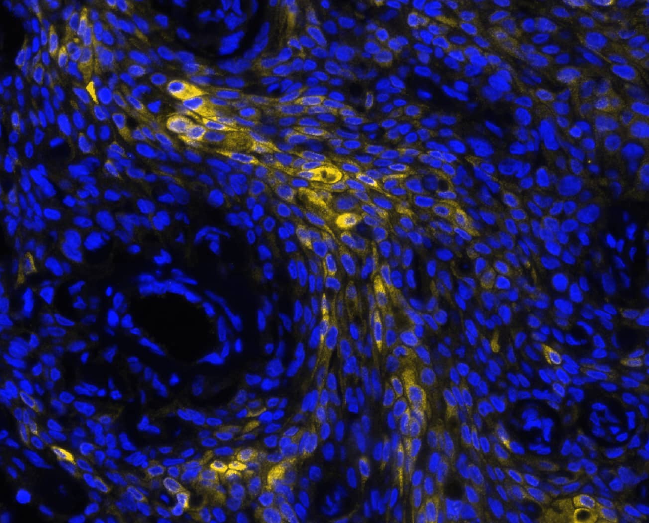
DR647MS) and counterstained with DAPI (blue; Lunaphore Catalog #
DR100). Specific staining was localized to the membrane. Protocol available in
COMET™ Panel Builder." alt="MUC1 was detected in immersion fixed paraffin-embedded sections of human pancreas tumor using Mouse Anti-Human MUC1, Monoclonal Antibody (Catalog # MAB6298) at 4ug/mL at 27 ° Celsius for 2 minutes. Before incubation with the primary antibody, tissue underwent an all-in-one dewaxing and antigen retrieval preprocessing using PreTreatment Module (PT Module) and Dewax and HIER Buffer H (pH 9; Epredia Catalog # TA-999-DHBH). Tissue was stained using the Alexa Fluor™ 647 Goat anti-Mouse IgG Secondary Antibody at 1:200 at 37 ° Celsius for 2 minutes. (Yellow; Lunaphore Catalog #
DR647MS) and counterstained with DAPI (blue; Lunaphore Catalog #
DR100). Specific staining was localized to the membrane. Protocol available in
COMET™ Panel Builder." class="big_thumb" />
DR647MS) and counterstained with DAPI (blue; Lunaphore Catalog #
DR100). Specific staining was localized to the membrane. Protocol available in
COMET™ Panel Builder." class="big_lightbox">
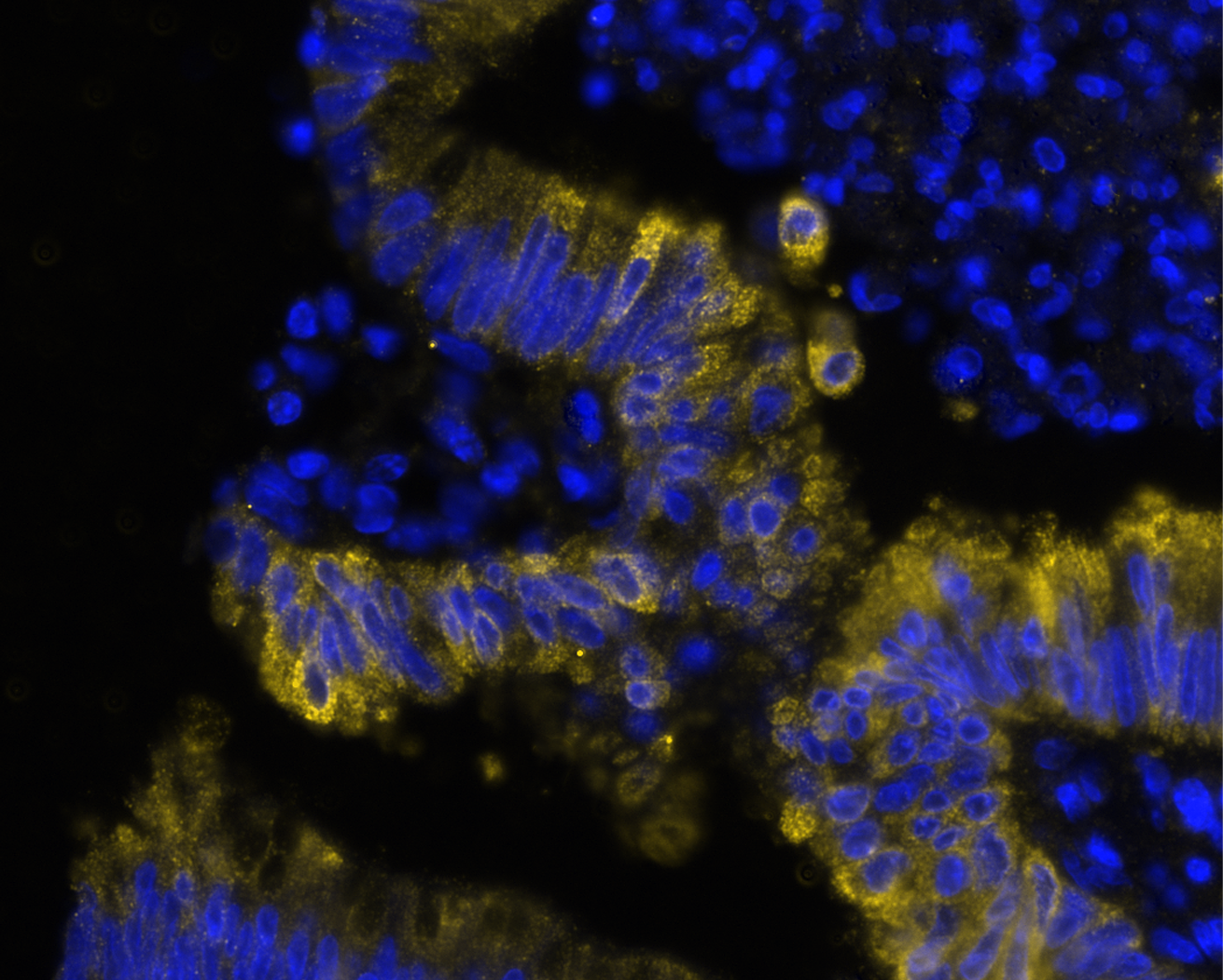
DR647MS) and counterstained with DAPI (blue; Lunaphore Catalog #
DR100). Specific staining was localized to the membrane. Protocol available in
COMET™ Panel Builder." class="big_thumb" />
DR647MS) and counterstained with DAPI (blue; Lunaphore Catalog #
DR100). Specific staining was localized to the membrane. Protocol available in
COMET™ Panel Builder." class="big_lightbox">
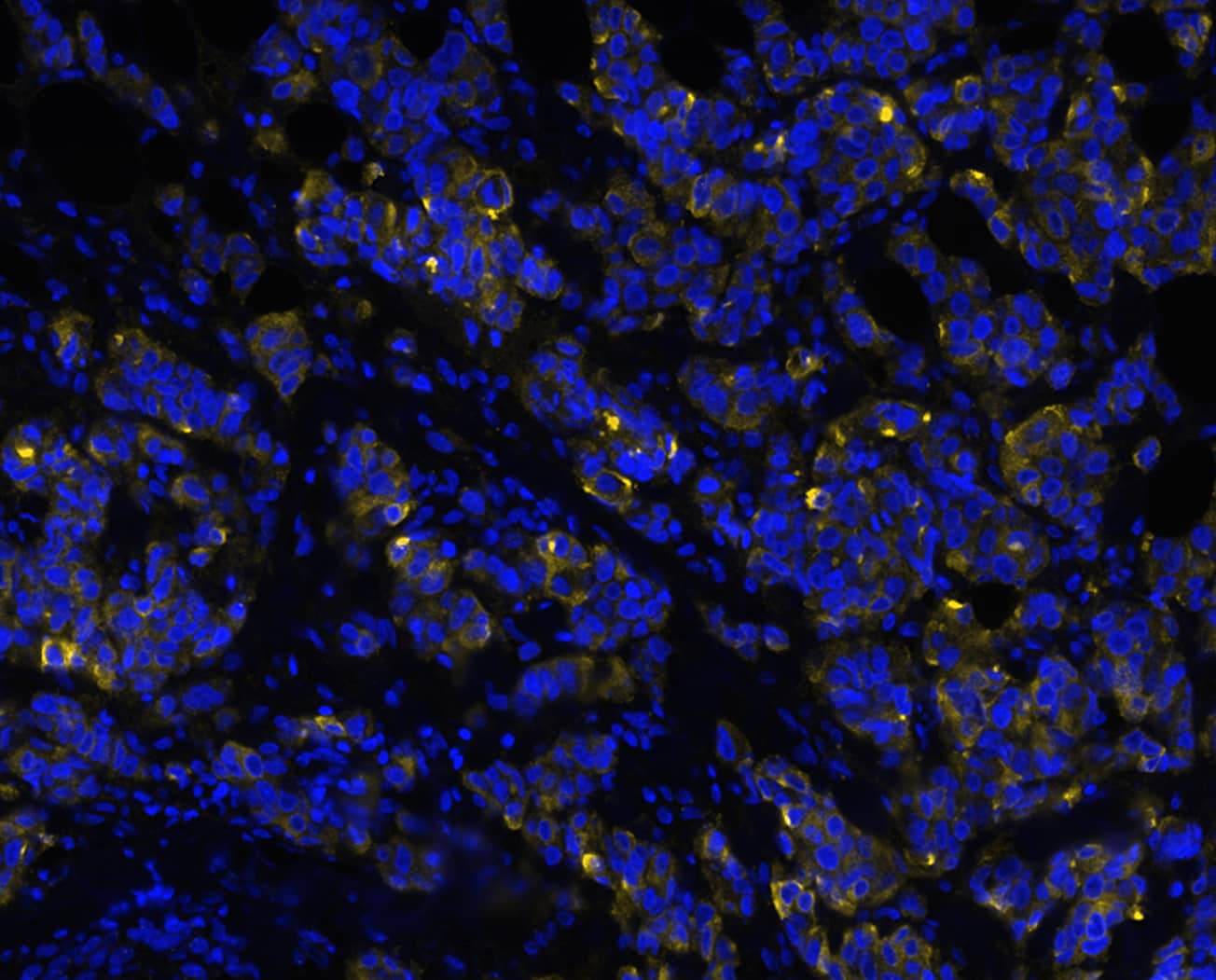
DR647MS) and counterstained with DAPI (blue; Lunaphore Catalog #
DR100). Specific staining was localized to the membrane. Protocol available in
COMET™ Panel Builder." class="big_thumb" />
HAF007). A specific band was detected for MUC-1 at approximately 300 kDa (as indicated). This experiment was conducted under non-reducing conditions and using
Immunoblot Buffer Group 1." class="big_lightbox">
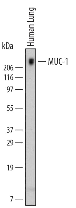
HAF007). A specific band was detected for MUC-1 at approximately 300 kDa (as indicated). This experiment was conducted under non-reducing conditions and using
Immunoblot Buffer Group 1." class="big_thumb" />
MAB0041, open histogram), followed by Allophycocyanin-conjugated Anti-Mouse IgG F(ab')
2Secondary Antibody (Catalog #
F0101B)." class="big_lightbox">
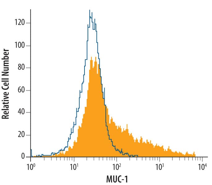
MAB0041, open histogram), followed by Allophycocyanin-conjugated Anti-Mouse IgG F(ab')
2Secondary Antibody (Catalog #
F0101B)." class="big_thumb" />
 DR647MS) and counterstained with DAPI (blue; Lunaphore Catalog # DR100). Specific staining was localized to the membrane. Protocol available in COMET™ Panel Builder." alt="MUC1 was detected in immersion fixed paraffin-embedded sections of human pancreas tumor using Mouse Anti-Human MUC1, Monoclonal Antibody (Catalog # MAB6298) at 4ug/mL at 27 ° Celsius for 2 minutes. Before incubation with the primary antibody, tissue underwent an all-in-one dewaxing and antigen retrieval preprocessing using PreTreatment Module (PT Module) and Dewax and HIER Buffer H (pH 9; Epredia Catalog # TA-999-DHBH). Tissue was stained using the Alexa Fluor™ 647 Goat anti-Mouse IgG Secondary Antibody at 1:200 at 37 ° Celsius for 2 minutes. (Yellow; Lunaphore Catalog # DR647MS) and counterstained with DAPI (blue; Lunaphore Catalog # DR100). Specific staining was localized to the membrane. Protocol available in COMET™ Panel Builder." class="big_thumb" />
 HAF016). A specific band was detected for MUC-1 at approximately 230 kDa (as indicated). This experiment was conducted under reducing conditions and using Immunoblot Buffer Group 1." alt="Western blot shows lysates of human lung tissue. PVDF Membrane was probed with 1 µg/mL of Sheep Anti-Human MUC-1 Antigen Affinity-purified Polyclonal Antibody (Catalog # AF6298) followed by HRP-conjugated Anti-Sheep IgG Secondary Antibody (Catalog # HAF016). A specific band was detected for MUC-1 at approximately 230 kDa (as indicated). This experiment was conducted under reducing conditions and using Immunoblot Buffer Group 1." class="big_thumb" />


![Immunocytochemistry/Immunofluorescence: MUC-1 Antibody (SM3) [NB120-22711] - MCF7 cells were fixed for 10 minutes using 10% formalin and then permeabilized for 5 minutes using 1X PBS + 0.05% Triton-X100. The cells were incubated with anti-MUC1 [SM3] at 5 ug/ml overnight at 4C and detected with an anti-mouse IgG Dylight 488 (Green) at a 1:500 dilution. Nuclei were counterstained with DAPI (Blue). Cells were imaged using a 40X objective.](http://images.novusbio.com/fullsize/MUC-1-Antibody-SM3-Immunocytochemistry-Immunofluorescence-NB120-22711-img0004.jpg "Immunocytochemistry/Immunofluorescence: MUC-1 Antibody (SM3) [NB120-22711] - MCF7 cells were fixed for 10 minutes using 10% formalin and then permeabilized for 5 minutes using 1X PBS + 0.05% Triton-X100. The cells were incubated with anti-MUC1 [SM3] at 5 ug/ml overnight at 4C and detected with an anti-mouse IgG Dylight 488 (Green) at a 1:500 dilution. Nuclei were counterstained with DAPI (Blue). Cells were imaged using a 40X objective.")
![Immunohistochemistry-Paraffin: MUC-1 Antibody (SM3) [NB120-22711] - IHC analysis of formalin fixed paraffin embedded tissue section of human breast cancer xenograft using MUC-1 antibody clone SM3 at 1:10 dilution. The xenograft section depicted a very specific and intense signal in the periphery of the cancer cells. The necrotic cells also developed a strong immune-positivity while the tumor stroma as well as the nuclei of cells were negative for immunostaining.](http://images.novusbio.com/fullsize/MUC-1-Antibody-SM3-Immunohistochemistry-Paraffin-NB120-22711-img0003.jpg)
![Flow Cytometry: MUC1 Antibody (SM3) - BSA Free [NB120-22711] - An intracellular stain was performed on MCF7 cells with MUC1 Antibody (SM3) NB120-22711AF700 (blue) and a matched isotype control (orange). Cells were fixed with 4% PFA and then permeabilized with 0.1% saponin. Cells were incubated in an antibody dilution of 5 ug/mL for 30 minutes at room temperature. Both antibodies were conjugated to Alexa Fluor 700.](http://images.novusbio.com/fullsize/MUC1-Antibody-SM3-BSA-Free-Flow-Cytometry-NB120-22711-img0005.jpg)
![Immunocytochemistry/Immunofluorescence: MUC-1 Antibody (SM3) [NB120-22711] - MUC-1 antibody was tested in MCF7 cells at 10 ug/ml using a Dylight 488 conjugated secondary antibody (Green). Actin (Red) and DNA (Blue) were counterstained using Phalloidin 568 and DAPI.](http://images.novusbio.com/fullsize/MUC-1-Antibody-SM3-Immunocytochemistry-Immunofluorescence-NB120-22711-img0001.jpg)
![Immunohistochemistry-Paraffin: MUC-1 Antibody (SM3) [NB120-22711] - IHC analysis of formalin fixed paraffin embedded tissue section of human breast cancer xenograft using MUC-1 antibody clone SM3 at 1:10 dilution. The xenograft section depicted a very specific and intense signal in the periphery of the cancer cells. The necrotic cells also developed a strong diffused immune-positivity while the tumor stroma as well as the nuclei of cells were negative for immunostaining.](http://images.novusbio.com/fullsize/MUC-1-Antibody-SM3-Immunohistochemistry-Paraffin-NB120-22711-img0002.jpg)


![Western Blot: MUC-1 Antibody [NBP1-60046] - Total protein from mouse 3T3 cells and human HeLa cells, stomach and colon was separated on a 4-15% gel by SDS-PAGE, transferred to PVDF membrane and blocked in 5% non-fat milk in TBST. The membrane was probed with 2.0 ug/ml anti-MUC-1 in 1% non-fat milk in TBST and detected with an anti-rabbit HRP secondary antibody using chemiluminescence.](http://images.novusbio.com/fullsize/MUC-1-Antibody-Western-Blot-NBP1-60046-img0011.jpg)
![Immunocytochemistry/Immunofluorescence: MUC1 Antibody [NBP1-60046] - Tested in HeLa cells at a 1:50 dilution against DyLight 488 (Green). Cells were counterstained for alpha-tubulin against DyLight 550 (Red) and mounted in DAPI fluoromount (Blue).](http://images.novusbio.com/fullsize/MUC-1-Antibody-Immunocytochemistry-Immunofluorescence-NBP1-60046-img0010.jpg)
![Immunohistochemistry-Paraffin: MUC1 Antibody [NBP1-60046] - Human kidney lysate tissue at an antibody concentration of 4-8ug/ml.](http://images.novusbio.com/fullsize/MUC1-Antibody-Immunohistochemistry-Paraffin-NBP1-60046-img0003.jpg)
![Immunohistochemistry-Paraffin: MUC1 Antibody [NBP1-60046] - Pig stomach.](http://images.novusbio.com/fullsize/MUC1-Antibody-Immunohistochemistry-Paraffin-NBP1-60046-img0007.jpg)
![Immunohistochemistry-Paraffin: MUC1 Antibody [NBP1-60046] - Pig stomach.](http://images.novusbio.com/fullsize/MUC1-Antibody-Immunohistochemistry-Paraffin-NBP1-60046-img0008.jpg)
![Immunohistochemistry-Paraffin: MUC1 Antibody [NBP1-60046] - Human stomach.](http://images.novusbio.com/fullsize/MUC1-Antibody-Immunohistochemistry-Paraffin-NBP1-60046-img0009.jpg)
![Immunocytochemistry/ Immunofluorescence: MUC1 Antibody - BSA Free [NBP1-60046] - Dexamethasone induces Muc1-CT nuclear translocation & co-localization with GR alpha in bronchial epithelium of WT mice but not in MUC1 KO mice. C57BL/6 Muc1 KO mice & WT mice were undergoing dexamethasone 10 mg/kg/day (orally) for 6 days. Animals were sacrificed at day 6 & lungs were fixed in paraformaldehyde (4%) for 48 h & embedded in Tissue-Tek® OCT™ cryosectioning compound. Lung slices were immunostained with MUC1-cytoplasmic tail (CT) & glucocorticoid receptor alpha (GR alpha ) antibodies with rhodamine/fluorescein secondary antibodies. DAPI was used to mark cell nucleus. Representative con-focal images are showed. Scale bar: 5 μm Image collected & cropped by CiteAb from the following publication (//pubmed.ncbi.nlm.nih.gov/30458870), licensed under a CC-BY license. Not internally tested by Novus Biologicals.](http://images.novusbio.com/fullsize/nbp1-60046_rabbit-polyclonal-muc1-antibody-310202415363741.jpg)
![Chromatin Immunoprecipitation: MUC1 Antibody - BSA Free [NBP1-60046] - MUC1 binds RIPK1 to mediate the RIPK1/RIPK3 pathway. (a,b) MUC1 knockdown by MUC1‐siRNA or NC‐siRNA were assayed in 16HBE cells following stimulation with TNF‐ alpha (300 ng/ml). After 24 hr of stimulation, RIPK3 & RIPK1 protein levels were measured by western blot analysis. (c,d) Normal 16HBE cells were treated with TNF‐ alpha for 24 hr, & the interaction between MUC1‐CT & RIPK1/RIPK3 were confirmed by immunoprecipitation. Data are expressed as means ± SD & were analyzed by one‐way analysis of variance, *p < 0.05. NC group (transfected with NC siRNA); MUC1‐siRNA group (transfected with MUC1‐siRNA). 16HBE: human bronchial epithelial; IB: immunoblotting; IP: immunoprecipitation; RIPK1: receptor‐interacting protein kinase‐1; RIPK3: receptor‐interacting protein kinase‐3; NC: negative control; siRNA: small interfering RNA; TNF‐ alpha : tumor necrosis factor‐ alpha Image collected & cropped by CiteAb from the following publication (//pubmed.ncbi.nlm.nih.gov/30666647), licensed under a CC-BY license. Not internally tested by Novus Biologicals.](http://images.novusbio.com/fullsize/nbp1-60046_rabbit-polyclonal-muc1-antibody-310202416161843.jpg)
![Western Blot: MUC1 Antibody - BSA Free [NBP1-60046] - MUC1 downregulation & TLR4 overexpression in lung tissue from heavy smokers & COPD patients. Lung tissue from healthy (n = 10), smokers (n = 11) & COPD patients (n = 13) were analysed. MUC1 mRNA gene expression in lung tissue homogenates (a), bronchial epithelial cells (b) & sputum neutrophils (c). d MUC1-CT protein expression in lung homogenates.TLR4 mRNA gene expression in lung tissue homogenates (e), bronchial epithelial cells (f) & sputum neutrophils (g). h MUC1-CT protein expression in lung homogenates. i Correlation of TL4 & MUC1 gene expression in lung tissue from smokers & COPD patients. j Correlation of FEV1% & MUC1 gene expression in lung tissue from smokers & COPD patients. Gene expression was analyzed by real time PCR using the 2-delta Ct as described in methods. Protein expression was analyzed by western blot. Representative western blot are showed. Data are presented as median with interquartile range. “P” exact values were obtained following Kruskal Wallis test. FEV1%: forced expiratory volume in 1 s Image collected & cropped by CiteAb from the following publication (//pubmed.ncbi.nlm.nih.gov/30458870), licensed under a CC-BY license. Not internally tested by Novus Biologicals.](http://images.novusbio.com/fullsize/nbp1-60046_rabbit-polyclonal-muc1-antibody-310202416165614.jpg)
![Chromatin Immunoprecipitation: MUC1 Antibody - BSA Free [NBP1-60046] - MUC1 binds RIPK1 to mediate the RIPK1/RIPK3 pathway. (a,b) MUC1 knockdown by MUC1‐siRNA or NC‐siRNA were assayed in 16HBE cells following stimulation with TNF‐ alpha (300 ng/ml). After 24 hr of stimulation, RIPK3 & RIPK1 protein levels were measured by western blot analysis. (c,d) Normal 16HBE cells were treated with TNF‐ alpha for 24 hr, & the interaction between MUC1‐CT & RIPK1/RIPK3 were confirmed by immunoprecipitation. Data are expressed as means ± SD & were analyzed by one‐way analysis of variance, *p < 0.05. NC group (transfected with NC siRNA); MUC1‐siRNA group (transfected with MUC1‐siRNA). 16HBE: human bronchial epithelial; IB: immunoblotting; IP: immunoprecipitation; RIPK1: receptor‐interacting protein kinase‐1; RIPK3: receptor‐interacting protein kinase‐3; NC: negative control; siRNA: small interfering RNA; TNF‐ alpha : tumor necrosis factor‐ alpha Image collected & cropped by CiteAb from the following publication (//pubmed.ncbi.nlm.nih.gov/30666647), licensed under a CC-BY license. Not internally tested by Novus Biologicals.](http://images.novusbio.com/fullsize/nbp1-60046_rabbit-polyclonal-muc1-antibody-310202416175235.jpg)
 DR647MS) and counterstained with DAPI (blue; Lunaphore Catalog #
  DR647MS) and counterstained with DAPI (blue; Lunaphore Catalog #
  HAF007). A specific band was detected for MUC-1 at approximately 300 kDa (as indicated). This experiment was conducted under non-reducing conditions and using
  MAB0041, open histogram), followed by Allophycocyanin-conjugated Anti-Mouse IgG F(ab')2Secondary Antibody (Catalog #
  NL007) and counterstained with DAPI (blue). Specific staining was localized to cell surfaces and cytoplasm. View our protocol for
  CTS013). Tissue was stained using the Anti-Mouse HRP-DAB Cell & Tissue Staining Kit (brown; Catalog #

![Western Blot: MUC1 Antibody (MUC1/955) [NBP2-44658] - Western Blot Analysis of human MCF-7 cell lysate using MUC1 antibody (MUC1/955).](http://images.novusbio.com/fullsize/MUC1-Antibody-MUC1-955-Western-Blot-NBP2-44658-img0005.jpg "Western Blot: MUC1 Antibody (MUC1/955) [NBP2-44658] - Western Blot Analysis of human MCF-7 cell lysate using MUC1 antibody (MUC1/955).")
![Immunohistochemistry-Paraffin: MUC1 Antibody (MUC1/955) [NBP2-44658] - Human Endometrial Carcinoma stained with EMA Monoclonal Antibody (MUC1/955).](http://images.novusbio.com/fullsize/MUC1-Antibody-MUC1-955-Immunohistochemistry-Paraffin-NBP2-44658-img0002.jpg)
![Western Blot: MUC1 Antibody (MUC1/955) [NBP2-44658] - Western Blot Analysis of human T47D cell lysate using MUC1 Antibody (MUC1/955).](http://images.novusbio.com/fullsize/MUC1-Antibody-MUC1-955-Western-Blot-NBP2-44658-img0004.jpg)
![Immunohistochemistry-Paraffin: MUC1 Antibody (MUC1/955) [NBP2-44658] - Human Breast Carcinoma stained with EMA Monoclonal Antibody (MUC1/955).](http://images.novusbio.com/fullsize/MUC1-Antibody-MUC1-955-Immunohistochemistry-Paraffin-NBP2-44658-img0001.jpg)
![Immunohistochemistry-Paraffin: MUC1 Antibody (MUC1/955) [NBP2-44658] - Human Lung Carcinoma stained with EMA Monoclonal Antibody (MUC1/955).](http://images.novusbio.com/fullsize/MUC1-Antibody-MUC1-955-Immunohistochemistry-Paraffin-NBP2-44658-img0003.jpg)
![Western Blot: MUC1 Antibody (139H2) [NBP2-44666] - Western Blot Analysis of human MCF-7 cell lysate using MUC1 antibody (139H2).](http://images.novusbio.com/fullsize/MUC1-Antibody-139H2-Western-Blot-NBP2-44666-img0005.jpg "Western Blot: MUC1 Antibody (139H2) [NBP2-44666] - Western Blot Analysis of human MCF-7 cell lysate using MUC1 antibody (139H2).")
![Immunohistochemistry-Paraffin: MUC1 Antibody (139H2) [NBP2-44666] - Human Endometrial Carcinoma stained with EMA Monoclonal Antibody (139H2).](http://images.novusbio.com/fullsize/MUC1-Antibody-139H2-Immunohistochemistry-Paraffin-NBP2-44666-img0002.jpg)
![Western Blot: MUC1 Antibody (139H2) [NBP2-44666] - Western Blot Analysis of human T47D cell lysate using MUC1 Antibody (139H2).](http://images.novusbio.com/fullsize/MUC1-Antibody-139H2-Western-Blot-NBP2-44666-img0004.jpg)
![Immunohistochemistry-Paraffin: MUC1 Antibody (139H2) [NBP2-44666] - Human Breast Cancer stained with EMA Monoclonal Antibody (139H2).](http://images.novusbio.com/fullsize/MUC1-Antibody-139H2-Immunohistochemistry-Paraffin-NBP2-44666-img0001.jpg)
![Immunohistochemistry-Paraffin: MUC1 Antibody (139H2) [NBP2-44666] - Human Lung Cancer stained with EMA Monoclonal Antibody (139H2).](http://images.novusbio.com/fullsize/MUC1-Antibody-139H2-Immunohistochemistry-Paraffin-NBP2-44666-img0003.jpg)
![Western Blot: MUC1 Antibody (GP1.4) [NBP2-29408] - Western Blot Analysis of MCF-7 cell lysate using MUC1 Mouse Monoclonal Antibody (GP1.4).](http://images.novusbio.com/fullsize/MUC1-Antibody-GP1-4-Western-Blot-NBP2-29408-img0005.jpg "Western Blot: MUC1 Antibody (GP1.4) [NBP2-29408] - Western Blot Analysis of MCF-7 cell lysate using MUC1 Mouse Monoclonal Antibody (GP1.4).")
![Immunohistochemistry-Paraffin: MUC1 Antibody (GP1.4) [NBP2-29408] - Formalin-paraffin human breast cancer stained with EMA Ab (GP1.4). Note cytoplasmic and membrane staining.](http://images.novusbio.com/fullsize/MUC1-Antibody-GP1-4-Immunohistochemistry-Paraffin-NBP2-29408-img0002.jpg)
![Western Blot: MUC1 Antibody (SN06-80) [NBP2-67650] - Western blot analysis of MUC1 on different lysates. Proteins were transferred to a PVDF membrane and blocked with 5% BSA in PBS for 1 hour at room temperature. The primary antibody (1/500) was used in 5% BSA at room temperature for 2 hours. Goat Anti-Rabb](http://images.novusbio.com/fullsize/MUC1-Antibody-SN06-80-Western-Blot-NBP2-67650-img0009.jpg "Western Blot: MUC1 Antibody (SN06-80) [NBP2-67650] - Western blot analysis of MUC1 on different lysates. Proteins were transferred to a PVDF membrane and blocked with 5% BSA in PBS for 1 hour at room temperature. The primary antibody (1/500) was used in 5% BSA at room temperature for 2 hours. Goat Anti-Rabb")
![Immunocytochemistry/Immunofluorescence: MUC1 Antibody (SN06-80) [NBP2-67650] - Staining MUC1 in B16-F1 cells (green). The nuclear counter stain is DAPI (blue). Cells were fixed in paraformaldehyde, permeabilised with 0.25% Triton X100/PBS.](http://images.novusbio.com/fullsize/MUC1-Antibody-SN06-80-Immunocytochemistry-Immunofluorescence-NBP2-67650-img0007.jpg)
![<b>Biological Strategies Validation. </b>Immunohistochemistry-Paraffin: MUC1 Antibody (SN06-80) [NBP2-67650] - Immunohistochemical analysis of paraffin-embedded human kidney tissue using anti-MUC1 antibody. The section was pre-treated using heat mediated antigen retrieval with Tris-EDTA buffer (pH 8.0-8.4) for 20 minutes. The tissues were blocked in 5% BSA for 30 minutes at room temperature, washed with ddH2O and PBS, and then probed with the primary antibody (1/50) for 30 minutes at room temperature. The detection was performed using an HRP conjugated compact polymer system. DAB was used as the chromogen. Tissues were counterstained with hematoxylin and mounted with DPX.](http://images.novusbio.com/fullsize/MUC1-Antibody-SN06-80-Immunohistochemistry-Paraffin-NBP2-67650-img0008.jpg)
![Flow Cytometry: MUC1 Antibody (SN06-80) [NBP2-67650] - Analysis of MCF-7 cells with MUC1 antibody at 1/50 dilution (red) compared with an unlabelled control (cells without incubation with primary antibody; black). Alexa Fluor 488-conjugated goat anti rabbit IgG was used as the secondary antibody.](http://images.novusbio.com/fullsize/MUC1-Antibody-SN06-80-Flow-Cytometry-NBP2-67650-img0001.jpg)
![Western Blot: MUC1 Antibody (SN06-80) [NBP2-67650] - Analysis of MUC1 on Hela cells lysates using anti-MUC1 antibody at 1/1,000 dilution.](http://images.novusbio.com/fullsize/MUC1-Antibody-SN06-80-Western-Blot-NBP2-67650-img0006.jpg)
![Immunocytochemistry/Immunofluorescence: MUC1 Antibody (SN06-80) [NBP2-67650] - Staining MUC1 in Hela cells (green). The nuclear counter stain is DAPI (blue). Cells were fixed in paraformaldehyde, permeabilised with 0.25% Triton X100/PBS.](http://images.novusbio.com/fullsize/MUC1-Antibody-SN06-80-Immunocytochemistry-Immunofluorescence-NBP2-67650-img0002.jpg)
![Immunohistochemistry-Paraffin: MUC1 Antibody (SN06-80) [NBP2-67650] - Analysis of paraffin-embedded human breast carcinoma tissue using anti-MUC1 antibody. Counter stained with hematoxylin.](http://images.novusbio.com/fullsize/MUC1-Antibody-SN06-80-Immunohistochemistry-Paraffin-NBP2-67650-img0003.jpg)
![Immunohistochemistry-Paraffin: MUC1 Antibody (SN06-80) [NBP2-67650] - Analysis of paraffin-embedded human breast tissue using anti-MUC1 antibody. Counter stained with hematoxylin.](http://images.novusbio.com/fullsize/MUC1-Antibody-SN06-80-Immunohistochemistry-Paraffin-NBP2-67650-img0004.jpg)
![Immunohistochemistry-Paraffin: MUC1 Antibody (SN06-80) [NBP2-67650] - Analysis of paraffin-embedded human kidney tissue using anti-MUC1 antibody. Counter stained with hematoxylin.](http://images.novusbio.com/fullsize/MUC1-Antibody-SN06-80-Immunohistochemistry-Paraffin-NBP2-67650-img0005.jpg)
![Western blot analysis on different lysates with NBP2-67650 at 1/2,000 dilution and competitor's antibody at 1/1,000 dilution. Lane 1: HeLa cell lysate (20 µg/Lane) Lane 2: HCT 116 cell lysate (negative) (20 µg/Lane) Lane 3: T-47D cell lysate (20 µg/Lane) Lane 4: Mouse lung tissue lysate (40 µg/Lane) Lane 5: Rat lung tissue lysate (40 µg/Lane) Predicted band size: 122 kDa Observed band size: 17~24 kDa Exposure time: 3 minutes 10 seconds; 4-20% SDS-PAGE gel. Proteins were transferred to a PVDF membrane and blocked with 5% NFDM/TBST for 1 hour at room temperature. NBP2-67650 at 1/2,000 dilution and competitor's antibody at 1/1,000 dilution were used in 5% NFDM/TBST at 4℃ overnight. Goat Anti-Rabbit IgG [HRP] Secondary Antibody at 1/50,000 dilution was used for 1 hour at room temperature.](http://images.novusbio.com/fullsize/nbp2-67650_-western-blot-639204586677213359.jpg)
![Recombinant Human MUC-1 Protein [H00004582-Q01] - 12.5% SDS-PAGE Stained with Coomassie Blue.](http://images.novusbio.com/fullsize/Recombinant-Human-MUC-1-Protein-H00004582-Q01-img0001.jpg "Recombinant Human MUC-1 Protein [H00004582-Q01] - 12.5% SDS-PAGE Stained with Coomassie Blue.")
![Western Blot: MUC-1 Antibody (2E3) [NBP2-45838] - Analysis of extracts (35ug) from 9 different cell lines by using MUC1 monoclonal antibody (HepG2: human; HeLa: human; SVT2: mouse; A549: human; COS7: monkey; Jurkat: human; MDCK: canine; PC12: rat; MCF7: human).](http://images.novusbio.com/fullsize/MUC-1-Antibody-2E3-Western-Blot-NBP2-45838-img0010.jpg "Western Blot: MUC-1 Antibody (2E3) [NBP2-45838] - Analysis of extracts (35ug) from 9 different cell lines by using MUC1 monoclonal antibody (HepG2: human; HeLa: human; SVT2: mouse; A549: human; COS7: monkey; Jurkat: human; MDCK: canine; PC12: rat; MCF7: human).")
![Immunohistochemistry: MUC-1 Antibody (2E3) [NBP2-45838] - Analysis of Human colon tissue. (Heat-induced epitope retrieval by 10mM citric buffer, pH6.0, 120C for 3min)](http://images.novusbio.com/fullsize/MUC-1-Antibody-2E3-Immunohistochemistry-NBP2-45838-img0009.jpg)
![Western Blot: MUC-1 Antibody (2E3) [NBP2-45838] - Analysis of HEK293T cells were transfected with the pCMV6-ENTRY control (Left lane) or pCMV6-ENTRY MUC-1.](http://images.novusbio.com/fullsize/MUC-1-Antibody-2E3-Western-Blot-NBP2-45838-img0011.jpg)
![Immunohistochemistry: MUC-1 Antibody (2E3) [NBP2-45838] - Analysis of Carcinoma of Human liver tissue.(Heat-induced epitope retrieval by 10mM citric buffer, pH6.0, 120C for 3min)](http://images.novusbio.com/fullsize/MUC-1-Antibody-2E3-Immunohistochemistry-NBP2-45838-img0001.jpg)
![Immunohistochemistry: MUC-1 Antibody (2E3) [NBP2-45838] - Analysis of Human prostate tissue. (Heat-induced epitope retrieval by 10mM citric buffer, pH6.0, 120C for 3min)](http://images.novusbio.com/fullsize/MUC-1-Antibody-2E3-Immunohistochemistry-NBP2-45838-img0002.jpg)
![Immunohistochemistry: MUC-1 Antibody (2E3) [NBP2-45838] - Analysis of Carcinoma of Human thyroid tissue. (Heat-induced epitope retrieval by 10mM citric buffer, pH6.0, 120C for 3min)](http://images.novusbio.com/fullsize/MUC-1-Antibody-2E3-Immunohistochemistry-NBP2-45838-img0003.jpg)
![Immunohistochemistry: MUC-1 Antibody (2E3) [NBP2-45838] - Analysis of Carcinoma of Human lung tissue.(Heat-induced epitope retrieval by 10mM citric buffer, pH6.0, 120C for 3min)](http://images.novusbio.com/fullsize/MUC-1-Antibody-2E3-Immunohistochemistry-NBP2-45838-img0004.jpg)
![Immunohistochemistry: MUC-1 Antibody (2E3) [NBP2-45838] - Analysis of Adenocarcinoma of Human colon tissue. (Heat-induced epitope retrieval by 10mM citric buffer, pH6.0, 120C for 3min)](http://images.novusbio.com/fullsize/MUC-1-Antibody-2E3-Immunohistochemistry-NBP2-45838-img0005.jpg)
![Immunohistochemistry: MUC-1 Antibody (2E3) [NBP2-45838] - Analysis of Human breast tissue. (Heat-induced epitope retrieval by 10mM citric buffer, pH6.0, 120C for 3min)](http://images.novusbio.com/fullsize/MUC-1-Antibody-2E3-Immunohistochemistry-NBP2-45838-img0006.jpg)
![Immunohistochemistry: MUC-1 Antibody (2E3) [NBP2-45838] - Analysis of Human Kidney tissue. (Heat-induced epitope retrieval by 10mM citric buffer, pH6.0, 120C for 3min)](http://images.novusbio.com/fullsize/MUC-1-Antibody-2E3-Immunohistochemistry-NBP2-45838-img0007.jpg)
![Immunohistochemistry: MUC-1 Antibody (2E3) [NBP2-45838] - Analysis of Adenocarcinoma of Human ovary tissue. (Heat-induced epitope retrieval by 10mM citric buffer, pH6.0, 120C for 3min)](http://images.novusbio.com/fullsize/MUC-1-Antibody-2E3-Immunohistochemistry-NBP2-45838-img0008.jpg)
![ELISA: Human MUC1 ELISA Kit (Colorimetric) [NBP2-76676] - Samples were spiked with high concentrations of Human MUC1 and diluted with Reference Standard & Sample Diluent to produce samples with values within the range of the assay.](http://images.novusbio.com/fullsize/nbp2-76676_human-muc1-elisa-kit-colorimetric-132202416215336.jpg "ELISA: Human MUC1 ELISA Kit (Colorimetric) [NBP2-76676] - Samples were spiked with high concentrations of Human MUC1 and diluted with Reference Standard & Sample Diluent to produce samples with values within the range of the assay.")
![ELISA: Human MUC1 ELISA Kit (Colorimetric) [NBP2-76676] - Standard Curve Reference](http://images.novusbio.com/fullsize/nbp2-76676_human-muc1-elisa-kit-colorimetric-4320249192880.jpg)
 CTS019) and counterstained with hematoxylin (blue). Specific staining was localized to nuclei and cytoplasm in epithelial cells. View our protocol for


