DR647RB) and counterstained with DAPI (blue; Lunaphore Catalog #
DR100). Specific staining was localized to the cytoplasm. Protocol available in
COMET™ Panel Builder." class="big_lightbox">
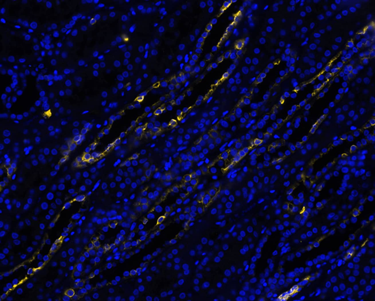
DR647RB) and counterstained with DAPI (blue; Lunaphore Catalog #
DR100). Specific staining was localized to the cytoplasm. Protocol available in
COMET™ Panel Builder." alt="Cytokeratin 7 was detected in immersion fixed paraffin-embedded sections of human kidney using Rabbit Anti-Human KRT7, Monoclonal Antibody (Catalog# MAB11701) at 1ug/mL at 37 ° Celsius for 4 minutes. Before incubation with the primary antibody, tissue underwent an all-in-one dewaxing and antigen retrieval preprocessing using PreTreatment Module (PT Module)and Dewax and HIER Buffer H (pH 9; Epredia Catalog #TA-999-DHBH). Tissue was stained using the Alexa Fluor™ Plus 647 Goat anti-Rabbit IgG Secondary Antibody at 1:200 at 37 ° Celsius for 2 minutes. (Yellow; Lunaphore Catalog #
DR647RB) and counterstained with DAPI (blue; Lunaphore Catalog #
DR100). Specific staining was localized to the cytoplasm. Protocol available in
COMET™ Panel Builder." class="big_thumb" />
DR647RB) and counterstained with DAPI (blue; Lunaphore Catalog #
DR100). Specific staining was localized to the cytoplasm. Protocol available in
COMET™ Panel Builder." class="big_lightbox">
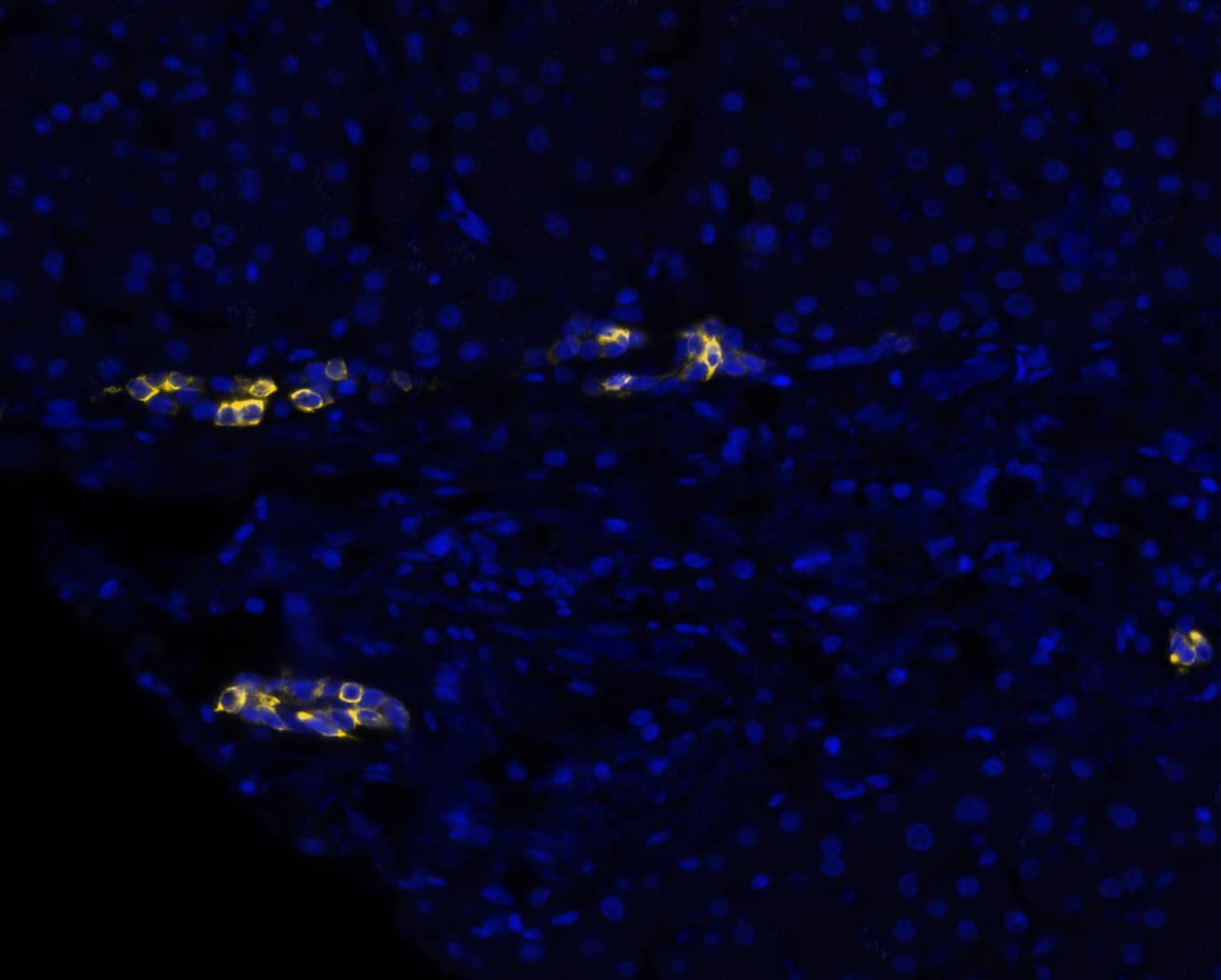
DR647RB) and counterstained with DAPI (blue; Lunaphore Catalog #
DR100). Specific staining was localized to the cytoplasm. Protocol available in
COMET™ Panel Builder." class="big_thumb" />
DR647RB) and counterstained with DAPI (blue; Lunaphore Catalog #
DR100). Specific staining was localized to the cytoplasm. Protocol available in
COMET™ Panel Builder." class="big_lightbox">
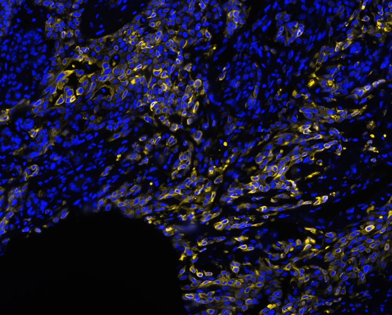
DR647RB) and counterstained with DAPI (blue; Lunaphore Catalog #
DR100). Specific staining was localized to the cytoplasm. Protocol available in
COMET™ Panel Builder." class="big_thumb" />
VC003) or the HRP-conjugated Anti-Rabbit IgG Secondary Antibody (Catalog #
HAF008). Before incubation with the primary antibody, tissue was subjected to heat-induced epitope retrieval using VisUCyte Antigen Retrieval Reagent-Basic (Catalog #
VCTS021). Tissue was stained using DAB (brown) and counterstained with hematoxylin (blue). Specific staining was localized to the cytoplasm. View our protocol for
IHC Staining with VisUCyte HRP Polymer Detection Reagents." class="big_lightbox">
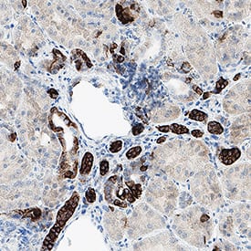
VC003) or the HRP-conjugated Anti-Rabbit IgG Secondary Antibody (Catalog #
HAF008). Before incubation with the primary antibody, tissue was subjected to heat-induced epitope retrieval using VisUCyte Antigen Retrieval Reagent-Basic (Catalog #
VCTS021). Tissue was stained using DAB (brown) and counterstained with hematoxylin (blue). Specific staining was localized to the cytoplasm. View our protocol for
IHC Staining with VisUCyte HRP Polymer Detection Reagents." class="big_thumb" />
VC003) or the HRP-conjugated Anti-Rabbit IgG Secondary Antibody (Catalog #
HAF008). Before incubation with the primary antibody, tissue was subjected to heat-induced epitope retrieval using VisUCyte Antigen Retrieval Reagent-Basic (Catalog #
VCTS021). Tissue was stained using DAB (brown) and counterstained with hematoxylin (blue). Specific staining was localized to the cytoplasm. View our protocol for
IHC Staining with VisUCyte HRP Polymer Detection Reagents." class="big_lightbox">

VC003) or the HRP-conjugated Anti-Rabbit IgG Secondary Antibody (Catalog #
HAF008). Before incubation with the primary antibody, tissue was subjected to heat-induced epitope retrieval using VisUCyte Antigen Retrieval Reagent-Basic (Catalog #
VCTS021). Tissue was stained using DAB (brown) and counterstained with hematoxylin (blue). Specific staining was localized to the cytoplasm. View our protocol for
IHC Staining with VisUCyte HRP Polymer Detection Reagents." class="big_thumb" />
HAF008). A specific band was detected for Cytokeratin 7 at approximately 52 kDa (as indicated). This experiment was conducted under reducing conditions and using
Western Blot Buffer Group 1." class="big_lightbox">

HAF008). A specific band was detected for Cytokeratin 7 at approximately 52 kDa (as indicated). This experiment was conducted under reducing conditions and using
Western Blot Buffer Group 1." class="big_thumb" />
042-206). This experiment was conducted under reducing conditions and using the 12-230kDa separation system." class="big_lightbox">

042-206). This experiment was conducted under reducing conditions and using the 12-230kDa separation system." class="big_thumb" />
 DR647RB) and counterstained with DAPI (blue; Lunaphore Catalog # DR100). Specific staining was localized to the cytoplasm. Protocol available in COMET™ Panel Builder." alt="Cytokeratin 7 was detected in immersion fixed paraffin-embedded sections of human kidney using Rabbit Anti-Human KRT7, Monoclonal Antibody (Catalog# MAB11701) at 1ug/mL at 37 ° Celsius for 4 minutes. Before incubation with the primary antibody, tissue underwent an all-in-one dewaxing and antigen retrieval preprocessing using PreTreatment Module (PT Module)and Dewax and HIER Buffer H (pH 9; Epredia Catalog #TA-999-DHBH). Tissue was stained using the Alexa Fluor™ Plus 647 Goat anti-Rabbit IgG Secondary Antibody at 1:200 at 37 ° Celsius for 2 minutes. (Yellow; Lunaphore Catalog # DR647RB) and counterstained with DAPI (blue; Lunaphore Catalog # DR100). Specific staining was localized to the cytoplasm. Protocol available in COMET™ Panel Builder." class="big_thumb" />
 DR647MS) and counterstained with DAPI (blue; Lunaphore Catalog # DR100). Specific staining was localized to the cytoplasm. Protocol available in COMET™ Panel Builder." alt="Cytokeratin 7 Peptide was detected in immersion fixed paraffin-embedded sections of human Pancreas Tumor using Mouse Anti-Human KRT7, Monoclonal Antibody (Catalog #MAB11626) at 25ug/mL at 37 ° Celsius for 4 minutes. Before incubation with the primary antibody, tissue underwent an all-in-one dewaxing and antigen retrieval preprocessing using PreTreatment Module (PT Module) and Dewax and HIER Buffer H (pH 9; Epredia Catalog # TA-999-DHBH). Tissue was stained using the Alexa Fluor™ 647 Goat anti-Mouse IgG Secondary Antibody at 1:200 at 37 ° Celsius for 2 minutes. (Yellow; Lunaphore Catalog # DR647MS) and counterstained with DAPI (blue; Lunaphore Catalog # DR100). Specific staining was localized to the cytoplasm. Protocol available in COMET™ Panel Builder." class="big_thumb" />


![Western Blot: Cytokeratin 7 Antibody (OV-TL12/30) [NBP2-44814] - Western Blot Analysis of human HeLa cell lysate using Cytokeratin 7 Antibody (OV-TL12/30).](http://images.novusbio.com/fullsize/Cytokeratin-7-Antibody-OV-TL12-30-Western-Blot-NBP2-44814-img0008.jpg "Western Blot: Cytokeratin 7 Antibody (OV-TL12/30) [NBP2-44814] - Western Blot Analysis of human HeLa cell lysate using Cytokeratin 7 Antibody (OV-TL12/30).")
![Immunocytochemistry/Immunofluorescence: Cytokeratin 7 Antibody (OV-TL12/30) [NBP2-44814] - Immunofluorescent staining of methanol-fixed HeLa cells. Cytokeratin 7 Antibody (OV-TL12/30) followed by goat anti-Mouse IgG-CF488 (Green). The nuclear counterstain is Red Dot.](http://images.novusbio.com/fullsize/Cytokeratin-7-Antibody-OV-TL12-30-Immunocytochemistry-Immunofluorescence-NBP2-44814-img0007.jpg)
![Immunohistochemistry-Paraffin: Cytokeratin 7 Antibody (OV-TL12/30) [NBP2-44814] - Paraffin-embedded human salivary gland tissue. Image from verified customer review. Image using the Azide and BSA Free format of this antibody.](http://images.novusbio.com/fullsize/Cytokeratin-7-Antibody-OV-TL12-30-Immunohistochemistry-Paraffin-NBP2-44814-img0005.jpg)
![Flow Cytometry: Cytokeratin 7 Antibody (OV-TL12/30) [NBP2-44814] - Flow Cytometric Analysis of HeLa cells using Cytokeratin 7 Antibody (OV-TL12/30) followed by Goat anti-Mouse IgG-CF488 (Blue); Isotype Control (Red).](http://images.novusbio.com/fullsize/Cytokeratin-7-Antibody-OV-TL12-30-Flow-Cytometry-NBP2-44814-img0006.jpg)
![Immunohistochemistry-Paraffin: Cytokeratin 7 Antibody (OV-TL12/30) [NBP2-44814] - Human Lung SCC stained with Cytokeratin 7 Monoclonal Antibody (OV-TL12/30)](http://images.novusbio.com/fullsize/Cytokeratin-7-Antibody-OV-TL12-30-Immunohistochemistry-Paraffin-NBP2-44814-img0001.jpg)
![Immunohistochemistry-Paraffin: Cytokeratin 7 Antibody (OV-TL12/30) [NBP2-44814] - Human Ovarian Carcinoma stained with Cytokeratin 7 Monoclonal Antibody (OV-TL12/30)](http://images.novusbio.com/fullsize/Cytokeratin-7-Antibody-OV-TL12-30-Immunohistochemistry-Paraffin-NBP2-44814-img0002.jpg)
![Simple Western: Cytokeratin 7 Antibody (OV-TL12/30) [NBP2-44814] - Simple Western lane view shows a specific band for Cytokeratin 7 in 0.1 mg/ml of HeLa lysate(s). This experiment was performed under reducing conditions using the 12-230 kDa separation system.](http://images.novusbio.com/fullsize/Cytokeratin-7-Antibody-OV-TL12-30-Simple-Western-NBP2-44814-img0003.jpg)
![Simple Western: Cytokeratin 7 Antibody (OV-TL12/30) [NBP2-44814] - Electropherogram image of the corresponding Simple Western lane. Cytokeratin 7 antibody was used at 1 ug/ml dilution of HeLa lysates(s) respectively.](http://images.novusbio.com/fullsize/Cytokeratin-7-Antibody-OV-TL12-30-Simple-Western-NBP2-44814-img0004.jpg)
![Immunohistochemistry-Paraffin: Cytokeratin 7 Antibody (OV-TL12/30) [NBP2-44814] - Formalin-fixed, paraffin-embedded human prsotatecarcinoma stained with Biotin-conjugated Cytokeratin 7 Mouse Monoclonal Antibody (OV-TL12/30).](http://images.novusbio.com/fullsize/nbp2-44814_mouse-monoclonal-cytokeratin-7-antibody-ov-tl12-30-17720249362618.jpg)
![Simple Western: Cytokeratin 7 Antibody (OV-TL12/30) [NBP2-44814] - Lane view shows a specific band for Cytokeratin 7 at 5 ug/mL in HeLa cell lysate. Electropherogram image of corresponding Simple Western lane view at WES molecular weight of 61 kDa. Image from an internal validation.](http://images.novusbio.com/fullsize/nbp2-44814_mouse-monoclonal-cytokeratin-7-antibody-ov-tl12-30-17720241003210.jpg)
![Immunohistochemistry-Paraffin: Cytokeratin 7 Antibody [NBP1-88080] - Staining in human placenta and skeletal muscle tissues . Corresponding KRT7 RNA-seq data are presented for the same tissues.](http://images.novusbio.com/fullsize/Cytokeratin-7-Antibody-Immunohistochemistry-Paraffin-NBP1-88080-img0021.jpg "<b>Orthogonal Strategies Validation. </b>Immunohistochemistry-Paraffin: Cytokeratin 7 Antibody [NBP1-88080] - Staining in human placenta and skeletal muscle tissues . Corresponding KRT7 RNA-seq data are presented for the same tissues.")
![<b>Orthogonal Strategies Validation. </b>Immunohistochemistry-Paraffin: Cytokeratin 7 Antibody [NBP1-88080] - Staining of human breast shows strong membranous positivity in glandular cells.](http://images.novusbio.com/fullsize/Cytokeratin-7-Antibody-Immunohistochemistry-Paraffin-NBP1-88080-img0017.jpg)
![Immunohistochemistry-Paraffin: Cytokeratin 7 Antibody [NBP1-88080] - Staining of human lung shows moderate membranous positivity in pneumocytes.](http://images.novusbio.com/fullsize/Cytokeratin-7-Antibody-Immunohistochemistry-Paraffin-NBP1-88080-img0018.jpg)
![Immunohistochemistry-Paraffin: Cytokeratin 7 Antibody [NBP1-88080] - Staining of human placenta shows strong membranous positivity in trophoblastic cells.](http://images.novusbio.com/fullsize/Cytokeratin-7-Antibody-Immunohistochemistry-Paraffin-NBP1-88080-img0019.jpg)
![Immunohistochemistry-Paraffin: Cytokeratin 7 Antibody [NBP1-88080] - Staining of human skeletal muscle shows no positivity in myocytes as expected.](http://images.novusbio.com/fullsize/Cytokeratin-7-Antibody-Immunohistochemistry-Paraffin-NBP1-88080-img0020.jpg)
![Simple Western: Cytokeratin 7 Antibody [NBP1-88080] - Electropherogram image(s) of corresponding Simple Western lane view. Cytokeratin 7 antibody was used at 1:50 dilution on Tonsil and RT-4 lysate(s).](http://images.novusbio.com/fullsize/Cytokeratin-7-Antibody-Simple-Western-NBP1-88080-img0007.jpg)
![Simple Western: Cytokeratin 7 Antibody [NBP1-88080] - Simple Western lane view shows a specific band for KRT7 in 0.2 mg/ml of Tonsil (left), RT-4 (right) lysate. This experiment was performed under reducing conditions using the 12-230 kDa separation system.](http://images.novusbio.com/fullsize/Cytokeratin-7-Antibody-Simple-Western-NBP1-88080-img0006.jpg)


![Western Blot: Cytokeratin 7/17 Antibody (C-46) [NB500-352] - Detection of Cytokeratin 7+17 in HeLa cell lysate with monoclonal antibody C-46.](http://images.novusbio.com/fullsize/Cytokeratin-7-17-Antibody-C-46-Western-Blot-NB500-352-img0002.jpg "Western Blot: Cytokeratin 7/17 Antibody (C-46) [NB500-352] - Detection of Cytokeratin 7+17 in HeLa cell lysate with monoclonal antibody C-46.")
![Immunohistochemistry-Paraffin: Cytokeratin 7/17 Antibody (C-46) [NB500-352] - Staining of human breast (paraffin-embedded sections) with anti-cytokeratin 7+17 (C-46).](http://images.novusbio.com/fullsize/Cytokeratin-7-17-Antibody-C-46-Immunohistochemistry-Paraffin-NB500-352-img0003.jpg)
![Flow (Intracellular): Cytokeratin 7/17 Antibody (C-46) [NB500-352] - An intracellular stain was performed on HeLa cells with Cytokeratin 7/17 (C-46) antibody NB500-352AF488 (blue) and a matched isotype control (orange). Cells were fixed with 4% PFA and then permeablized with 0.1% saponin. Cells were incubated in an antibody dilution of 5 ug/mL for 30 minutes at room temperature. Both antibodies were conjugated to Alexa Fluor 488.](http://images.novusbio.com/fullsize/Cytokeratin-7-17-Antibody-C-46-Flow-Intracellular-NB500-352-img0006.jpg)
![Immunohistochemistry: Cytokeratin 7/17 Antibody (C-46) [NB500-352] - Fig. 1. Immunohistochemistry staining of pancreas (paraffin-embedded sections) with anti-Cytokeratin 7/17 Antibody (C-46).](http://images.novusbio.com/fullsize/Cytokeratin-7-17-Antibody-C-46-Immunohistochemistry-NB500-352-img0001.jpg)
![Flow Cytometry: Cytokeratin 7/17 Antibody (C-46) [NB500-352] - An intracellular stain was performed on HeLa cells with Cytokeratin 7/17 antibody (C-46) NB500-352PE (blue) and a matched isotype control (orange). Cells were fixed with 4% PFA and then permeablized with 0.1% saponin. Cells were incubated in an antibody dilution of 1 ug/mL for 30 minutes at room temperature. Both antibodies were conjugated to Phycoerythrin.](http://images.novusbio.com/fullsize/Cytokeratin-7-17-Antibody-C-46-Flow-Cytometry-NB500-352-img0004.jpg)
![Flow Cytometry: Cytokeratin 7/17 Antibody (C-46) [NB500-352] - Using the Alexa Fluor 488 direct conjugate An intracellular stain was performed on A549 cells with Cytokeratin 7/17 (C-46) antibody NB500-352AF488 (blue) and a matched isotype control (orange). Cells were fixed with 4% PFA and then permeablized with 0.1% saponin. Cells were incubated in an antibody dilution of 5 ug/mL for 30 minutes at room temperature. Both antibodies were conjugated to Alexa Fluor 488.](http://images.novusbio.com/fullsize/Cytokeratin-7-17-Antibody-C-46-Flow-Cytometry-NB500-352-img0005.jpg)
 DR647RB) and counterstained with DAPI (blue; Lunaphore Catalog #
  DR647RB) and counterstained with DAPI (blue; Lunaphore Catalog #
  VC003) or the HRP-conjugated Anti-Rabbit IgG Secondary Antibody (Catalog #
  VC003) or the HRP-conjugated Anti-Rabbit IgG Secondary Antibody (Catalog #
  NL004) and counterstained with DAPI (blue). Specific staining was localized to the cytoskeleton. View our protocol for
  HAF008). A specific band was detected for Cytokeratin 7 at approximately 52 kDa (as indicated). This experiment was conducted under reducing conditions and using
  042-206). This experiment was conducted under reducing conditions and using the 12-230kDa separation system." class="big_thumb" />


![Immunohistochemistry-Paraffin: Cytokeratin 7 Antibody (R17-S) [NBP1-30152] - Pulmonary adenocarcinoma (A), ductal pancreatic adenocarcinoma (B) and ductal carcinoma of the breast (C) showing diffuse CK7 positivity. Formalin fixed, paraffin embedded human tissues (4 um sections) stained with anti - Cytokeratin 7 monospecific clonal antibody.](http://images.novusbio.com/fullsize/Cytokeratin-7-Antibody-R17-S-Immunohistochemistry-Paraffin-NBP1-30152-img0005.jpg)
![Western Blot: Cytokeratin 7 Antibody (R17-S) [NBP1-30152] - Collagen type I.The total amount of collagen type I quantified as percent of total area in both control (n = 5 tumors) & HBOT (n = 5 tumors) primary tumors (A) is shown. The total amount of ITGB1 as percent of total area in both control (n = 5 tumors) & HBOT (n = 5 tumors) primary tumors (B). A representative immunofluorescence (IF) staining picture from a control (C) & HBOT (D) tumor is shown. Green represents stroma cells (stained with antibody to mouse integrin-beta 1) red represents collagen type I secreted by the stromal cells (stained with antibody to mouse collagen type I), & blue shows the nuclei stained with DAPI. Scale bar represents 100 μm. Representative IF pictures are also shown to illustrate the compostion/proportion of the tumor cells (red, stained with cytokeratin 7 antibody) & the stromal cells (green, stained with integrin beta 1 antibody) from the control (E) & and HBOT (F) tumors, respectively. Scale bar represents 100 μm. Image collected & cropped by CiteAb from the following publication (//pubmed.ncbi.nlm.nih.gov/28832662), licensed under a CC-BY license. Not internally tested by Novus Biologicals.](http://images.novusbio.com/fullsize/nbp1-30152_rabbit-monoclonal-cytokeratin-7-antibody-r17-s-310202416175258.jpg)
![Immunocytochemistry/ Immunofluorescence: Cytokeratin 7 Antibody (R17-S) [NBP1-30152] - Collagen type I.The total amount of collagen type I quantified as percent of total area in both control (n = 5 tumors) & HBOT (n = 5 tumors) primary tumors (A) is shown. The total amount of ITGB1 as percent of total area in both control (n = 5 tumors) & HBOT (n = 5 tumors) primary tumors (B). A representative immunofluorescence (IF) staining picture from a control (C) & HBOT (D) tumor is shown. Green represents stroma cells (stained with antibody to mouse integrin-beta 1) red represents collagen type I secreted by the stromal cells (stained with antibody to mouse collagen type I), & blue shows the nuclei stained with DAPI. Scale bar represents 100 μm. Representative IF pictures are also shown to illustrate the compostion/proportion of the tumor cells (red, stained with cytokeratin 7 antibody) & the stromal cells (green, stained with integrin beta 1 antibody) from the control (E) & and HBOT (F) tumors, respectively. Scale bar represents 100 μm. Image collected & cropped by CiteAb from the following publication (//pubmed.ncbi.nlm.nih.gov/28832662), licensed under a CC-BY license. Not internally tested by Novus Biologicals.](http://images.novusbio.com/fullsize/nbp1-30152_rabbit-monoclonal-cytokeratin-7-antibody-r17-s-310202416175257.jpg)
![Immunocytochemistry/ Immunofluorescence: Cytokeratin 7 Antibody (R17-S) [NBP1-30152] - Collagen type I.The total amount of collagen type I quantified as percent of total area in both control (n = 5 tumors) & HBOT (n = 5 tumors) primary tumors (A) is shown. The total amount of ITGB1 as percent of total area in both control (n = 5 tumors) & HBOT (n = 5 tumors) primary tumors (B). A representative immunofluorescence (IF) staining picture from a control (C) & HBOT (D) tumor is shown. Green represents stroma cells (stained with antibody to mouse integrin-beta 1) red represents collagen type I secreted by the stromal cells (stained with antibody to mouse collagen type I), & blue shows the nuclei stained with DAPI. Scale bar represents 100 μm. Representative IF pictures are also shown to illustrate the compostion/proportion of the tumor cells (red, stained with cytokeratin 7 antibody) & the stromal cells (green, stained with integrin beta 1 antibody) from the control (E) & and HBOT (F) tumors, respectively. Scale bar represents 100 μm. Image collected & cropped by CiteAb from the following publication (//pubmed.ncbi.nlm.nih.gov/28832662), licensed under a CC-BY license. Not internally tested by Novus Biologicals.](http://images.novusbio.com/fullsize/nbp1-30152_rabbit-monoclonal-cytokeratin-7-antibody-r17-s-310202416163774.jpg)
 DR647MS) and counterstained with DAPI (blue; Lunaphore Catalog #
  HAF018). A specific band was detected for Cytokeratin 7 at approximately 52 kDa (as indicated). This experiment was conducted under reducing conditions and using
  VC001). Before incubation with the primary antibody, tissue was subjected to heat-induced epitope retrieval using VisUCyte Antigen Retrieval Reagent-Basic (Catalog #
  VC001). Before incubation with the primary antibody, tissue was subjected to heat-induced epitope retrieval using VisUCyte Antigen Retrieval Reagent-Basic (Catalog #
  NL007) and counterstained with DAPI (blue). Specific staining was localized to the cytoplasm. View our protocol for
 ![Western Blot: Cytokeratin 7/13/17/18 Antibody (ST50-05) [NBP2-67533] - Analysis of Cytokeratin 7 on different lysates using anti-Cytokeratin 7 antibody at 1/1,000 dilution. Positive control: Lane 1: A549 Lane 2: HepG2 Lane 3: SK-BR-3 Lane 4: SW1990](http://images.novusbio.com/fullsize/Cytokeratin-7-13-17-18-Antibody-ST50-05-Western-Blot-NBP2-67533-img0007.jpg "Western Blot: Cytokeratin 7/13/17/18 Antibody (ST50-05) [NBP2-67533] - Analysis of Cytokeratin 7 on different lysates using anti-Cytokeratin 7 antibody at 1/1,000 dilution. Positive control: Lane 1: A549 Lane 2: HepG2 Lane 3: SK-BR-3 Lane 4: SW1990")
![Immunocytochemistry/Immunofluorescence: Cytokeratin 7/13/17/18 Antibody (ST50-05) [NBP2-67533] - Analysis of SKBR-3 cells labeling Cytokeratin 7. Cells were fixed in 4% paraformaldehyde and permeabilized with 0.1% Triton X-100 in PBS for 10 minutes and blocked with 1% BSA for 15 minutes at room temperature. Cells were probed with the primary antibodies Cytokeratin 7 (red) at 1/100 dilution for overnight at 4 . Goat anti rabbit IgG (iFluorTM 594) was used as the secondary antibody at 1/1,000 dilution dilution. DAPI was used as nuclear counterstain.](http://images.novusbio.com/fullsize/Cytokeratin-7-13-17-18-Antibody-ST50-05-Immunocytochemistry-Immunofluorescence-NBP2-67533-img0011.jpg)
![Immunohistochemistry-Paraffin: Cytokeratin 7/13/17/18 Antibody (ST50-05) [NBP2-67533] - Immunofluorescence analysis of paraffin-embedded human liver tissue labeling Cytokeratin 7 ( washed with PBS. And then probed with the primary antibodies Cytokeratin 7 (red) at 1/400 dilution at +4 overnight, washed with PBS. Goat Anti-Rabbit IgG H&L (iFluor(TM) 594) was used as the secondary antibodies at 1/1,000 dilution. Nuclei were counterstained with DAPI (blue).](http://images.novusbio.com/fullsize/Cytokeratin-7-13-17-18-Antibody-ST50-05-Immunohistochemistry-Paraffin-NBP2-67533-img0016.jpg)
![Flow Cytometry: Cytokeratin 7/13/17/18 Antibody (ST50-05) [NBP2-67533] - Flow cytometric analysis of Cytokeratin 7/13/17/18 was done on Hela cells. The cells were fixed, permeabilized and stained with the primary antibody (1/50) (red). After incubation of the primary antibody at room temperature for an hour, the cells were stained with a Alexa Fluor 488-conjugated Goat anti-Rabbit IgG Secondary antibody at 1/1000 dilution for 30 minutes. Unlabeled sample was used as a control (cells without incubation with primary antibody; black).](http://images.novusbio.com/fullsize/Cytokeratin-7-13-17-18-Antibody-ST50-05-Flow-Cytometry-NBP2-67533-img0008.jpg)
![Immunocytochemistry/Immunofluorescence: Cytokeratin 7/13/17/18 Antibody (ST50-05) [NBP2-67533] - Staining Cytokeratin 7 in BT-20 cells (green). The nuclear counter stain is DAPI (blue). Cells were fixed in paraformaldehyde, permeabilised with 0.25% Triton X100/PBS.](http://images.novusbio.com/fullsize/Cytokeratin-7-13-17-18-Antibody-ST50-05-Immunocytochemistry-Immunofluorescence-NBP2-67533-img0002.jpg)
![Immunocytochemistry/Immunofluorescence: Cytokeratin 7/13/17/18 Antibody (ST50-05) [NBP2-67533] - Staining Cytokeratin 7 in A549 cells (green). The nuclear counter stain is DAPI (blue). Cells were fixed in paraformaldehyde, permeabilised with 0.25% Triton X100/PBS.](http://images.novusbio.com/fullsize/Cytokeratin-7-13-17-18-Antibody-ST50-05-Immunocytochemistry-Immunofluorescence-NBP2-67533-img0003.jpg)
![Immunocytochemistry/Immunofluorescence: Cytokeratin 7/13/17/18 Antibody (ST50-05) [NBP2-67533] - Staining Cytokeratin 7 in Hela cells (green). The nuclear counter stain is DAPI (blue). Cells were fixed in paraformaldehyde, permeabilised with 0.25% Triton X100/PBS.](http://images.novusbio.com/fullsize/Cytokeratin-7-13-17-18-Antibody-ST50-05-Immunocytochemistry-Immunofluorescence-NBP2-67533-img0004.jpg)
![Immunohistochemistry-Paraffin: Cytokeratin 7/13/17/18 Antibody (ST50-05) [NBP2-67533] - Analysis of paraffin-embedded human breast carcinoma tissue using anti-Cytokeratin 7 antibody. Counter stained with hematoxylin.](http://images.novusbio.com/fullsize/Cytokeratin-7-13-17-18-Antibody-ST50-05-Immunohistochemistry-Paraffin-NBP2-67533-img0005.jpg)
![Immunohistochemistry-Paraffin: Cytokeratin 7/13/17/18 Antibody (ST50-05) [NBP2-67533] - Analysis of paraffin-embedded human liver tissue using anti-Cytokeratin 7 antibody. Counter stained with hematoxylin.](http://images.novusbio.com/fullsize/Cytokeratin-7-13-17-18-Antibody-ST50-05-Immunohistochemistry-Paraffin-NBP2-67533-img0006.jpg)
![<b>Biological Strategies Validation. </b>Immunohistochemistry-Paraffin: Cytokeratin 7/13/17/18 Antibody (ST50-05) [NBP2-67533] - Immunohistochemical analysis of paraffin-embedded mouse colon tissue using anti-Cytokeratin 7/13/17/18 antibody. The section was pre-treated using heat mediated antigen retrieval with Tris-EDTA buffer (pH 8.0-8.4) for 20 minutes. The tissues were blocked in 5% BSA for 30 minutes at room temperature, washed with ddH2O and PBS, and then probed with the primary antibody (ET1609-62, 1/50) for 30 minutes at room temperature. The detection was performed using an HRP conjugated compact polymer system. DAB was used as the chromogen. Tissues were counterstained with hematoxylin and mounted with DPX.](http://images.novusbio.com/fullsize/Cytokeratin-7-13-17-18-Antibody-ST50-05-Immunohistochemistry-Paraffin-NBP2-67533-img0009.jpg)
![<b>Biological Strategies Validation. </b>Immunohistochemistry-Paraffin: Cytokeratin 7/13/17/18 Antibody (ST50-05) [NBP2-67533] - Immunohistochemical analysis of paraffin-embedded mouse liver tissue using anti-Cytokeratin 7/13/17/18 antibody. The section was pre-treated using heat mediated antigen retrieval with Tris-EDTA buffer (pH 8.0-8.4) for 20 minutes. The tissues were blocked in 5% BSA for 30 minutes at room temperature, washed with ddH2O and PBS, and then probed with the primary antibody (ET1609-62, 1/50) for 30 minutes at room temperature. The detection was performed using an HRP conjugated compact polymer system. DAB was used as the chromogen. Tissues were counterstained with hematoxylin and mounted with DPX.](http://images.novusbio.com/fullsize/Cytokeratin-7-13-17-18-Antibody-ST50-05-Immunohistochemistry-Paraffin-NBP2-67533-img0010.jpg)
![<b>Biological Strategies Validation. </b>Immunohistochemistry-Paraffin: Cytokeratin 7/13/17/18 Antibody (ST50-05) [NBP2-67533] - Analysis of paraffin-embedded human liver tissue using anti-Cytokeratin 7 antibody. The section was pre-treated using heat mediated antigen retrieval with Tris-EDTA buffer (pH 9.0) for 20 minutes.The tissues were blocked in 5% BSA for 30 minutes at room temperature, washed with ddH2O and PBS, and then probed with the primary antibody (1/200) for 30 minutes at room temperature. The detection was performed using an HRP conjugated compact polymer system. DAB was used as the chromogen. Tissues were counterstained with hematoxylin and mounted with DPX.](http://images.novusbio.com/fullsize/Cytokeratin-7-13-17-18-Antibody-ST50-05-Immunohistochemistry-Paraffin-NBP2-67533-img0012.jpg)
![<b>Biological Strategies Validation. </b>Immunohistochemistry-Paraffin: Cytokeratin 7/13/17/18 Antibody (ST50-05) [NBP2-67533] - Analysis of paraffin-embedded human breast tissue using anti-Cytokeratin 7 antibody. The section was pre-treated using heat mediated antigen retrieval with Tris-EDTA buffer (pH 9.0) for 20 minutes.The tissues were blocked in 5% BSA for 30 minutes at room temperature, washed with ddH2O and PBS, and then probed with the primary antibody (1/50) for 30 minutes at room temperature. The detection was performed using an HRP conjugated compact polymer system. DAB was used as the chromogen. Tissues were counterstained with hematoxylin and mounted with DPX.](http://images.novusbio.com/fullsize/Cytokeratin-7-13-17-18-Antibody-ST50-05-Immunohistochemistry-Paraffin-NBP2-67533-img0013.jpg)
![<b>Biological Strategies Validation. </b>Immunohistochemistry-Paraffin: Cytokeratin 7/13/17/18 Antibody (ST50-05) [NBP2-67533] - Analysis of paraffin-embedded human placenta tissue using anti-Cytokeratin 7 antibody. The section was pre-treated using heat mediated antigen retrieval with Tris-EDTA buffer (pH 9.0) for 20 minutes.The tissues were blocked in 5% BSA for 30 minutes at room temperature, washed with ddH2O and PBS, and then probed with the primary antibody 1/50) for 30 minutes at room temperature. The detection was performed using an HRP conjugated compact polymer system. DAB was used as the chromogen. Tissues were counterstained with hematoxylin and mounted with DPX.](http://images.novusbio.com/fullsize/Cytokeratin-7-13-17-18-Antibody-ST50-05-Immunohistochemistry-Paraffin-NBP2-67533-img0014.jpg)
![Immunohistochemistry-Paraffin: Cytokeratin 7/13/17/18 Antibody (ST50-05) [NBP2-67533] - Immunofluorescence analysis of paraffin-embedded human breast tissue labeling Cytokeratin 7 (washed with PBS. And then probed with the primary antibodies Cytokeratin 7 (red) at 1/400 dilution at +4 overnight, washed with PBS. Goat Anti-Rabbit IgG H&L (iFluor(TM) 594) was used as the secondary antibodies at 1/1,000 dilution. Nuclei were counterstained with DAPI (blue).](http://images.novusbio.com/fullsize/Cytokeratin-7-13-17-18-Antibody-ST50-05-Immunohistochemistry-Paraffin-NBP2-67533-img0015.jpg)
![Flow Cytometry: Cytokeratin 7/13/17/18 Antibody (ST50-05) [NBP2-67533] - Analysis of Hela cells with Cytokeratin 7 antibody at 1/50 dilution (red) compared with an unlabelled control (cells without incubation with primary antibody; black). Alexa Fluor 488-conjugated goat anti rabbit IgG was used as the secondary antibody.](http://images.novusbio.com/fullsize/Cytokeratin-7-13-17-18-Antibody-ST50-05-Flow-Cytometry-NBP2-67533-img0001.jpg)
![Western blot analysis on different lysates with NBP2-67533 at 1/5,000 dilution. Lane 1: HeLa cell lysate Lane 2: A549 cell lysate Lane 3: MCF7 cell lysate (negative) Lysates/proteins at 15 µg/Lane. Predicted band size: 51 kDa Observed band size: 55 kDa Exposure time: 20 seconds; 4-20% SDS-PAGE gel. Proteins were transferred to a PVDF membrane and blocked with 5% NFDM/TBST for 1 hour at room temperature. NBP2-67533 at 1/5,000 dilution was used in 5% NFDM/TBST at 4℃ overnight. Goat Anti-Rabbit IgG [HRP] Secondary Antibody at 1/50,000 dilution was used for 1 hour at room temperature.](http://images.novusbio.com/fullsize/nbp2-67533_-western-blot-639204597490624365.jpg)
![Western Blot: Cytokeratin 7 Antibody (KRT7/760) [NBP2-44813] - Western Blot Analysis of human HeLa cell lysate using Cytokeratin 7 Antibody (KRT7/760).](http://images.novusbio.com/fullsize/Cytokeratin-7-Antibody-KRT7-760-Western-Blot-NBP2-44813-img0006.jpg "Western Blot: Cytokeratin 7 Antibody (KRT7/760) [NBP2-44813] - Western Blot Analysis of human HeLa cell lysate using Cytokeratin 7 Antibody (KRT7/760).")
![Immunocytochemistry/Immunofluorescence: Cytokeratin 7 Antibody (KRT7/760) [NBP2-44813] - Immunofluorescent staining of methanol-fixed HeLa cells. Cytokeratin 7 Antibody (KRT7/760) followed by goat anti-Mouse IgG-CF488 (Green). The nuclear counterstain is Red Dot.](http://images.novusbio.com/fullsize/Cytokeratin-7-Antibody-KRT7-760-Immunocytochemistry-Immunofluorescence-NBP2-44813-img0005.jpg)
![Immunohistochemistry-Paraffin: Cytokeratin 7 Antibody (KRT7/760) [NBP2-44813] - Human Ovarian Carcinoma stained with Cytokeratin 7 Monoclonal Antibody (KRT7/760)](http://images.novusbio.com/fullsize/Cytokeratin-7-Antibody-KRT7-760-Immunohistochemistry-Paraffin-NBP2-44813-img0003.jpg)
![Flow Cytometry: Cytokeratin 7 Antibody (KRT7/760) [NBP2-44813] - Flow Cytometric Analysis of paraformaldehyde-fixed HeLa cells. Cytokeratin 7 Antibody (KRT7/760) followed by goat anti-Mouse IgG-CF488 (Blue); Isotype Control (Red).](http://images.novusbio.com/fullsize/Cytokeratin-7-Antibody-KRT7-760-Flow-Cytometry-NBP2-44813-img0004.jpg)
![Immunohistochemistry-Paraffin: Cytokeratin 7 Antibody (KRT7/760) [NBP2-44813] - Human Endometrial Carcinoma stained with Cytokeratin 7 Monoclonal Antibody (KRT7/760)](http://images.novusbio.com/fullsize/Cytokeratin-7-Antibody-KRT7-760-Immunohistochemistry-Paraffin-NBP2-44813-img0001.jpg)
![Immunohistochemistry-Paraffin: Cytokeratin 7 Antibody (KRT7/760) [NBP2-44813] - Human Lung Carcinoma stained with Cytokeratin 7 Monoclonal Antibody (KRT7/760)](http://images.novusbio.com/fullsize/Cytokeratin-7-Antibody-KRT7-760-Immunohistochemistry-Paraffin-NBP2-44813-img0002.jpg)
![Western Blot: Cytokeratin 7 Antibody (KRT7/1198) [NBP2-44820] - Western Blot Analysis of human HeLa cell lysate using Cytokeratin 7 Antibody (KRT7/1198).](http://images.novusbio.com/fullsize/Cytokeratin-7-Antibody-KRT7-1198-Western-Blot-NBP2-44820-img0005.jpg "Western Blot: Cytokeratin 7 Antibody (KRT7/1198) [NBP2-44820] - Western Blot Analysis of human HeLa cell lysate using Cytokeratin 7 Antibody (KRT7/1198).")
![Immunocytochemistry/Immunofluorescence: Cytokeratin 7 Antibody (KRT7/1198) [NBP2-44820] - Immunofluorescence Analysis of MeOH-fixed HeLa cells labeling KRT7 with Cytokeratin 7 Antibody (KRT7/1198) followed by Goat anti-Mouse IgG-CF488 (Green). The nuclear counterstain is Red Dot.](http://images.novusbio.com/fullsize/Cytokeratin-7-Antibody-KRT7-1198-Immunocytochemistry-Immunofluorescence-NBP2-44820-img0004.jpg)
![Immunohistochemistry-Paraffin: Cytokeratin 7 Antibody (KRT7/1198) [NBP2-44820] - Rat Lung stained with Cytokeratin 7 Monoclonal Antibody (KRT7/1198)](http://images.novusbio.com/fullsize/Cytokeratin-7-Antibody-KRT7-1198-Immunohistochemistry-Paraffin-NBP2-44820-img0003.jpg)
![Immunohistochemistry-Paraffin: Cytokeratin 7 Antibody (KRT7/1198) [NBP2-44820] - Human Lung Carcinoma stained with Cytokeratin 7 Monoclonal Antibody (KRT7/1198)](http://images.novusbio.com/fullsize/Cytokeratin-7-Antibody-KRT7-1198-Immunohistochemistry-Paraffin-NBP2-44820-img0001.jpg)
![Immunohistochemistry-Paraffin: Cytokeratin 7 Antibody (KRT7/1198) [NBP2-44820] - Human Ovarian Carcinoma stained with Cytokeratin 7 Monoclonal Antibody (KRT7/1198)](http://images.novusbio.com/fullsize/Cytokeratin-7-Antibody-KRT7-1198-Immunohistochemistry-Paraffin-NBP2-44820-img0002.jpg)
![Western Blot: Cytokeratin 7 Antibody (5V4M10) [NBP3-16388] - Western blot analysis of various lysates using Cytokeratin 7 Rabbit mAb at 1:20000 dilution incubated at room temperature for 1.5 hours.<br>Secondary antibody: HRP-conjugated Goat anti-Rabbit IgG (H+L) at 1:10000 dilution.<br>Lysates/proteins: 25 ug per lane.<br>Blocking buffer: 3% nonfat dry milk in TBST.<br>Detection: ECL Basic Kit.<br>Negative control (NC): MCF-7<br>Exposure time: 1s.](http://images.novusbio.com/fullsize/nbp3-16388_rabbit-cytokeratin-7-mab-5v4m10-522025125454.jpg "Western Blot: Cytokeratin 7 Antibody (5V4M10) [NBP3-16388] - Western blot analysis of various lysates using Cytokeratin 7 Rabbit mAb at 1:20000 dilution incubated at room temperature for 1.5 hours.<br>Secondary antibody: HRP-conjugated Goat anti-Rabbit IgG (H+L) at 1:10000 dilution.<br>Lysates/proteins: 25 ug per lane.<br>Blocking buffer: 3% nonfat dry milk in TBST.<br>Detection: ECL Basic Kit.<br>Negative control (NC): MCF-7<br>Exposure time: 1s.")
![Immunocytochemistry/ Immunofluorescence: Cytokeratin 7 Antibody (5V4M10) [NBP3-16388] - Immunofluorescence analysis of paraffin-embedded Human lung tissue using Cytokeratin 7 Rabbit mAb at dilution of 1:50 (40x lens). Secondary antibody: Cy3-conjugated Goat anti-Rabbit IgG (H+L) at 1:500 dilution. Blue: DAPI for nuclear staining.](http://images.novusbio.com/fullsize/nbp3-16388_rabbit-cytokeratin-7-mab-5v4m10-52202511411483.jpg)
![Immunohistochemistry: Cytokeratin 7 Antibody (5V4M10) [NBP3-16388] - Immunohistochemistry analysis of paraffin-embedded Human lung adenocarcinoma using Cytokeratin 7 Rabbit mAb at dilution of 1:100 (40x lens). Microwave antigen retrieval performed with 0.01M PBS Buffer (pH 7.2) prior to IHC staining.](http://images.novusbio.com/fullsize/nbp3-16388_rabbit-cytokeratin-7-mab-5v4m10-5220251242545.jpg)
![Immunohistochemistry: Cytokeratin 7 Antibody (5V4M10) [NBP3-16388] - Immunohistochemistry analysis of paraffin-embedded Human breast using Cytokeratin 7 Rabbit mAb at dilution of 1:100 (40x lens). Microwave antigen retrieval performed with 0.01M PBS Buffer (pH 7.2) prior to IHC staining.](http://images.novusbio.com/fullsize/nbp3-16388_rabbit-cytokeratin-7-mab-5v4m10-522025113898.jpg)
![Immunohistochemistry: Cytokeratin 7 Antibody (5V4M10) [NBP3-16388] - Immunohistochemistry analysis of paraffin-embedded Human lung using Cytokeratin 7 Rabbit mAb at dilution of 1:100 (40x lens). Microwave antigen retrieval performed with 0.01M PBS Buffer (pH 7.2) prior to IHC staining.](http://images.novusbio.com/fullsize/nbp3-16388_rabbit-cytokeratin-7-mab-5v4m10-5220251265313.jpg)
![Immunocytochemistry/ Immunofluorescence: Cytokeratin 7 Antibody (5V4M10) [NBP3-16388] - Immunofluorescence analysis of paraffin-embedded Rat lung tissue using Cytokeratin 7 Rabbit mAb at dilution of 1:50 (40x lens). Secondary antibody: Cy3-conjugated Goat anti-Rabbit IgG (H+L) at 1:500 dilution. Blue: DAPI for nuclear staining.](http://images.novusbio.com/fullsize/nbp3-16388_rabbit-cytokeratin-7-mab-5v4m10-52202511411491.jpg)
![Immunocytochemistry/ Immunofluorescence: Cytokeratin 7 Antibody (5V4M10) [NBP3-16388] - Immunofluorescence analysis of paraffin-embedded Mouse lung tissue using Cytokeratin 7 Rabbit mAb at dilution of 1:50 (40x lens). Secondary antibody: Cy3-conjugated Goat anti-Rabbit IgG (H+L) at 1:500 dilution. Blue: DAPI for nuclear staining.](http://images.novusbio.com/fullsize/nbp3-16388_rabbit-cytokeratin-7-mab-5v4m10-52202511392235.jpg)
![Western Blot: Cytokeratin 7 Antibody (2A2) [NBP2-45578] - Analysis of HEK293T cells were transfected with the pCMV6-ENTRY control (Left lane) or pCMV6-ENTRY Cytokeratin 7.](http://images.novusbio.com/fullsize/Cytokeratin-7-Antibody-2A2-Western-Blot-NBP2-45578-img0026.jpg "Western Blot: Cytokeratin 7 Antibody (2A2) [NBP2-45578] - Analysis of HEK293T cells were transfected with the pCMV6-ENTRY control (Left lane) or pCMV6-ENTRY Cytokeratin 7.")
![Immunohistochemistry: Cytokeratin 7 Antibody (2A2) [NBP2-45578] - Analysis of Human tonsil tissue. (Heat-induced epitope retrieval by 1 mM EDTA in 10mM Tris, pH8.5, 120C for 3min)](http://images.novusbio.com/fullsize/Cytokeratin-7-Antibody-2A2-Immunohistochemistry-NBP2-45578-img0025.jpg)
![Western Blot: Cytokeratin 7 Antibody (2A2) [NBP2-45578] - Analysis of A549 cell lysate (35ug).](http://images.novusbio.com/fullsize/Cytokeratin-7-Antibody-2A2-Western-Blot-NBP2-45578-img0027.jpg)
![Immunohistochemistry: Cytokeratin 7 Antibody (2A2) [NBP2-45578] - Analysis of Carcinoma of Human thyroid tissue.](http://images.novusbio.com/fullsize/Cytokeratin-7-Antibody-2A2-Immunohistochemistry-NBP2-45578-img0001.jpg)
![Immunohistochemistry: Cytokeratin 7 Antibody (2A2) [NBP2-45578] - Analysis of Human breast tissue. (Heat-induced epitope retrieval by 1 mM EDTA in 10mM Tris, pH8.5, 120C for 3min)](http://images.novusbio.com/fullsize/Cytokeratin-7-Antibody-2A2-Immunohistochemistry-NBP2-45578-img0002.jpg)
![Immunohistochemistry: Cytokeratin 7 Antibody (2A2) [NBP2-45578] - Analysis of Adenocarcinoma of Human breast tissue. (Heat-induced epitope retrieval by 1 mM EDTA in 10mM Tris, pH8.5, 120C for 3min)](http://images.novusbio.com/fullsize/Cytokeratin-7-Antibody-2A2-Immunohistochemistry-NBP2-45578-img0003.jpg)
![Immunohistochemistry: Cytokeratin 7 Antibody (2A2) [NBP2-45578] - Analysis of Human colon tissue. (Heat-induced epitope retrieval by 1 mM EDTA in 10mM Tris, pH8.5, 120C for 3min)](http://images.novusbio.com/fullsize/Cytokeratin-7-Antibody-2A2-Immunohistochemistry-NBP2-45578-img0004.jpg)
![Immunohistochemistry: Cytokeratin 7 Antibody (2A2) [NBP2-45578] - Analysis of Adenocarcinoma of Human colon tissue. (Heat-induced epitope retrieval by 1 mM EDTA in 10mM Tris, pH8.5, 120C for 3min)](http://images.novusbio.com/fullsize/Cytokeratin-7-Antibody-2A2-Immunohistochemistry-NBP2-45578-img0005.jpg)
![Immunohistochemistry: Cytokeratin 7 Antibody (2A2) [NBP2-45578] - Analysis of Human Kidney tissue. (Heat-induced epitope retrieval by 1 mM EDTA in 10mM Tris, pH8.5, 120C for 3min)](http://images.novusbio.com/fullsize/Cytokeratin-7-Antibody-2A2-Immunohistochemistry-NBP2-45578-img0006.jpg)
![Immunohistochemistry: Cytokeratin 7 Antibody (2A2) [NBP2-45578] - Analysis of Carcinoma of Human kidney tissue. (Heat-induced epitope retrieval by 1 mM EDTA in 10mM Tris, pH8.5, 120C for 3min)](http://images.novusbio.com/fullsize/Cytokeratin-7-Antibody-2A2-Immunohistochemistry-NBP2-45578-img0007.jpg)
![Immunohistochemistry: Cytokeratin 7 Antibody (2A2) [NBP2-45578] - Analysis of Human liver tissue. (Heat-induced epitope retrieval by 1 mM EDTA in 10mM Tris, pH8.5, 120C for 3min)](http://images.novusbio.com/fullsize/Cytokeratin-7-Antibody-2A2-Immunohistochemistry-NBP2-45578-img0008.jpg)
![Immunohistochemistry: Cytokeratin 7 Antibody (2A2) [NBP2-45578] - Analysis of Carcinoma of Human liver tissue.(Heat-induced epitope retrieval by 1 mM EDTA in 10mM Tris, pH8.5, 120C for 3min)](http://images.novusbio.com/fullsize/Cytokeratin-7-Antibody-2A2-Immunohistochemistry-NBP2-45578-img0009.jpg)
![Immunohistochemistry: Cytokeratin 7 Antibody (2A2) [NBP2-45578] - Analysis of Human lung tissue. (Heat-induced epitope retrieval by 1 mM EDTA in 10mM Tris, pH8.5, 120C for 3min)](http://images.novusbio.com/fullsize/Cytokeratin-7-Antibody-2A2-Immunohistochemistry-NBP2-45578-img0010.jpg)
![Immunohistochemistry: Cytokeratin 7 Antibody (2A2) [NBP2-45578] - Analysis of Carcinoma of Human lung tissue.(Heat-induced epitope retrieval by 1 mM EDTA in 10mM Tris, pH8.5, 120C for 3min)](http://images.novusbio.com/fullsize/Cytokeratin-7-Antibody-2A2-Immunohistochemistry-NBP2-45578-img0011.jpg)
![Immunohistochemistry: Cytokeratin 7 Antibody (2A2) [NBP2-45578] - Analysis of Human Ovary tissue. (Heat-induced epitope retrieval by 1 mM EDTA in 10mM Tris, pH8.5, 120C for 3min)](http://images.novusbio.com/fullsize/Cytokeratin-7-Antibody-2A2-Immunohistochemistry-NBP2-45578-img0012.jpg)
![Immunohistochemistry: Cytokeratin 7 Antibody (2A2) [NBP2-45578] - Analysis of Adenocarcinoma of Human ovary tissue. (Heat-induced epitope retrieval by 1 mM EDTA in 10mM Tris, pH8.5, 120C for 3min)](http://images.novusbio.com/fullsize/Cytokeratin-7-Antibody-2A2-Immunohistochemistry-NBP2-45578-img0013.jpg)
![Immunohistochemistry: Cytokeratin 7 Antibody (2A2) [NBP2-45578] - Analysis of Human pancreas tissue. (Heat-induced epitope retrieval by 1 mM EDTA in 10mM Tris, pH8.5, 120C for 3min)](http://images.novusbio.com/fullsize/Cytokeratin-7-Antibody-2A2-Immunohistochemistry-NBP2-45578-img0014.jpg)
![Immunohistochemistry: Cytokeratin 7 Antibody (2A2) [NBP2-45578] - Analysis of Carcinoma of Human pancreas tissue. (Heat-induced epitope retrieval by 1 mM EDTA in 10mM Tris, pH8.5, 120C for 3min)](http://images.novusbio.com/fullsize/Cytokeratin-7-Antibody-2A2-Immunohistochemistry-NBP2-45578-img0015.jpg)
![Immunohistochemistry: Cytokeratin 7 Antibody (2A2) [NBP2-45578] - Analysis of Human thyroid tissue. (Heat-induced epitope retrieval by 1 mM EDTA in 10mM Tris, pH8.5, 120C for 3min)](http://images.novusbio.com/fullsize/Cytokeratin-7-Antibody-2A2-Immunohistochemistry-NBP2-45578-img0016.jpg)
![Immunohistochemistry: Cytokeratin 7 Antibody (2A2) [NBP2-45578] - Analysis of Human endometrium tissue. (Heat-induced epitope retrieval by 1 mM EDTA in 10mM Tris, pH8.5, 120C for 3min)](http://images.novusbio.com/fullsize/Cytokeratin-7-Antibody-2A2-Immunohistochemistry-NBP2-45578-img0017.jpg)
![Immunohistochemistry: Cytokeratin 7 Antibody (2A2) [NBP2-45578] - Analysis of Adenocarcinoma of Human endometrium tissue. (Heat-induced epitope retrieval by 1 mM EDTA in 10mM Tris, pH8.5, 120C for 3min)](http://images.novusbio.com/fullsize/Cytokeratin-7-Antibody-2A2-Immunohistochemistry-NBP2-45578-img0018.jpg)
![Immunohistochemistry: Cytokeratin 7 Antibody (2A2) [NBP2-45578] - Analysis of Human prostate tissue. (Heat-induced epitope retrieval by 1 mM EDTA in 10mM Tris, pH8.5, 120C for 3min)](http://images.novusbio.com/fullsize/Cytokeratin-7-Antibody-2A2-Immunohistochemistry-NBP2-45578-img0019.jpg)
![Immunohistochemistry: Cytokeratin 7 Antibody (2A2) [NBP2-45578] - Analysis of Carcinoma of Human prostate tissue . (Heat-induced epitope retrieval by 1 mM EDTA in 10mM Tris, pH8.5, 120C for 3min)](http://images.novusbio.com/fullsize/Cytokeratin-7-Antibody-2A2-Immunohistochemistry-NBP2-45578-img0020.jpg)
![Immunohistochemistry: Cytokeratin 7 Antibody (2A2) [NBP2-45578] - Analysis of Human bladder tissue. (Heat-induced epitope retrieval by 1 mM EDTA in 10mM Tris, pH8.5, 120C for 3min)](http://images.novusbio.com/fullsize/Cytokeratin-7-Antibody-2A2-Immunohistochemistry-NBP2-45578-img0021.jpg)
![Immunohistochemistry: Cytokeratin 7 Antibody (2A2) [NBP2-45578] - Analysis of Carcinoma of Human bladder tissue. (Heat-induced epitope retrieval by 1 mM EDTA in 10mM Tris, pH8.5, 120C for 3min)](http://images.novusbio.com/fullsize/Cytokeratin-7-Antibody-2A2-Immunohistochemistry-NBP2-45578-img0022.jpg)
![Immunohistochemistry: Cytokeratin 7 Antibody (2A2) [NBP2-45578] - Analysis of Human lymph node tissue. (Heat-induced epitope retrieval by 1 mM EDTA in 10mM Tris, pH8.5, 120C for 3min)](http://images.novusbio.com/fullsize/Cytokeratin-7-Antibody-2A2-Immunohistochemistry-NBP2-45578-img0023.jpg)
![Immunohistochemistry: Cytokeratin 7 Antibody (2A2) [NBP2-45578] - Analysis of Human lymphoma tissue. (Heat-induced epitope retrieval by 1 mM EDTA in 10mM Tris, pH8.5, 120C for 3min)](http://images.novusbio.com/fullsize/Cytokeratin-7-Antibody-2A2-Immunohistochemistry-NBP2-45578-img0024.jpg)
![Western Blot: Cytokeratin 7 Antibody (K72.7) [NBP2-44816] - Western Blot Analysis of human HeLa cell lysate using Cytokeratin 7 Antibody (K72.7).](http://images.novusbio.com/fullsize/Cytokeratin-7-Antibody-K72-7-Western-Blot-NBP2-44816-img0006.jpg "Western Blot: Cytokeratin 7 Antibody (K72.7) [NBP2-44816] - Western Blot Analysis of human HeLa cell lysate using Cytokeratin 7 Antibody (K72.7).")
![Immunocytochemistry/Immunofluorescence: Cytokeratin 7 Antibody (K72.7) [NBP2-44816] - Immunofluorescence Analysis of MeOH-fixed HeLa cells labeling KRT7 with Cytokeratin 7 Antibody (K72.7) followed by Goat anti-Mouse IgG-CF488 (Green). The nuclear counterstain is Red Dot](http://images.novusbio.com/fullsize/Cytokeratin-7-Antibody-K72-7-Immunocytochemistry-Immunofluorescence-NBP2-44816-img0005.jpg)
![Immunohistochemistry-Paraffin: Cytokeratin 7 Antibody (K72.7) [NBP2-44816] - Ovarian Carcinoma stained with Cytokeratin 7 Monoclonal Antibody (K72.7)](http://images.novusbio.com/fullsize/Cytokeratin-7-Antibody-K72-7-Immunohistochemistry-Paraffin-NBP2-44816-img0003.jpg)
![Flow Cytometry: Cytokeratin 7 Antibody (K72.7) [NBP2-44816] - Flow Cytometric Analysis of PFA-fixed HeLa cells using Cytokeratin 7 Antibody (K72.7)followed by Goat anti-Mouse IgG-CF488 (blue); Isotype Control (red).](http://images.novusbio.com/fullsize/Cytokeratin-7-Antibody-K72-7-Flow-Cytometry-NBP2-44816-img0004.jpg)
![Immunohistochemistry-Paraffin: Cytokeratin 7 Antibody (K72.7) [NBP2-44816] - Human Lung SCC stained with Cytokeratin 7 Monoclonal Antibody (K72.7)](http://images.novusbio.com/fullsize/Cytokeratin-7-Antibody-K72-7-Immunohistochemistry-Paraffin-NBP2-44816-img0001.jpg)


