FAB24086P) and either (A) Mouse Anti-Human CD57 Monoclonal Antibody (Catalog # MAB11708) or (B) isotype control antibody (Catalog #
MAB002) followed by Allophycocyanin-conjugated Anti-Mouse IgG Secondary Antibody (Catalog #
F0101B). View our protocol for
Staining Membrane-associated Proteins. " class="big_lightbox">

FAB24086P) and either (A) Mouse Anti-Human CD57 Monoclonal Antibody (Catalog # MAB11708) or (B) isotype control antibody (Catalog #
MAB002) followed by Allophycocyanin-conjugated Anti-Mouse IgG Secondary Antibody (Catalog #
F0101B). View our protocol for
Staining Membrane-associated Proteins. " alt="PBMC were stained with Rabbit Anti-Human NCAM‑1/CD56 PE‑conjugated Monoclonal Antibody (Catalog #
FAB24086P) and either (A) Mouse Anti-Human CD57 Monoclonal Antibody (Catalog # MAB11708) or (B) isotype control antibody (Catalog #
MAB002) followed by Allophycocyanin-conjugated Anti-Mouse IgG Secondary Antibody (Catalog #
F0101B). View our protocol for
Staining Membrane-associated Proteins. " class="big_thumb" />
VC001) or the HRP-conjugated Anti-Mouse IgG Secondary Antibody (Catalog #
HAF007). Before incubation with the primary antibody, tissue was subjected to heat-induced epitope retrieval using VisUCyte Antigen Retrieval Reagent-Basic (Catalog #
VCTS021). Tissue was stained using DAB (brown) and counterstained with hematoxylin (blue). Specific staining was localized to the cell surface. View our protocol for
IHC Staining with VisUCyte HRP Polymer Detection Reagents." class="big_lightbox">
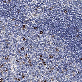
VC001) or the HRP-conjugated Anti-Mouse IgG Secondary Antibody (Catalog #
HAF007). Before incubation with the primary antibody, tissue was subjected to heat-induced epitope retrieval using VisUCyte Antigen Retrieval Reagent-Basic (Catalog #
VCTS021). Tissue was stained using DAB (brown) and counterstained with hematoxylin (blue). Specific staining was localized to the cell surface. View our protocol for
IHC Staining with VisUCyte HRP Polymer Detection Reagents." class="big_thumb" />
VC001) or the HRP-conjugated Anti-Mouse IgG Secondary Antibody (Catalog #
HAF007). Before incubation with the primary antibody, tissue was subjected to heat-induced epitope retrieval using VisUCyte Antigen Retrieval Reagent-Basic (Catalog #
VCTS021). Tissue was stained using DAB (brown) and counterstained with hematoxylin (blue). Specific staining was localized to the cell surface. View our protocol for
IHC Staining with VisUCyte HRP Polymer Detection Reagents." class="big_lightbox">
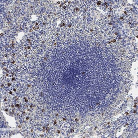
VC001) or the HRP-conjugated Anti-Mouse IgG Secondary Antibody (Catalog #
HAF007). Before incubation with the primary antibody, tissue was subjected to heat-induced epitope retrieval using VisUCyte Antigen Retrieval Reagent-Basic (Catalog #
VCTS021). Tissue was stained using DAB (brown) and counterstained with hematoxylin (blue). Specific staining was localized to the cell surface. View our protocol for
IHC Staining with VisUCyte HRP Polymer Detection Reagents." class="big_thumb" />
VC001) or the HRP-conjugated Anti-Mouse IgG Secondary Antibody (Catalog #
HAF007). Before incubation with the primary antibody, tissue was subjected to heat-induced epitope retrieval using VisUCyte Antigen Retrieval Reagent-Basic (Catalog #
VCTS021). Tissue was stained using DAB (brown) and counterstained with hematoxylin (blue). Specific staining was localized to the cell surface. View our protocol for
IHC Staining with VisUCyte HRP Polymer Detection Reagents." class="big_lightbox">
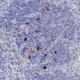
VC001) or the HRP-conjugated Anti-Mouse IgG Secondary Antibody (Catalog #
HAF007). Before incubation with the primary antibody, tissue was subjected to heat-induced epitope retrieval using VisUCyte Antigen Retrieval Reagent-Basic (Catalog #
VCTS021). Tissue was stained using DAB (brown) and counterstained with hematoxylin (blue). Specific staining was localized to the cell surface. View our protocol for
IHC Staining with VisUCyte HRP Polymer Detection Reagents." class="big_thumb" />
 FAB24086P) and either (A) Mouse Anti-Human CD57 Monoclonal Antibody (Catalog # MAB11708) or (B) isotype control antibody (Catalog # MAB002) followed by Allophycocyanin-conjugated Anti-Mouse IgG Secondary Antibody (Catalog # F0101B). View our protocol for Staining Membrane-associated Proteins. " alt="PBMC were stained with Rabbit Anti-Human NCAM‑1/CD56 PE‑conjugated Monoclonal Antibody (Catalog # FAB24086P) and either (A) Mouse Anti-Human CD57 Monoclonal Antibody (Catalog # MAB11708) or (B) isotype control antibody (Catalog # MAB002) followed by Allophycocyanin-conjugated Anti-Mouse IgG Secondary Antibody (Catalog # F0101B). View our protocol for Staining Membrane-associated Proteins. " class="big_thumb" />
 HAF018). A specific band was detected for beta -1,3-Glucuronyltransferase 1/B3GAT1 at approximately 38 kDa (as indicated). This experiment was conducted under reducing conditions and using Immunoblot Buffer Group 1." alt="Western blot shows lysates of human cerebellum tissue. PVDF membrane was probed with 2 µg/mL of Mouse Anti-Human beta -1,3-Glucuronyltransferase 1/B3GAT1 Monoclonal Antibody (Catalog # MAB85602) followed by HRP-conjugated Anti-Mouse IgG Secondary Antibody (Catalog # HAF018). A specific band was detected for beta -1,3-Glucuronyltransferase 1/B3GAT1 at approximately 38 kDa (as indicated). This experiment was conducted under reducing conditions and using Immunoblot Buffer Group 1." class="big_thumb" />
 VC001). Before incubation with the primary antibody, tissue was subjected to heat-induced epitope retrieval using VisUCyte Antigen Retrieval Reagent-Basic (Catalog # VCTS021). Tissue was stained using DAB (brown) and counterstained with hematoxylin (blue). Specific staining was localized to cell surface and cytoplasm in neurons. View our protocol for IHC Staining with VisUCyte HRP Polymer Detection Reagents." alt=" beta ‑1,3‑Glucuronyltransferase 1/B3GAT1 was detected in immersion fixed paraffin-embedded sections of Human Brain Hippocampus using Mouse Anti-Human beta ‑1,3‑Glucuronyltransferase 1/B3GAT1 Monoclonal Antibody (Catalog # MAB85603) at 5 µg/mL for 1 hour at room temperature followed by incubation with the Anti-Mouse IgG VisUCyte™ HRP Polymer Antibody (Catalog # VC001). Before incubation with the primary antibody, tissue was subjected to heat-induced epitope retrieval using VisUCyte Antigen Retrieval Reagent-Basic (Catalog # VCTS021). Tissue was stained using DAB (brown) and counterstained with hematoxylin (blue). Specific staining was localized to cell surface and cytoplasm in neurons. View our protocol for IHC Staining with VisUCyte HRP Polymer Detection Reagents." class="big_thumb" />
 VC001). Before incubation with the primary antibody, tissue was subjected to heat-induced epitope retrieval using Antigen Retrieval Reagent-Basic (Catalog # CTS013). Tissue was stained using DAB (brown) and counterstained with hematoxylin (blue). Specific staining was localized to cytoplasm in neurons. View our protocol for IHC Staining with VisUCyte HRP Polymer Detection Reagents." alt=" beta -1,3-Glucuronyltransferase 1/B3GAT1 was detected in immersion fixed paraffin-embedded sections of human brain (cortex) using Mouse Anti-Human beta -1,3-Glucuronyltransferase 1/B3GAT1 Monoclonal Antibody (Catalog # MAB8560) at 5 µg/mL for 1 hour at room temperature followed by incubation with the Anti-Mouse IgG VisUCyte™ HRP Polymer Antibody (Catalog # VC001). Before incubation with the primary antibody, tissue was subjected to heat-induced epitope retrieval using Antigen Retrieval Reagent-Basic (Catalog # CTS013). Tissue was stained using DAB (brown) and counterstained with hematoxylin (blue). Specific staining was localized to cytoplasm in neurons. View our protocol for IHC Staining with VisUCyte HRP Polymer Detection Reagents." class="big_thumb" />


![Immunohistochemistry-Paraffin: CD57 Antibody (NK/804) [NBP2-44355] - Human Tonsil stained with CD57 Monoclonal Antibody (NK/804).](http://images.novusbio.com/fullsize/beta-1-3-Glucuronyltransferase-1-B3GAT1-Antibody-NK-804-Immunohistochemistry-Paraffin-NBP2-44355-img0001.jpg "Immunohistochemistry-Paraffin: CD57 Antibody (NK/804) [NBP2-44355] - Human Tonsil stained with CD57 Monoclonal Antibody (NK/804).")
![Flow Cytometry: CD57 Antibody (TB01) [NBP2-62203] - Staining of human peripheral blood with anti-human CD57 (TB01) APC.](http://images.novusbio.com/fullsize/beta-1-3-Glucuronyltransferase-1-B3GAT1-Antibody-TB01-Flow-Cytometry-NBP2-62203-img0004.jpg "Flow Cytometry: CD57 Antibody (TB01) [NBP2-62203] - Staining of human peripheral blood with anti-human CD57 (TB01) APC.")
![Flow (Cell Surface): CD57 Antibody (TB01) [NBP2-62203] - Surface staining of human buffy coat cells with anti-human CD57 (TB01) purified, GAM-APC.](http://images.novusbio.com/fullsize/beta-1-3-Glucuronyltransferase-1-B3GAT1-Antibody-TB01-Flow-Cell-Surface-NBP2-62203-img0001.jpg)
![Flow (Cell Surface): CD57 Antibody (TB01) [NBP2-62203] - Surface staining of human peripheral blood with anti-human CD57 (TB01) PE.](http://images.novusbio.com/fullsize/beta-1-3-Glucuronyltransferase-1-B3GAT1-Antibody-TB01-Flow-Cell-Surface-NBP2-62203-img0002.jpg)
![Flow (Cell Surface): CD57 Antibody (TB01) [NBP2-62203] - Surface staining of human peripheral blood with anti-human CD57 (TB01) FITC.](http://images.novusbio.com/fullsize/beta-1-3-Glucuronyltransferase-1-B3GAT1-Antibody-TB01-Flow-Cell-Surface-NBP2-62203-img0003.jpg)
![Immunocytochemistry/Immunofluorescence: beta-1,3-Glucuronyltransferase 1/B3GAT1 Antibody [NBP1-19788] - Antibody was tested in U2OS cells with FITC (green). Nuclei and alpha-tubulin were counterstained with DAPI (blue) and Dylight 550 (red).](http://images.novusbio.com/fullsize/beta-1-3-Glucuronyltransferase-1-B3GAT1-Antibody-Immunocytochemistry-Immunofluorescence-NBP1-19788-img0005.jpg "Immunocytochemistry/Immunofluorescence: beta-1,3-Glucuronyltransferase 1/B3GAT1 Antibody [NBP1-19788] - Antibody was tested in U2OS cells with FITC (green). Nuclei and alpha-tubulin were counterstained with DAPI (blue) and Dylight 550 (red).")
![Immunohistochemistry: beta-1,3-Glucuronyltransferase 1/B3GAT1 Antibody [NBP1-19788] - Analysis in mouse brain using DAB with hematoxylin counterstain.](http://images.novusbio.com/fullsize/beta-1-3-Glucuronyltransferase-1-B3GAT1-Antibody-Immunohistochemistry-NBP1-19788-img0006.jpg)
 VC001) or the HRP-conjugated Anti-Mouse IgG Secondary Antibody (Catalog #
  VC001) or the HRP-conjugated Anti-Mouse IgG Secondary Antibody (Catalog #
  VC001) or the HRP-conjugated Anti-Mouse IgG Secondary Antibody (Catalog #
 VC001). Before incubation with the primary antibody, tissue was subjected to heat-induced epitope retrieval using Antigen Retrieval Reagent-Basic (Catalog #
 ![Immunocytochemistry/Immunofluorescence: beta-1,3-Glucuronyltransferase 1/B3GAT1 Antibody [NBP1-78982] - Antibody was tested in U2OS cells with FITC (green). Nuclei and alpha-tubulin were counterstained with DAPI (blue) and Dylight 550 (red).](http://images.novusbio.com/fullsize/beta-1-3-Glucuronyltransferase-1-B3GAT1-Antibody-Immunocytochemistry-Immunofluorescence-NBP1-78982-img0006.jpg "Immunocytochemistry/Immunofluorescence: beta-1,3-Glucuronyltransferase 1/B3GAT1 Antibody [NBP1-78982] - Antibody was tested in U2OS cells with FITC (green). Nuclei and alpha-tubulin were counterstained with DAPI (blue) and Dylight 550 (red).")
![Immunohistochemistry: beta-1,3-Glucuronyltransferase 1/B3GAT1 Antibody [NBP1-78982] - Analysis of CD57 in mouse brain using DAB with hematoxylin counterstain.](http://images.novusbio.com/fullsize/beta-1-3-Glucuronyltransferase-1-B3GAT1-Antibody-Immunohistochemistry-NBP1-78982-img0007.jpg)


 NL007) and counterstained with DAPI (blue). Specific staining was localized to cell surface and cytoplasm. View our protocol for
 ![Immunohistochemistry-Paraffin: CD57 Antibody (SPM129) [NBP2-44353] - Analysis of CD57 antibody (SPM129) on human spleen. Image from verified customer review.](http://images.novusbio.com/fullsize/CD57-Antibody-SPM129-Immunohistochemistry-Paraffin-NBP2-44353-img0002.jpg "Immunohistochemistry-Paraffin: CD57 Antibody (SPM129) [NBP2-44353] - Analysis of CD57 antibody (SPM129) on human spleen. Image from verified customer review.")
![Immunohistochemistry-Paraffin: CD57 Antibody (SPM129) [NBP2-44353] - Human Spleen stained with CD57 Monoclonal Antibody (SPM129).](http://images.novusbio.com/fullsize/beta-1-3-Glucuronyltransferase-1-B3GAT1-Antibody-SPM129-Immunohistochemistry-Paraffin-NBP2-44353-img0001.jpg)
![Immunohistochemistry-Paraffin: CD57 Antibody (NK-1) [NBP2-44352] - Human spleen stained with CD57 Monoclonal Antibody (NK-1).](http://images.novusbio.com/fullsize/beta-1-3-Glucuronyltransferase-1-B3GAT1-Antibody-NK-1-Immunohistochemistry-Paraffin-NBP2-44352-img0001.jpg "Immunohistochemistry-Paraffin: CD57 Antibody (NK-1) [NBP2-44352] - Human spleen stained with CD57 Monoclonal Antibody (NK-1).")
![Western Blot: beta-1,3-Glucuronyltransferase 1/B3GAT1 Antibody (3B8B10) [NBP2-61708] - Analysis using B3GAT1 mAb against HEK293 (1) and B3GAT1 (AA: 193-334)-hIgGFc transfected HEK293 (2) cell lysate.](http://images.novusbio.com/fullsize/beta-1-3-Glucuronyltransferase-1-B3GAT1-Antibody-3B8B10-Western-Blot-NBP2-61708-img0004.jpg "Western Blot: beta-1,3-Glucuronyltransferase 1/B3GAT1 Antibody (3B8B10) [NBP2-61708] - Analysis using B3GAT1 mAb against HEK293 (1) and B3GAT1 (AA: 193-334)-hIgGFc transfected HEK293 (2) cell lysate.")
![Flow Cytometry: beta-1,3-Glucuronyltransferase 1/B3GAT1 Antibody (3B8B10) [NBP2-61708] - Analysis of Hela cells using B3GAT1 mouse mAb (green) and negative control (red).](http://images.novusbio.com/fullsize/beta-1-3-Glucuronyltransferase-1-B3GAT1-Antibody-3B8B10-Flow-Cytometry-NBP2-61708-img0002.jpg)
![Western Blot: beta-1,3-Glucuronyltransferase 1/B3GAT1 Antibody (3B8B10) [NBP2-61708] - Analysis using B3GAT1 mAb against human B3GAT1 (AA: 193-334) recombinant protein. (Expected MW is 41.5 kDa)](http://images.novusbio.com/fullsize/beta-1-3-Glucuronyltransferase-1-B3GAT1-Antibody-3B8B10-Western-Blot-NBP2-61708-img0003.jpg)
![Western Blot: beta-1,3-Glucuronyltransferase 1/B3GAT1 Antibody (3B8B10) [NBP2-61708] - Analysis using B3GAT1 mouse mAb against MOLT4 (1), Raji (2), and Ramos (3) cell lysate.](http://images.novusbio.com/fullsize/beta-1-3-Glucuronyltransferase-1-B3GAT1-Antibody-3B8B10-Western-Blot-NBP2-61708-img0005.jpg)
![ELISA: beta-1,3-Glucuronyltransferase 1/B3GAT1 Antibody (3B8B10) [NBP2-61708] - Black line: Control Antigen (100 ng);Purple line: Antigen (10ng); Blue line: Antigen (50 ng); Red line:Antigen (100 ng)](http://images.novusbio.com/fullsize/beta-1-3-Glucuronyltransferase-1-B3GAT1-Antibody-3B8B10-ELISA-NBP2-61708-img0001.jpg)


