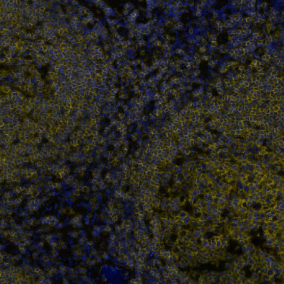
MAB14303) at 5µg/mL at 37 ° Celsius for 4 minutes. Before incubation with the primary antibody, tissue underwent an all-in-one dewaxing and antigen retrieval preprocessing using PreTreatment Module (PT Module) and Dewax and HIER Buffer H (pH 9). Tissue was stained using the Alexa Fluor™ 555 Goat anti-Mouse IgG Secondary Antibody at 1:100 at 37 ° Celsius for 2 minutes. (Yellow; Lunaphore Catalog #
DR555MS) and counterstained with DAPI (blue; Lunaphore Catalog #
DR100). Specific staining was localized to the membrane. Protocol available in
COMET™ Panel Builder." class="big_lightbox">
MAB14303) at 5µg/mL at 37 ° Celsius for 4 minutes. Before incubation with the primary antibody, tissue underwent an all-in-one dewaxing and antigen retrieval preprocessing using PreTreatment Module (PT Module) and Dewax and HIER Buffer H (pH 9). Tissue was stained using the Alexa Fluor™ 555 Goat anti-Mouse IgG Secondary Antibody at 1:100 at 37 ° Celsius for 2 minutes. (Yellow; Lunaphore Catalog #
DR555MS) and counterstained with DAPI (blue; Lunaphore Catalog #
DR100). Specific staining was localized to the membrane. Protocol available in
COMET™ Panel Builder." alt="CD45 was detected in immersion fixed paraffin-embedded sections of human lymph node using Mouse Anti-Human CD45 Monoclonal Antibody (
MAB14303) at 5µg/mL at 37 ° Celsius for 4 minutes. Before incubation with the primary antibody, tissue underwent an all-in-one dewaxing and antigen retrieval preprocessing using PreTreatment Module (PT Module) and Dewax and HIER Buffer H (pH 9). Tissue was stained using the Alexa Fluor™ 555 Goat anti-Mouse IgG Secondary Antibody at 1:100 at 37 ° Celsius for 2 minutes. (Yellow; Lunaphore Catalog #
DR555MS) and counterstained with DAPI (blue; Lunaphore Catalog #
DR100). Specific staining was localized to the membrane. Protocol available in
COMET™ Panel Builder." class="big_thumb" />
HAF018). A specific band was detected for CD45 at approximately 255 kDa (as indicated). GAPDH (
MAB5718) is shown as a loading control. This experiment was conducted under reducing conditions and using Western Blot Buffer Group 1." class="big_lightbox">

HAF018). A specific band was detected for CD45 at approximately 255 kDa (as indicated). GAPDH (
MAB5718) is shown as a loading control. This experiment was conducted under reducing conditions and using Western Blot Buffer Group 1." class="big_thumb" />
VC001). Before incubation with the primary antibody, tissue was subjected to heat-induced epitope retrieval using Antigen Retrieval Reagent-Basic (
CTS013). Tissue was stained using DAB (brown) and counterstained with hematoxylin (blue). Specific staining was localized to lymphocytes. Staining was performed using our protocol for IHC Staining with VisUCyte HRP Polymer Detection Reagents." class="big_lightbox">

VC001). Before incubation with the primary antibody, tissue was subjected to heat-induced epitope retrieval using Antigen Retrieval Reagent-Basic (
CTS013). Tissue was stained using DAB (brown) and counterstained with hematoxylin (blue). Specific staining was localized to lymphocytes. Staining was performed using our protocol for IHC Staining with VisUCyte HRP Polymer Detection Reagents." class="big_thumb" />
HAF018). A specific band was detected for CD45 at approximately 255 kDa (as indicated) in the parental THP‑1 human acute monocytic leukemia cell line, but is not detectable in knockout THP‑1 human acute monocytic leukemia cell line. GAPDH (
MAB5718) is shown as a loading control. This experiment was conducted under reducing conditions and using Western Blot Buffer Group 1." class="big_lightbox">

HAF018). A specific band was detected for CD45 at approximately 255 kDa (as indicated) in the parental THP‑1 human acute monocytic leukemia cell line, but is not detectable in knockout THP‑1 human acute monocytic leukemia cell line. GAPDH (
MAB5718) is shown as a loading control. This experiment was conducted under reducing conditions and using Western Blot Buffer Group 1." class="big_thumb" />

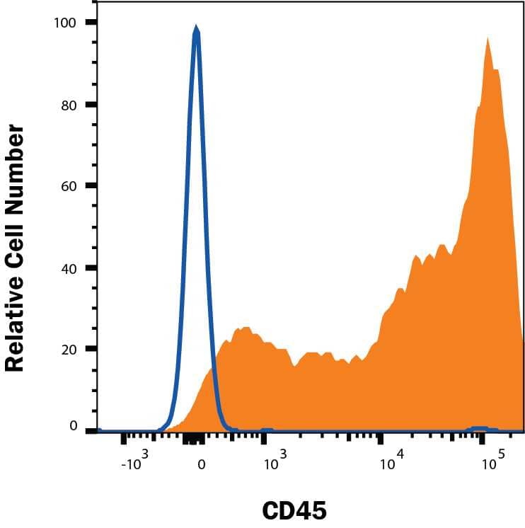
MAB002, open histogram), followed by Phycoerythrin-conjugated Anti-Mouse IgG Secondary Antibody (
F0102B). View our protocol for Staining Membrane-associated Proteins." class="big_lightbox">
MAB002, open histogram), followed by Phycoerythrin-conjugated Anti-Mouse IgG Secondary Antibody (
F0102B). View our protocol for Staining Membrane-associated Proteins." class="big_thumb" />
 NL001) and counterstained with DAPI (blue). Specific staining was localized to cell surface. Staining was performed using our protocol for Fluorescent ICC Staining of Non-adherent Cells." alt="CD45 was detected in immersion fixed RAW 264.7 mouse monocyte/macrophage cell line (positive staining) and Neuro‑2A mouse neuroblastoma cell line (negative staining) using Goat Anti-Mouse CD45 Antigen Affinity-purified Polyclonal Antibody (Catalog # AF114) at 5 µg/mL for 3 hours at room temperature. Cells were stained using the NorthernLights™ 557-conjugated Anti-Goat IgG Secondary Antibody (red; NL001) and counterstained with DAPI (blue). Specific staining was localized to cell surface. Staining was performed using our protocol for Fluorescent ICC Staining of Non-adherent Cells." class="big_thumb" />
 MAB002, open histogram), followed by Phycoerythrin-conjugated Anti-Mouse IgG Secondary Antibody (Catalog # F0102B). View our protocol for Staining Membrane-associated Proteins." alt="Human peripheral blood lymphocytes were stained with Mouse Anti-Human CD45 Monoclonal Antibody (Catalog # MAB1430, filled histogram) or isotype control antibody (Catalog # MAB002, open histogram), followed by Phycoerythrin-conjugated Anti-Mouse IgG Secondary Antibody (Catalog # F0102B). View our protocol for Staining Membrane-associated Proteins." class="big_thumb" />
 MAB14303) at 5µg/mL at 37 ° Celsius for 4 minutes. Before incubation with the primary antibody, tissue underwent an all-in-one dewaxing and antigen retrieval preprocessing using PreTreatment Module (PT Module) and Dewax and HIER Buffer H (pH 9). Tissue was stained using the Alexa Fluor™ 555 Goat anti-Mouse IgG Secondary Antibody at 1:100 at 37 ° Celsius for 2 minutes. (Yellow; Lunaphore Catalog # DR555MS) and counterstained with DAPI (blue; Lunaphore Catalog # DR100). Specific staining was localized to the membrane. Protocol available in COMET™ Panel Builder." alt="CD45 was detected in immersion fixed paraffin-embedded sections of human lymph node using Mouse Anti-Human CD45 Monoclonal Antibody (MAB14303) at 5µg/mL at 37 ° Celsius for 4 minutes. Before incubation with the primary antibody, tissue underwent an all-in-one dewaxing and antigen retrieval preprocessing using PreTreatment Module (PT Module) and Dewax and HIER Buffer H (pH 9). Tissue was stained using the Alexa Fluor™ 555 Goat anti-Mouse IgG Secondary Antibody at 1:100 at 37 ° Celsius for 2 minutes. (Yellow; Lunaphore Catalog # DR555MS) and counterstained with DAPI (blue; Lunaphore Catalog # DR100). Specific staining was localized to the membrane. Protocol available in COMET™ Panel Builder." class="big_thumb" />


 VC004). Before incubation with the primary antibody, tissue was subjected to heat-induced epitope retrieval using Antigen Retrieval Reagent-Basic (
![Flow Cytometry: CD45 Antibody (30-F11) [NB100-77417] - Analysis of CD45 (30F11) in freshly isolated mouse (Balb C) splenocytes (1x10^6) were stained with [NB100-77417] at 1:1000 dilution and detected with FITC conjugated donkey anti-rat IgG secondary (orange). Shown with rat IgG isotype control (blue).](http://images.novusbio.com/fullsize/CD45-Antibody-30-F11-Flow-Cytometry-NB100-77417-img0004.jpg "Flow Cytometry: CD45 Antibody (30-F11) [NB100-77417] - Analysis of CD45 (30F11) in freshly isolated mouse (Balb C) splenocytes (1x10^6) were stained with [NB100-77417] at 1:1000 dilution and detected with FITC conjugated donkey anti-rat IgG secondary (orange). Shown with rat IgG isotype control (blue).")
![Immunohistochemistry-Paraffin: CD45 Antibody (30-F11) [NB100-77417] - Analysis of FFPE section of mouse spleen using rat monoclonal CD45 antibody (30-F11) [NB100-77417] at 1:50 dilution. The antibody generated an expected membrane staining in the red and white pulp spleenocytes of the tested spleen section.](http://images.novusbio.com/fullsize/CD45-Antibody-30-F11-Immunohistochemistry-Paraffin-NB100-77417-img0006.jpg)
![Immunohistochemistry-Paraffin: CD45 Antibody (30-F11) [NB100-77417] - Mouse glomeruli stained with CD45 antibody. 4% formaldehyde fixed paraffin sections. EDTA was used for antigen retrieval. IHC-P image submitted by a verified customer review.](http://images.novusbio.com/fullsize/CD45-Antibody-30-F11-Immunohistochemistry-Paraffin-NB100-77417-img0013.jpg)
![Immunohistochemistry: CD45 Antibody (30-F11) [NB100-77417] - Analysis of a FFPE embedded tissue section of mouse liver using 1:100 dilution of CD45 antibody (30-F11) [NB100-77417]. The signal was developed using AEC reagent based method with hematoxylin counterstaining.](http://images.novusbio.com/fullsize/CD45-Antibody-30-F11-Immunohistochemistry-NB100-77417-img0003.jpg)
![Immunohistochemistry-Frozen: CD45 Antibody (30-F11) [NB100-77417] - Analysis of a mouse spleen cryosection using CD45 antibody (30-F11) [NB100-77417] at 1:50 dilution. The signal was detected using goat anti-rat Alexa fluor 488 secondary with DAPI counterstaining. IHC-P image submitted by a verified customer review.](http://images.novusbio.com/fullsize/CD45-Antibody-30-F11-Immunohistochemistry-Frozen-NB100-77417-img0008.jpg)
![Immunohistochemistry-Paraffin: CD45 Antibody (30-F11) [NB100-77417] - Staining of a FFPE mouse liver tissue section using 1:100 dilution of CD45 antibody (30-F11) [NB100-77417]. Signal was detected using AF488 conjugated donkey anti-rat secondary antibody at 1:500 dilution. IHC-P image submitted by a verified customer review.](http://images.novusbio.com/fullsize/CD45-Antibody-30-F11-Immunohistochemistry-Paraffin-NB100-77417-img0002.jpg)


![Flow Cytometry: CD45 Antibody (OX-1) [NB100-64895] - Analysis using Alexa Fluor (R) 488 conjugate of CD45 antibody (OX-1). Rat Splenocytes were stained with CD45 antibody (OX-1) [NB100-64895] (blue) and a matched isotype control [NBP2-27287] (orange). Cells were incubated in an antibody dilution of 1 ug/mL for 20 minutes at room temperature. Both antibodies were conjugated to Alexa Fluor 488.](http://images.novusbio.com/fullsize/CD45-Antibody-OX-1-Flow-Cytometry-NB100-64895-img0005.jpg "Flow Cytometry: CD45 Antibody (OX-1) [NB100-64895] - Analysis using Alexa Fluor (R) 488 conjugate of CD45 antibody (OX-1). Rat Splenocytes were stained with CD45 antibody (OX-1) [NB100-64895] (blue) and a matched isotype control [NBP2-27287] (orange). Cells were incubated in an antibody dilution of 1 ug/mL for 20 minutes at room temperature. Both antibodies were conjugated to Alexa Fluor 488.")
![Flow Cytometry: CD45 Antibody (OX-1) [NB100-64895] - Analysis using the FITC conjugate of CD45 antibody (OX-1). Staining of rat spleen cells.](http://images.novusbio.com/fullsize/CD45-Antibody-OX-1-Flow-Cytometry-NB100-64895-img0004.jpg)

![Flow Cytometry: CD45 Antibody (OX-1) [NB100-64895] - Staining of rat peripheral blood lymphocytes with mouse anti-rat CD45 antibody (OX-1) [NB100-64895].](http://images.novusbio.com/fullsize/CD45-Antibody-OX-1-Flow-Cytometry-NB100-64895-img0001.jpg)
 NL007) and counter-stained with DAPI (blue). View our protocol for
  MAB002), followed by Phycoerythrin-conjugated Anti-Mouse IgG Secondary Antibody (
 MAB002) followed by PE-conjugated anti-Mouse IgG Secondary Antibody (
![Flow Cytometry: CD45 Antibody (MEM-28) [NB500-319] - Staining of human peripheral blood lymphocytes with anti-human CD45 antibody (MEM-28) Alexa Fluor 700.](http://images.novusbio.com/fullsize/CD45-Antibody-MEM-28-Flow-Cytometry-NB500-319-img0021.jpg "Flow Cytometry: CD45 Antibody (MEM-28) [NB500-319] - Staining of human peripheral blood lymphocytes with anti-human CD45 antibody (MEM-28) Alexa Fluor 700.")
![Immunocytochemistry/Immunofluorescence: CD45 Antibody (MEM-28) [NB500-319] - staining of human peripheral blood mononuclear cell using anti-human CD45 antibody (MEM-28) (green). DNA visualized by DAPI (blue).](http://images.novusbio.com/fullsize/CD45-Antibody-MEM-28-Immunocytochemistry-NB500-319-img0001.jpg)
![Flow Cytometry: CD45 Antibody (MEM-28) [NB500-319] - Staining of human peripheral blood cells with anti-human CD45 antibody (MEM-28) PE-CyTM5.](http://images.novusbio.com/fullsize/CD45-Antibody-MEM-28-Flow-Cytometry-NB500-319-img0018.jpg)
![Immunohistochemistry-Paraffin: CD45 Antibody (MEM-28) [NB500-319] - Staining of human small intestine (paraffin-embedded sections) with CD45 antibody (MEM-28), 10 ug/ml.](http://images.novusbio.com/fullsize/CD45-Antibody-MEM-28-Immunohistochemistry-Paraffin-NB500-319-img0022.jpg)
![Flow Cytometry: CD45 Antibody (MEM-28) [NB500-319] - Surface staining of human peripheral blood cells with anti-human CD45 antibody (MEM-28) PerCP-CyTM5.5.](http://images.novusbio.com/fullsize/CD45-Antibody-MEM-28-Flow-Cytometry-NB500-319-img0007.jpg)
![Flow Cytometry: CD45 Antibody (MEM-28) [NB500-319] - Surface staining of human peripheral blood cells with anti-human CD45 antibody (MEM-28) purified, GAM-APC.](http://images.novusbio.com/fullsize/CD45-Antibody-MEM-28-Flow-Cytometry-NB500-319-img0008.jpg)
![Flow Cytometry: CD45 Antibody (MEM-28) [NB500-319] - Surface staining of human peripheral blood cells with anti-human CD45 antibody (MEM-28) APC-CyTM7.](http://images.novusbio.com/fullsize/CD45-Antibody-MEM-28-Flow-Cytometry-NB500-319-img0010.jpg)
![Flow Cytometry: CD45 Antibody (MEM-28) [NB500-319] - Staining of human peripheral blood cells with anti-human CD45 antibody (MEM-28) Pacific BlueTM.](http://images.novusbio.com/fullsize/CD45-Antibody-MEM-28-Flow-Cytometry-NB500-319-img0016.jpg)
![Flow Cytometry: CD45 Antibody (MEM-28) [NB500-319] - Staining of human peripheral blood cells with anti-human CD45 antibody (MEM-28) PE.](http://images.novusbio.com/fullsize/CD45-Antibody-MEM-28-Flow-Cytometry-NB500-319-img0017.jpg)
![Flow Cytometry: CD45 Antibody (MEM-28) [NB500-319] - Staining of human peripheral blood cells with anti-human CD45 antibody (MEM-28) PE-CyTM7.](http://images.novusbio.com/fullsize/CD45-Antibody-MEM-28-Flow-Cytometry-NB500-319-img0019.jpg)
![Flow Cytometry: CD45 Antibody (MEM-28) [NB500-319] - Staining of human peripheral blood cells with anti-human CD45 antibody (MEM-28) PE-DyLight (R) 594.](http://images.novusbio.com/fullsize/CD45-Antibody-MEM-28-Flow-Cytometry-NB500-319-img0020.jpg)
![Formalin-fixed, paraffin-embedded human tonsil (20X) stained with CD45 antibody (2B11 + PD7/26) [NBP2-34287].](http://images.novusbio.com/fullsize/CD45-Antibody-2B11-+-PD7-26-Immunohistochemistry-Paraffin-NBP2-34287-img0003.jpg "Formalin-fixed, paraffin-embedded human tonsil (20X) stained with CD45 antibody (2B11 + PD7/26) [NBP2-34287].")
![Western Blot Analysis of human Spleen tissue lysates using CD45 antibody (2B11 + PD7/26) [NBP2-34287]. Observed molecular weight ~200 kDa.](http://images.novusbio.com/fullsize/CD45-Antibody-2B11-+-PD7-26-Western-Blot-NBP2-34287-img0007.jpg)

![Formalin-fixed, paraffin-embedded human tonsil (10X) stained with CD45 antibody(2B11 + PD7/26) [NBP2-34287].](http://images.novusbio.com/fullsize/CD45-Antibody-2B11-+-PD7-26-Immunohistochemistry-Paraffin-NBP2-34287-img0001.jpg)
![Flow Cytometric Analysis of PFA-fixed Jurkat cells using CD45 antibody (2B11 + PD7/26) [NBP2-34287] followed by Goat anti-Mouse-IgG-CF488 (Blue); Isotype Control (Red).](http://images.novusbio.com/fullsize/CD45-Antibody-2B11-+-PD7-26-Flow-Cytometry-NBP2-34287-img0006.jpg)

![Immunohistochemistry-Paraffin: CD45 Antibody (2B11 + PD7/26) [NBP2-34287] - Staining of human tonsil with Alexa Fluor 488 conjugate of CD45 Antibody (2B11 + PD7/26), pH 9 antigen retrieval. Image from verified customer review.](http://images.novusbio.com/fullsize/nbp2-34287_mouse-monoclonal-cd45-antibody-2b11-pd7-26-17720241003243.jpg)
![Flow Cytometry: CD45 Antibody (HI30) [NBP1-79127] - Surface staining of human peripheral blood cells with anti-human CD45 antibody (HI30) [NBP1-79127] Pacific BlueTM.](http://images.novusbio.com/fullsize/CD45-Antibody-HI30-Flow-Cytometry-NBP1-79127-img0007.jpg "Flow Cytometry: CD45 Antibody (HI30) [NBP1-79127] - Surface staining of human peripheral blood cells with anti-human CD45 antibody (HI30) [NBP1-79127] Pacific BlueTM.")
![Flow (Cell Surface): CD45 Antibody (HI30) [NBP1-79127] - hPBMC cells were stained with CD45 antibody (HI30) [NBP1-79127] (blue) and a matched isotype control [NBP2-27287] (orange). Cells were incubated in an antibody dilution of 1 ug/mL for 20 minutes at room temperature. The antibodies were directly conjugated to Alexa Flour 488.](http://images.novusbio.com/fullsize/CD45-Antibody-HI30-Flow-Cell-Surface-NBP1-79127-img0005.jpg)
![Flow Cytometry: CD45 Antibody (HI30) [NBP1-79127] - Surface staining of human peripheral blood cells with anti-human CD45 antibody (HI30) [NBP1-79127] Pacific OrangeTM.](http://images.novusbio.com/fullsize/CD45-Antibody-HI30-Flow-Cytometry-NBP1-79127-img0008.jpg)
![Flow Cytometry: CD45 Antibody (HI30) [NBP1-79127] - Surface staining of human peripheral blood cells with anti-human CD45 antibody (HI30) [NBP1-79127] purified, GAM-APC.](http://images.novusbio.com/fullsize/CD45-Antibody-HI30-Flow-Cytometry-NBP1-79127-img0009.jpg)
![Flow Cytometry: CD45 Antibody (HI30) [NBP1-79127] - Analysis of PE/Cy7 conjugate of CD45 antibody (HI30) [NBP1-79127]. Staining of normal human peripheral blood cells with Mouse IgG1 K Isotype Control PE-Cy7 (blue histogram) or Anti-Human CD45 PE-Cy7 (purple histogram). Cells in the lymphocyte gate were used for analysis.](http://images.novusbio.com/fullsize/CD45-Antibody-HI30-Flow-Cytometry-NBP1-79127-img0010.jpg)
![Flow (Cell Surface): CD45 Antibody (HI30) [NBP1-79127] - Flow (Cell Surface): Cell surface staining of PBMC's (Lymphocyte gate at 1 x 10^6 cells/ml) with CD45 antibody (HI30) [Alexa Fluor (R) 647] [NBP1-79127AF647] (orange) stained at a dilution of 1:500. Shown with mIgG1 (AF647 conjugated) Isotype control (blue). Image using the Alexa Fluor 647 form of this antibody.](http://images.novusbio.com/fullsize/CD45-Antibody-HI30-Flow-Cell-Surface-NBP1-79127-img0011.jpg)
 HAF018). A specific band was detected for CD45 at approximately 255 kDa (as indicated). GAPDH (
 VC001). Before incubation with the primary antibody, tissue was subjected to heat-induced epitope retrieval using Antigen Retrieval Reagent-Basic (
 HAF018). A specific band was detected for CD45 at approximately 255 kDa (as indicated) in the parental THP‑1 human acute monocytic leukemia cell line, but is not detectable in knockout THP‑1 human acute monocytic leukemia cell line. GAPDH (
 MAB002, open histogram), followed by Phycoerythrin-conjugated Anti-Mouse IgG Secondary Antibody (
![Flow Cytometry: CD45 Antibody [NBP2-15811] - THP-1 cell. Black: Unlabelled sample was used as a control. Red: CD45 antibody [NBP2-15811], dilution: 1:50.](http://images.novusbio.com/fullsize/CD45-Antibody-Flow-Cytometry-NBP2-15811-img0006.jpg "Flow Cytometry: CD45 Antibody [NBP2-15811] - THP-1 cell. Black: Unlabelled sample was used as a control. Red: CD45 antibody [NBP2-15811], dilution: 1:50.")
![Flow Cytometry: CD45 Antibody [NBP2-15811] - Mouse splenocytes cell fixed in 4% paraformaldehyde at 4 C for 15 min. Black: Unlabelled sample was used as a control. Red: CD45 antibody [NBP2-15811], dilution: 1:50. Acquisition of 20,000 events were collected using a Dylight 488- Conjugated secondary antibody for FACS analysis.](http://images.novusbio.com/fullsize/CD45-Antibody-Flow-Cytometry-NBP2-15811-img0005.jpg)

![Western Blot: CD45 Antibody (BL-178-12C7) [NBP3-14668] - Whole cell lysate (50 ug) from Jurkat, HEK293T, MOLT-4, HeLa, and SR cells prepared using NETN lysis buffer.Antibody: Rabbit anti-CD45 recombinant monoclonal antibody [BL-178-12C7] used at 1:1000. Secondary: HRP-conjugated goat anti-rabbit IgG. Chemiluminescence with an exposure timeof 1 second. Lower Panel: Rabbit anti-COPB2 antibody.](http://images.novusbio.com/fullsize/CD45-Antibody-BL-178-12C7-Western-Blot-NBP3-14668-img0013.jpg "Western Blot: CD45 Antibody (BL-178-12C7) [NBP3-14668] - Whole cell lysate (50 ug) from Jurkat, HEK293T, MOLT-4, HeLa, and SR cells prepared using NETN lysis buffer.Antibody: Rabbit anti-CD45 recombinant monoclonal antibody [BL-178-12C7] used at 1:1000. Secondary: HRP-conjugated goat anti-rabbit IgG. Chemiluminescence with an exposure timeof 1 second. Lower Panel: Rabbit anti-COPB2 antibody.")
![Immunocytochemistry/Immunofluorescence: CD45 Antibody (BL-178-12C7) [NBP3-14668] - CD45 Antibody [NBP3-14668] - Antibody: Rabbit anti-CD45recombinant monoclonal antibody [BL-178-12C7](A700-012). Secondary: HRP-conjugated goat anti-rabbitIgG.Substrate: DAB.](http://images.novusbio.com/fullsize/CD45-Antibody-Immunohistochemistry-NBP3-14668-img0006.jpg)
![Immunohistochemistry: CD45 Antibody [NBP3-14668] - Sample: FFPE section of human coloncarcinoma. Antibody: Rabbit anti-CD45 recombinant monoclonal antibody used at 1:250. Secondary: HRP-conjugated goat anti-rabbit IgG. Substrate: Opal™.Counterstain: DAPI (blue).](http://images.novusbio.com/fullsize/CD45-Antibody-Immunohistochemistry-NBP3-14668-img0007.jpg)
![Flow Cytometry: CD45 Antibody [NBP3-14668] - Sample: Jurkat cells were fixed in 4% formaldehyde andpermeabilized with 90% methanol. Antibody:1E6 cells were incubated with 1 ul of rabbit anti-CD45recombinant monoclonal antibody (red shaded) or isotype control(unshaded). Secondary: DyLight® 650-conjugated goat anti-rabbit IgG.](http://images.novusbio.com/fullsize/CD45-Antibody-Flow-Cytometry-NBP3-14668-img0008.jpg)
![Immunocytochemistry/Immunofluorescence: CD45 Antibody (BL-178-12C7) [NBP3-14668] - CD45 Antibody [NBP3-14668] - Antibody: Rabbit anti-CD45recombinant monoclonal antibody. Secondary: HRP-conjugated goat antirabbit IgG.Substrate: DAB.](http://images.novusbio.com/fullsize/CD45-Antibody-Immunohistochemistry-NBP3-14668-img0005.jpg)
![Immunohistochemistry-Paraffin: CD45 Antibody (BL-178-12C7) [NBP3-14668] - CD45 Antibody [NBP3-14668] - Antibody: Rabbit anti-CD45recombinant monoclonal antibody. Secondary: HRP-conjugated goat antirabbit IgG .Substrate: DAB.](http://images.novusbio.com/fullsize/CD45-Antibody-Immunohistochemistry-NBP3-14668-img0003.jpg)
![Immunohistochemistry-Paraffin: CD45 Antibody (BL-178-12C7) [NBP3-14668] - CD45 Antibody [NBP3-14668] - Antibody: Rabbit anti-CD45recombinant monoclonal antibody. Secondary: HRP-conjugated goat antirabbit IgG.Substrate: DAB.](http://images.novusbio.com/fullsize/CD45-Antibody-Immunohistochemistry-NBP3-14668-img0004.jpg)
![Immunoprecipitation: CD45 Antibody [NBP3-14668] - Samples: Whole cell lysate (1.0 mgper IP reaction; 20% of IP loaded) from Jurkat cells preparedusing NETN lysis buffer. Antibodies: Rabbit anti-CD45recombinant monoclonal antibody used for IP at 20 ul/mg lysate. CD45 wasalso immunoprecipitated by a previous lot of this antibodyand rabbit anti-CD45 antibody. For blotting immunoprecipitated CD45,was used at 1:1000. Chemiluminescence withan exposure time of 250 milliseconds.](http://images.novusbio.com/fullsize/CD45-Antibody-Immunoprecipitation-NBP3-14668-img0002.jpg)
![Immunohistochemistry-Paraffin: CD45 Antibody (101) [NBP2-89218] - Staining of human LCA in human lymphoma with rabbit monoclonal CD45 antibody (101) [NBP2-89218] at 1:200 dilution.](http://images.novusbio.com/fullsize/CD45-Antibody-101-Immunohistochemistry-Paraffin-NBP2-89218-img0002.jpg "Immunohistochemistry-Paraffin: CD45 Antibody (101) [NBP2-89218] - Staining of human LCA in human lymphoma with rabbit monoclonal CD45 antibody (101) [NBP2-89218] at 1:200 dilution.")
![Immunohistochemistry-Paraffin: CD45 Antibody (101) [NBP2-89218] - Staining of human LCA in human spleen with rabbit monoclonal CD45 antibody (101) [NBP2-89218] at 1:200 dilution.](http://images.novusbio.com/fullsize/CD45-Antibody-101-Immunohistochemistry-Paraffin-NBP2-89218-img0001.jpg)


