![Western Blot: Luciferase Antibody [NB110-17348] - Lysates of HEK-293T cells overexpressing luciferase was separated on SDS-PAGE and probed with Anti-Luciferase The antibody was developed using Goat Anti-Rabbit IgG-Alkaline Phosphatase and a colorimetric substrate. Lanes 1. Antibody dilution 1:1,000 2. Antibody dilution 1:1,000 + luciferase immunizing peptide](http://images.novusbio.com/fullsize/Luciferase-Antibody-Western-Blot-NB110-17348-img0003.jpg "Western Blot: Luciferase Antibody [NB110-17348] - Lysates of HEK-293T cells overexpressing luciferase was separated on SDS-PAGE and probed with Anti-Luciferase The antibody was developed using Goat Anti-Rabbit IgG-Alkaline Phosphatase and a colorimetric substrate. Lanes 1. Antibody dilution 1:1,000 2. Antibody dilution 1:1,000 + luciferase immunizing peptide")
| Reactivity | PpSpecies Glossary |
| Applications | WB, ICC/IF |
| Clonality | Polyclonal |
| Host | Rabbit |
| Conjugate | Unconjugated |
| Format | BSA Free |
| Immunogen | This Luciferase Antibody was developed against full length native protein (purified) (Firefly (Photinus pyralis)). |
| Localization | Peroxisomal |
| Specificity | This Luciferase Antibody identifies recombinant luciferase in eukaryotic cells transfected with a plasmid bearing the luciferase gene. |
| Isotype | IgG |
| Clonality | Polyclonal |
| Host | Rabbit |
| Purity | IgG purified |
| Innovator's Reward | Test in a species/application not listed above to receive a full credit towards a future purchase. |
| Dilutions |
|
|
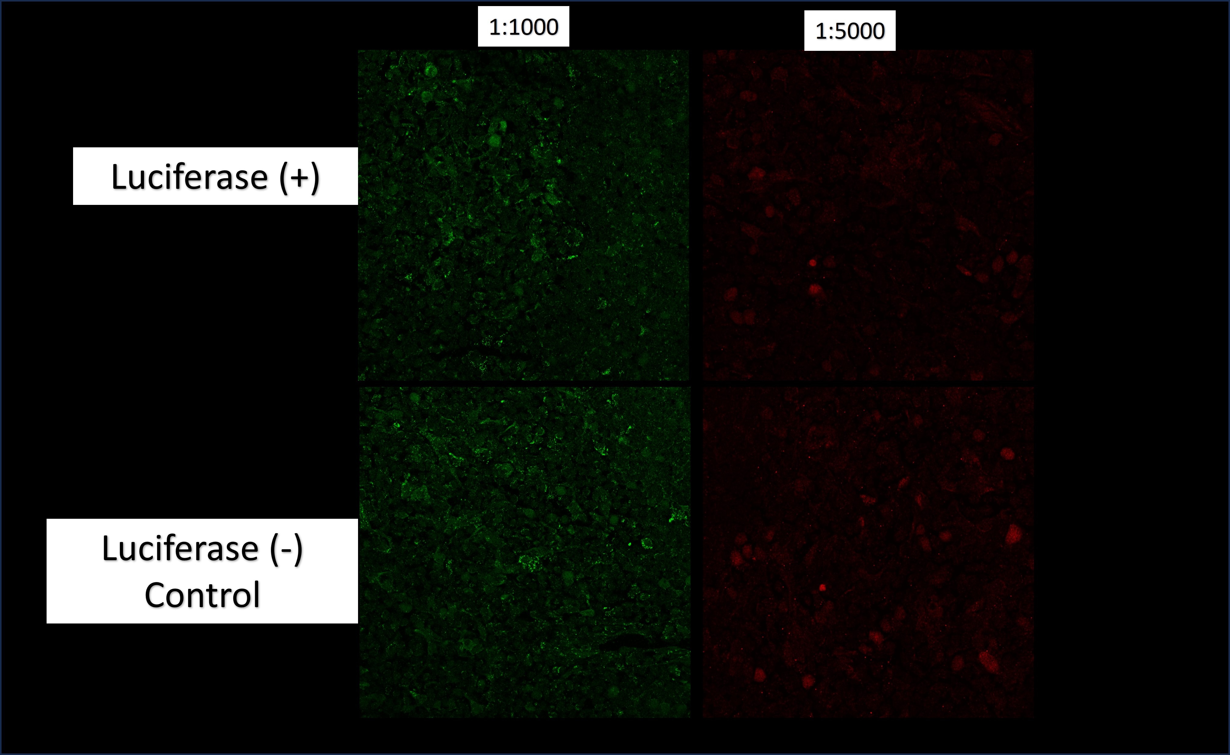
| Application Notes | ICC/IF: A working dilution of 1:5001:1,000 is obtained on methanol-acetone fixed transfected cells using an FITC conjugated secondary antibody. Cells were transfected with a reporter plasmid containing the gene for luciferase. |
|
| Reviewed Applications |
|
| Storage | Store at 4C short term. Aliquot and store at -20C long term. Avoid freeze-thaw cycles. |
| Buffer | 10mM PBS (pH 7.4) |
| Preservative | 0.09% Sodium Azide |
| Purity | IgG purified |
| Images | Ratings | Applications | Species | Date | Details | ||||||||||
|---|---|---|---|---|---|---|---|---|---|---|---|---|---|---|---|
Enlarge |
reviewed by:
Verified Customer |
Immunofluorescence-Frozen | Mouse | 01/31/2024 |
Summary
Comments
|
Secondary Antibodies |
Isotype Controls |
Research Areas for Luciferase Antibody (NB110-17348)Find related products by research area.
|
|
The use of a GFP antibody for research applications in transgenic C. elegans, GFP tagged yeast and porcine model GFP, or green fluorescent protein, is a chemiluminescent protein derived from Aequorea jellyfish that was first discovered by Osamu Shimomura. It was soon after established that the emission spectra of GFP was right around 509nm, or the ultraviol... Read full blog post. |
|
Luciferase: Shining a Light to See Inside Living Animal Models The luciferase reporter is a valuable tool for research into physiology and disease. Light emitted from luciferase enables the monitoring of xenografted tumors, specific cell types, gene expression and pathogens within live animals over time using bio... Read full blog post. |
The concentration calculator allows you to quickly calculate the volume, mass or concentration of your vial. Simply enter your mass, volume, or concentration values for your reagent and the calculator will determine the rest.
5 | |
4 | |
3 | |
2 | |
1 |
| Verified Customer 01/31/2024 |
||
| Application: | Immunofluorescence-Frozen | |
| Species: | Mouse |


![Immunocytochemistry/Immunofluorescence: Luciferase Antibody [NB110-17348] - HEK-293T cells overexpressing luciferase were fixed and stained 10 ug/mL Anti-Luciferase. The antibody was developed using Goat Anti-Rabbit IgG, FITC conjugate.](http://images.novusbio.com/fullsize/Luciferase-Antibody-Immunocytochemistry-Immunofluorescence-NB110-17348-img0004.jpg "Immunocytochemistry/Immunofluorescence: Luciferase Antibody [NB110-17348] - HEK-293T cells overexpressing luciferase were fixed and stained 10 ug/mL Anti-Luciferase. The antibody was developed using Goat Anti-Rabbit IgG, FITC conjugate.")
![Western Blot: Goat anti-Rabbit IgG (H+L) Secondary Antibody [HRP] [NB7160] - Western blot showing vemurafenib treatment in BRAFV600E CRC cells inhibits fission mediator DRP1 with no significant effect on fusion proteins (Mfn1 & 2) using MFN-1 antibody (NBP1-51841) and corresponding secondary antibody, goat anti-rabbit IgG-HRP (NB7160). Image collected and cropped by CiteAb from the following publication (https://pubmed.ncbi.nlm.nih.gov/33738242).](https://images.novusbio.com/images/Goat-anti-Rabbit-IgG-H+L-Secondary-Antibody-HRP-Western-Blot-NB7160-img0001.jpg "Western Blot: Goat anti-Rabbit IgG (H+L) Secondary Antibody [HRP] [NB7160] - Western blot showing vemurafenib treatment in BRAFV600E CRC cells inhibits fission mediator DRP1 with no significant effect on fusion proteins (Mfn1 & 2) using MFN-1 antibody (NBP1-51841) and corresponding secondary antibody, goat anti-rabbit IgG-HRP (NB7160). Image collected and cropped by CiteAb from the following publication (https://pubmed.ncbi.nlm.nih.gov/33738242).")
 followed by 30 min incubation with Goat anti Rabbit HRP conjugated secondary antibodies (Catalog # HAF008) at 1:20 dilution + DAB chromogen (brown). The tissue was counterstained with Hematoxylin (blue). Control was done by omitting primary antibody.")
![Flow Cytometry: Rabbit IgG Isotype Control [NBP2-24891] - An intracellular stain was performed on Raji cells with Adiponectin antibody NB100-65810 (blue) and a matched isotype control NBP2-24893 (orange). Cells were fixed with 4% PFA and then permeablized with 0.1% saponin. Cells were incubated in an antibody dilution of 1 ug/mL for 30 minutes at room temperature, followed by Dylight488-conjugated anti-rabbit secondary antibody. Image using the Azide Free form of this antibody.](https://images.novusbio.com/images/Rabbit--Mouse-IgG-Isotype-Control-Flow-Cytometry-NBP2-24891-img0006.jpg "Flow Cytometry: Rabbit IgG Isotype Control [NBP2-24891] - An intracellular stain was performed on Raji cells with Adiponectin antibody NB100-65810 (blue) and a matched isotype control NBP2-24893 (orange). Cells were fixed with 4% PFA and then permeablized with 0.1% saponin. Cells were incubated in an antibody dilution of 1 ug/mL for 30 minutes at room temperature, followed by Dylight488-conjugated anti-rabbit secondary antibody. Image using the Azide Free form of this antibody.")
