HAF007). A specific band was detected for STING/TMEM173 at approximately 40 kDa (as indicated). This experiment was conducted under reducing conditions and using
Immunoblot Buffer Group 1." class="big_lightbox">
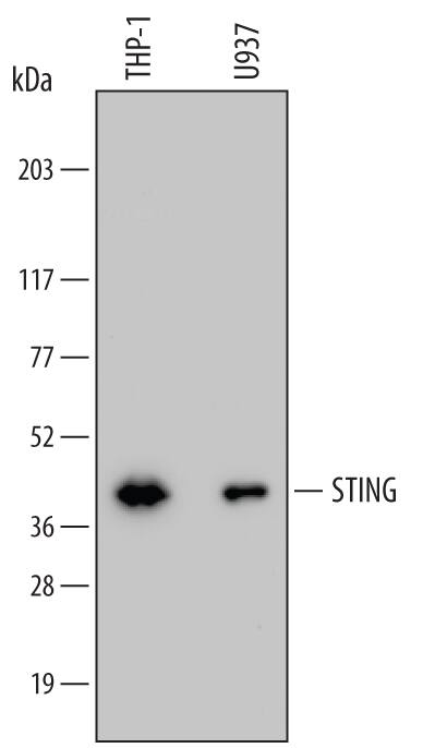
HAF007). A specific band was detected for STING/TMEM173 at approximately 40 kDa (as indicated). This experiment was conducted under reducing conditions and using
Immunoblot Buffer Group 1." alt="Western blot shows lysates of THP-1 human acute monocytic leukemia cell line and U937 human histiocytic lymphoma cell line. PVDF membrane was probed with 0.2 µg/mL of Mouse Anti-Human STING/TMEM173 Monoclonal Antibody (Catalog # MAB7169) followed by HRP-conjugated Anti-Mouse IgG Secondary Antibody (
HAF007). A specific band was detected for STING/TMEM173 at approximately 40 kDa (as indicated). This experiment was conducted under reducing conditions and using
Immunoblot Buffer Group 1." class="big_thumb" />
MAB0041, open histogram), followed by Allophycocyanin-conjugated Anti-Mouse IgG Secondary Antibody (
F0101B). To facilitate intracellular staining, cells were fixed with paraformaldehyde and permeabilized with saponin." class="big_lightbox">
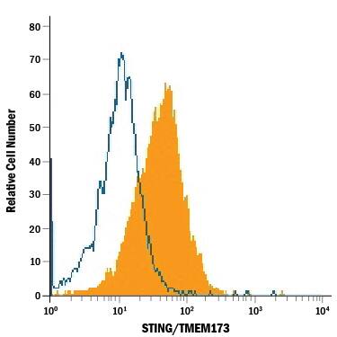
MAB0041, open histogram), followed by Allophycocyanin-conjugated Anti-Mouse IgG Secondary Antibody (
F0101B). To facilitate intracellular staining, cells were fixed with paraformaldehyde and permeabilized with saponin." class="big_thumb" />
MAB0041, open histogram), followed by Allophycocyanin-conjugated Anti-Mouse IgG Secondary Antibody (
F0101B). To facilitate intracellular staining, cells were fixed with paraformaldehyde and permeabilized with saponin." class="big_lightbox">
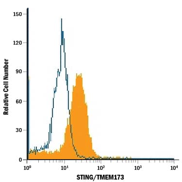
MAB0041, open histogram), followed by Allophycocyanin-conjugated Anti-Mouse IgG Secondary Antibody (
F0101B). To facilitate intracellular staining, cells were fixed with paraformaldehyde and permeabilized with saponin." class="big_thumb" />
MAB0041, open histogram), followed by Allophycocyanin-conjugated Anti-Mouse IgG Secondary Antibody (
F0101B). To facilitate intracellular staining, cells were fixed with paraformaldehyde and permeabilized with saponin." class="big_lightbox">
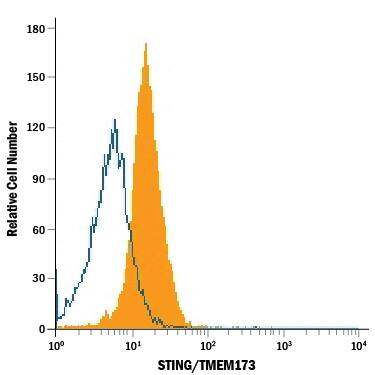
MAB0041, open histogram), followed by Allophycocyanin-conjugated Anti-Mouse IgG Secondary Antibody (
F0101B). To facilitate intracellular staining, cells were fixed with paraformaldehyde and permeabilized with saponin." class="big_thumb" />
 HAF007). A specific band was detected for STING/TMEM173 at approximately 40 kDa (as indicated). This experiment was conducted under reducing conditions and using Immunoblot Buffer Group 1." alt="Western blot shows lysates of THP-1 human acute monocytic leukemia cell line and U937 human histiocytic lymphoma cell line. PVDF membrane was probed with 0.2 µg/mL of Mouse Anti-Human STING/TMEM173 Monoclonal Antibody (Catalog # MAB7169) followed by HRP-conjugated Anti-Mouse IgG Secondary Antibody (HAF007). A specific band was detected for STING/TMEM173 at approximately 40 kDa (as indicated). This experiment was conducted under reducing conditions and using Immunoblot Buffer Group 1." class="big_thumb" />
 DR647RB) and counterstained with DAPI (blue; Lunaphore Catalog # DR100). Specific staining was localized to the membrane. Protocol available in COMET™ Panel Builder.The image is attached." alt="STING/TMEM173 was detected in immersion fixed paraffin-embedded sections of human brain Cortex (Cerebrum) using Rabbit Anti-Human STING/TMEM173, Monoclonal Antibody (Catalog #NBP3-18816) at 10ug/mL at 37 ° Celsius for 4 minutes. Before incubation with the primary antibody, tissue underwent an all-in-one dewaxing and antigen retrieval preprocessing using PreTreatment Module (PT Module) and Dewax and HIER Buffer H (pH 9; Epredia Catalog # TA-999-DHBH). Tissue was stained using the Alexa Fluor™ Plus 647 Goat anti-Rabbit IgG Secondary Antibody at 1:200 at 37 ° Celsius for 2 minutes. (Yellow; Lunaphore Catalog # DR647RB) and counterstained with DAPI (blue; Lunaphore Catalog # DR100). Specific staining was localized to the membrane. Protocol available in COMET™ Panel Builder.The image is attached." class="big_thumb" />
 HAF016). A specific band was detected for STING/TMEM173 at approximately 42 kDa (as indicated). This experiment was conducted under reducing conditions and using Immunoblot Buffer Group 8." alt="Western blot shows lysates of human peripheral blood lymphocytes (PBL). PVDF Membrane was probed with 1 µg/mL of Human/Mouse STING/TMEM173 Antigen Affinity-purified Polyclonal Antibody (Catalog # AF6516) followed by HRP-conjugated Anti-Sheep IgG Secondary Antibody (Catalog # HAF016). A specific band was detected for STING/TMEM173 at approximately 42 kDa (as indicated). This experiment was conducted under reducing conditions and using Immunoblot Buffer Group 8." class="big_thumb" />


 MAB0041, open histogram), followed by Allophycocyanin-conjugated Anti-Mouse IgG Secondary Antibody (
 MAB0041, open histogram), followed by Allophycocyanin-conjugated Anti-Mouse IgG Secondary Antibody (
 MAB0041, open histogram), followed by Allophycocyanin-conjugated Anti-Mouse IgG Secondary Antibody (
 NL007) and counterstained with DAPI (blue). Specific staining was localized to cytoplasm. View our protocol for


![Western Blot: STING/TMEM173 Antibody [NBP2-24683] - STING/TMEM173 expression was induced in human breast MDA-MB-231 cells followed by Western blotting using STING/TMEM173 Antibody antibody (1:1000). Only one specific band at an apparent molecular mass of 37 kDa was observed. Image from verified customer review.](http://images.novusbio.com/fullsize/STING-TMEM173-Antibody-Western-Blot-NBP2-24683-img0014.jpg "Western Blot: STING/TMEM173 Antibody [NBP2-24683] - STING/TMEM173 expression was induced in human breast MDA-MB-231 cells followed by Western blotting using STING/TMEM173 Antibody antibody (1:1000). Only one specific band at an apparent molecular mass of 37 kDa was observed. Image from verified customer review.")
![Immunocytochemistry/Immunofluorescence: STING/TMEM173 Antibody [NBP2-24683] - RH-30 cells were fixed for 10 minutes using 10% formalin and then permeabilized for 5 minutes using 1X PBS + 0.05% Triton X-100. The cells were incubated with STING/TMEM173 Antibody at 2 ug/mL overnight at 4C and detected with an anti-rabbit DyLight 488 (Green) at a 1:500 dilution. Alpha Tubulin Antibody (DM1A) (NB100-690) was used as a co-stain at a 1:1000 dilution and detected with an anti-mouse Dylight 550 (Red) at a 1:500 dilution. Nuclei were counterstained with DAPI (Blue). Cells were imaged using a 40X objective.](http://images.novusbio.com/fullsize/STING-TMEM173-Antibody-Immunocytochemistry-Immunofluorescence-NBP2-24683-img0017.jpg)
![Western Blot: STING/TMEM173 Antibody [NBP2-24683] - Total protein from THP-1, HT-29, U2OS cells and human spleen was separated on a 12% gel by SDS-PAGE, transferred to PVDF membrane and blocked in 5% non-fat milk in TBST. The membrane was probed with 2.0 ug/ml STING/TMEM173 Antibody in 1% non-fat milk in TBST and detected with an anti-rabbit HRP secondary antibody using chemiluminescence.](http://images.novusbio.com/fullsize/STING-TMEM173-Antibody-Western-Blot-NBP2-24683-img0011.jpg)
![Immunohistochemistry-Paraffin: STING/TMEM173 Antibody [NBP2-24683] - Human colon cancer tissue section using STING/TMEM173 Antibody at 1:100 dilution with detection employing HRP-conjugated secondary antibody. The signal was developed using DAB reagent and the nuclei were counterstained with hematoxylin. The antibody generated very weak cytoplasmic staining in columnar epithelial cells with a very strong signal in the secretory/goblet cells.](http://images.novusbio.com/fullsize/STING-TMEM173-Antibody-Immunohistochemistry-Paraffin-NBP2-24683-img0006.jpg)
![Immunohistochemistry-Paraffin: STING/TMEM173 Antibody [NBP2-24683] - Mouse lung tissue section using STING/TMEM173 Antibody at 1:150 dilution with detection employing HRP-conjugated secondary antibody. The signal was developed using DAB reagent and the nuclei were counterstained with hematoxylin. The antibody generated mainly a cytoplasmic staining in the bronchiolar and alveolar epithelial cells.](http://images.novusbio.com/fullsize/STING-TMEM173-Antibody-Immunohistochemistry-Paraffin-NBP2-24683-img0007.jpg)
![Flow Cytometry: STING/TMEM173 Antibody [NBP2-24683] - An intracellular stain was performed on U937 cells with NBP2-24683AF594 (blue) and a matched isotype control (orange). Cells were fixed with 4% PFA and then permeabilized with 0.1% saponin. Cells were incubated in an antibody dilution of 2.5 ug/mL for 30 minutes at room temperature. Both antibodies were conjugated to Alexa Fluor 594.](http://images.novusbio.com/fullsize/STING-TMEM173-Antibody-Flow-Cytometry-NBP2-24683-img0020.jpg)
![Flow Cytometry: STING/TMEM173 Antibody - BSA Free [NBP2-24683] - An intracellular stain was performed on THP-1 cells with STING/TMEM173 Antibody NBP2-24683AF647 (blue) and a matched isotype control NBP2-24891 (orange). Cells were fixed with 4% PFA and then permeabilized with 0.1% saponin. Cells were incubated in an antibody dilution of 2.5 ug/mL for 30 minutes at room temperature. Both antibodies were conjugated to Alexa Fluor 647.](http://images.novusbio.com/fullsize/STING-TMEM173-Antibody---BSA-Free-Flow-Cytometry-NBP2-24683-img0021.jpg)
![Immunocytochemistry/Immunofluorescence: STING/TMEM173 Antibody [NBP2-24683] - HT-29 cells were fixed for 10 minutes using 10% formalin and then permeabilized for 5 minutes using 1X PBS + 0.05% Triton-X100. The cells were incubated with STING/TMEM173 Antibody at 10 ug/ml overnight at 4C and detected with an anti-rabbit Dylight 488 (Green) at a 1:500 dilution. Nuclei were counterstained with DAPI (Blue). Cells were imaged using a 40X objective.](http://images.novusbio.com/fullsize/STING-TMEM173-Antibody-Immunocytochemistry-Immunofluorescence-NBP2-24683-img0015.jpg)
![Flow Cytometry: STING/TMEM173 Antibody [NBP2-24683] - An intracellular stain was performed on THP-1 cells with STING/TMEM173 Antibody and a matched isotype control. Cells were fixed with 4% PFA and then permeablized with 0.1% saponin. Cells were incubated in an antibody dilution of 2.5 ug/mL for 30 minutes at room temperature, followed by Rabbit IgG (H+L) Cross-Adsorbed Secondary Antibody.](http://images.novusbio.com/fullsize/STING-TMEM173-Antibody-Flow-Cytometry-NBP2-24683-img0012.jpg)
![Flow Cytometry: STING/TMEM173 Antibody [NBP2-24683] - An intracellular stain was performed on RH30 cells with STING/TMEM173 Antibody (blue) and a matched isotype control (orange). Cells were fixed with 4% PFA and then permeabilized with 0.1% saponin. Cells were incubated in an antibody dilution of 2.5 ug/mL for 30 minutes at room temperature, followed by Rabbit IgG (H+L) Cross-Adsorbed Secondary Antibody.](http://images.novusbio.com/fullsize/STING-TMEM173-Antibody-Flow-Cytometry-NBP2-24683-img0016.jpg)


![Expression of STING and cGAS mRNA and protein in adipocytes. (A) Venn diagram summarizing the number of equally and differently expressed mRNA transcripts of young and adult mouse iAT. A gene network associated with Sting1 was equally expressed by young and adult iAT. A protein–protein interaction map, generated by STRING [30] is shown below the Venn diagram. Extended analysis presented in [24]. (B) Immunofluorescence of in vitro cultured adipocytes from young and adult mouse iAT; nc: nucleus, scale bar 20 μm. (C) Immunostaining of STING and cGAS proteins in the iAT of young mice, showing a region containing both multilocular and unilocular adipocytes. Arrowheads label nuclei; lp: lipid droplet; cyt: cytoplasm; scale bar: 50 μm. (D) Top: Expression of STING1 and CGAS mRNA in human inguinal and abdominal adipose tissue specimens. Linear regression analysis indicates a significant positive correlation between STING1 and CGAS mRNA levels. Each data point represents one tissue donor patient. Bottom: Correlation of donor age and the adipose tissue expression levels of STING1 and CGAS. (E) Immunohistochemistry of STING and cGAS proteins in human adipose tissue, collected from the inguinal-low abdominal region. Nineteen-month-old male infant; arrowheads label nuclei; lp: lipid droplet; cyt: cytoplasm; scale bar: 25 μm. Inlet shows nuclear STING labeling of an in vitro cultured human adipocyte. Scale bar: 20 μm. (F) Body mass index z-score (BMI z-score) and BMI standard deviation score (BMI-SDS) of adipose tissue donors involved in this study. Correlation of BMI z-score with adipose tissue STING1 and CGAS mRNA levels. Image collected and cropped by CiteAb from the following open publication (//pubmed.ncbi.nlm.nih.gov/37830559), licensed under a CC-BY license. Not internally tested by Novus Biologicals.](http://images.novusbio.com/fullsize/nbp2-24683_rabbit-polyclonal-sting-tmem173-antibody-immunocytochemistry-immunofluorescence-13220261944235.jpg)

![Expression of STING and cGAS mRNA and protein in adipocytes. (A) Venn diagram summarizing the number of equally and differently expressed mRNA transcripts of young and adult mouse iAT. A gene network associated with Sting1 was equally expressed by young and adult iAT. A protein–protein interaction map, generated by STRING [30] is shown below the Venn diagram. Extended analysis presented in [24]. (B) Immunofluorescence of in vitro cultured adipocytes from young and adult mouse iAT; nc: nucleus, scale bar 20 μm. (C) Immunostaining of STING and cGAS proteins in the iAT of young mice, showing a region containing both multilocular and unilocular adipocytes. Arrowheads label nuclei; lp: lipid droplet; cyt: cytoplasm; scale bar: 50 μm. (D) Top: Expression of STING1 and CGAS mRNA in human inguinal and abdominal adipose tissue specimens. Linear regression analysis indicates a significant positive correlation between STING1 and CGAS mRNA levels. Each data point represents one tissue donor patient. Bottom: Correlation of donor age and the adipose tissue expression levels of STING1 and CGAS. (E) Immunohistochemistry of STING and cGAS proteins in human adipose tissue, collected from the inguinal-low abdominal region. Nineteen-month-old male infant; arrowheads label nuclei; lp: lipid droplet; cyt: cytoplasm; scale bar: 25 μm. Inlet shows nuclear STING labeling of an in vitro cultured human adipocyte. Scale bar: 20 μm. (F) Body mass index z-score (BMI z-score) and BMI standard deviation score (BMI-SDS) of adipose tissue donors involved in this study. Correlation of BMI z-score with adipose tissue STING1 and CGAS mRNA levels. Image collected and cropped by CiteAb from the following open publication (//pubmed.ncbi.nlm.nih.gov/37830559), licensed under a CC-BY license. Not internally tested by Novus Biologicals.](http://images.novusbio.com/fullsize/nbp2-24683_rabbit-polyclonal-sting-tmem173-antibody-immunohistochemistry-132202619354822.jpg)
![Expression of STING and cGAS mRNA and protein in adipocytes. (A) Venn diagram summarizing the number of equally and differently expressed mRNA transcripts of young and adult mouse iAT. A gene network associated with Sting1 was equally expressed by young and adult iAT. A protein–protein interaction map, generated by STRING [30] is shown below the Venn diagram. Extended analysis presented in [24]. (B) Immunofluorescence of in vitro cultured adipocytes from young and adult mouse iAT; nc: nucleus, scale bar 20 μm. (C) Immunostaining of STING and cGAS proteins in the iAT of young mice, showing a region containing both multilocular and unilocular adipocytes. Arrowheads label nuclei; lp: lipid droplet; cyt: cytoplasm; scale bar: 50 μm. (D) Top: Expression of STING1 and CGAS mRNA in human inguinal and abdominal adipose tissue specimens. Linear regression analysis indicates a significant positive correlation between STING1 and CGAS mRNA levels. Each data point represents one tissue donor patient. Bottom: Correlation of donor age and the adipose tissue expression levels of STING1 and CGAS. (E) Immunohistochemistry of STING and cGAS proteins in human adipose tissue, collected from the inguinal-low abdominal region. Nineteen-month-old male infant; arrowheads label nuclei; lp: lipid droplet; cyt: cytoplasm; scale bar: 25 μm. Inlet shows nuclear STING labeling of an in vitro cultured human adipocyte. Scale bar: 20 μm. (F) Body mass index z-score (BMI z-score) and BMI standard deviation score (BMI-SDS) of adipose tissue donors involved in this study. Correlation of BMI z-score with adipose tissue STING1 and CGAS mRNA levels. Image collected and cropped by CiteAb from the following open publication (//pubmed.ncbi.nlm.nih.gov/37830559), licensed under a CC-BY license. Not internally tested by Novus Biologicals.](http://images.novusbio.com/fullsize/nbp2-24683_rabbit-polyclonal-sting-tmem173-antibody-immunohistochemistry-132202619213316.jpg)
 DR647RB) and counterstained with DAPI (blue; Lunaphore Catalog #
  DR647RB) and counterstained with DAPI (blue; Lunaphore Catalog #
  DR647RB) and counterstained with DAPI (blue; Lunaphore Catalog #
 ![Western Blot: STING/TMEM173 Antibody (2922D) [NBP3-18816] - Western blot shows cell lysates K562, THP-1, C2C12, and C6. Membrane was probed with 1 ug/mL of (Catalog # NBP3-18816) followed by HRP-conjugated Anti-Rabbit IgG Secondary Antibody (Catalog # HAF008). A specific band was detected for STING/TMEM173 at approximately 42 kDa (as indicated). This experiment was conducted under reducing conditions and using Immunoblot Buffer Group 1. Internally validated Western blot.](http://images.novusbio.com/fullsize/STING-TMEM173-Antibody-2922D-Western-Blot-NBP3-18816-img0003.jpg)
![Immunocytochemistry/Immunofluorescence: STING/TMEM173 Antibody (2292D) [NBP3-18816] - STING/TMEM173 was detected in immersion fixed U937 human myeloid leukaemia cell line but is not detected in Daudi cell line using Rabbit Anti-Human STING/TMEM173 Monoclonal Antibody (Catalog # NBP3-18816) at 1 ug/mL for 3 hours at room temperature. Cells were stained using the NorthernLights(TM) 557-conjugated Anti-Rabbit IgG Secondary Antibody (red; Catalog # NL004) and counterstained with DAPI (blue).](http://images.novusbio.com/fullsize/STING-TMEM173-Antibody-2292D-Immunocytochemistry-Immunofluorescence-NBP3-18816-img0002.jpg)
![Immunohistochemistry: STING/TMEM173 Antibody (2292D) [NBP3-18816] - STING/TMEM173 was detected in immersion fixed paraffin-embedded sections of mouse lung tissue using Rabbit Anti-Human STING/TMEM173 Monoclonal Antibody at 0.5 ug/mL for 1 hour at room temperature followed by incubation with the Anti-Rabbit IgG VisUCyte™ HRP Polymer Antibody. Tissue was stained using DAB (brown) and counterstained with hematoxylin (blue). Specific staining was localized to pneumocytes and alveolar cells. Staining with VisUCyte HRP Polymer Detection Reagents.](http://images.novusbio.com/fullsize/STING-TMEM173-Antibody-2292D-Immunohistochemistry-NBP3-18816-img0001.jpg)
![Simple Western: STING/TMEM173 Antibody (2922D) [NBP3-18816] - Simple Western Lane view shows K562 and THP-1 whole cell lysate (WCL). A specific band was detected for hSTING/TMEM173 antibody (NBP3-18816) at approximately 44 kDa (as indicated) using 10 ug/mL of hSTING/TMEM173 antibody. This experiment was conducted under reducing conditions.](http://images.novusbio.com/fullsize/STING-TMEM173-Antibody-2922D-Simple-Western-NBP3-18816-img0004.jpg)


 HAF016). This experiment was conducted under reducing conditions and using the 12-230 kDa separation system." class="big_thumb" />


![Western Blot: STING/TMEM173 Antibody [NBP2-48684] - Analysis in human cell line HDLM-2 using STING/TMEM173 Antibody.](http://images.novusbio.com/fullsize/STING-TMEM173-Antibody-Western-Blot-NBP2-48684-img0003.jpg "Western Blot: STING/TMEM173 Antibody [NBP2-48684] - Analysis in human cell line HDLM-2 using STING/TMEM173 Antibody.")


![Western Blot: STING/TMEM173 Antibody (OTI4H1) [NBP2-45998] - Analysis of HEK293T cells were transfected with the pCMV6-ENTRY control (Left lane) or pCMV6-ENTRY TMEM173.](http://images.novusbio.com/fullsize/STING-TMEM173-Antibody-OTI4H1-Western-Blot-NBP2-45998-img0012.jpg "Western Blot: STING/TMEM173 Antibody (OTI4H1) [NBP2-45998] - Analysis of HEK293T cells were transfected with the pCMV6-ENTRY control (Left lane) or pCMV6-ENTRY TMEM173.")
![Immunohistochemistry: STING/TMEM173 Antibody (OTI4H1) [NBP2-45998] - Analysis of Adenocarcinoma of Human breast tissue using STING/TMEM173 Antibody (OTI4H1). (Heat-induced epitope retrieval by 10mM citric buffer, pH6.0, 120C for 3min)](http://images.novusbio.com/fullsize/STING-TMEM173-Antibody-OTI4H1-Immunohistochemistry-NBP2-45998-img0011.jpg)
![Western Blot: STING/TMEM173 Antibody (OTI4H1) [NBP2-45998] - Analysis of extracts (35ug) from 9 different cell lines (HepG2: human; HeLa: human; SVT2: mouse; A549: human; COS7: monkey; Jurkat: human; MDCK: canine; PC12: rat; MCF7: human).](http://images.novusbio.com/fullsize/STING-TMEM173-Antibody-OTI4H1-Western-Blot-NBP2-45998-img0013.jpg)
![Immunohistochemistry: STING/TMEM173 Antibody (OTI4H1) [NBP2-45998] - Analysis of Human pancreas tissue using STING/TMEM173 Antibody (OTI4H1). (Heat-induced epitope retrieval by 10mM citric buffer, pH6.0, 120C for 3min)](http://images.novusbio.com/fullsize/STING-TMEM173-Antibody-OTI4H1-Immunohistochemistry-NBP2-45998-img0001.jpg)
![Immunohistochemistry: STING/TMEM173 Antibody (OTI4H1) [NBP2-45998] - Analysis of Carcinoma of Human bladder tissue using STING/TMEM173 Antibody (OTI4H1). (Heat-induced epitope retrieval by 10mM citric buffer, pH6.0, 120C for 3min)](http://images.novusbio.com/fullsize/STING-TMEM173-Antibody-OTI4H1-Immunohistochemistry-NBP2-45998-img0002.jpg)
![Immunohistochemistry: STING/TMEM173 Antibody (OTI4H1) [NBP2-45998] - Analysis of Human thyroid tissue using STING/TMEM173 Antibody (OTI4H1). (Heat-induced epitope retrieval by 10mM citric buffer, pH6.0, 120C for 3min)](http://images.novusbio.com/fullsize/STING-TMEM173-Antibody-OTI4H1-Immunohistochemistry-NBP2-45998-img0003.jpg)
![Immunohistochemistry: STING/TMEM173 Antibody (OTI4H1) [NBP2-45998] - Analysis of Carcinoma of Human thyroid tissue using STING/TMEM173 Antibody (OTI4H1). (Heat-induced epitope retrieval by 10mM citric buffer, pH6.0, 120C for 3min)](http://images.novusbio.com/fullsize/STING-TMEM173-Antibody-OTI4H1-Immunohistochemistry-NBP2-45998-img0004.jpg)
![Immunohistochemistry: STING/TMEM173 Antibody (OTI4H1) [NBP2-45998] - Analysis of Adenocarcinoma of Human ovary tissue using STING/TMEM173 Antibody (OTI4H1). (Heat-induced epitope retrieval by 10mM citric buffer, pH6.0, 120C for 3min)](http://images.novusbio.com/fullsize/STING-TMEM173-Antibody-OTI4H1-Immunohistochemistry-NBP2-45998-img0005.jpg)
![Immunohistochemistry: STING/TMEM173 Antibody (OTI4H1) [NBP2-45998] - Analysis of Human prostate tissue using STING/TMEM173 Antibody (OTI4H1). (Heat-induced epitope retrieval by 10mM citric buffer, pH6.0, 120C for 3min)](http://images.novusbio.com/fullsize/STING-TMEM173-Antibody-OTI4H1-Immunohistochemistry-NBP2-45998-img0006.jpg)
![Immunohistochemistry: STING/TMEM173 Antibody (OTI4H1) [NBP2-45998] - Analysis of Carcinoma of Human pancreas tissue using STING/TMEM173 Antibody (OTI4H1). (Heat-induced epitope retrieval by 10mM citric buffer, pH6.0, 120C for 3min)](http://images.novusbio.com/fullsize/STING-TMEM173-Antibody-OTI4H1-Immunohistochemistry-NBP2-45998-img0007.jpg)
![Immunohistochemistry: STING/TMEM173 Antibody (OTI4H1) [NBP2-45998] - Analysis of Carcinoma of Human lung tissue using STING/TMEM173 Antibody (OTI4H1). (Heat-induced epitope retrieval by 10mM citric buffer, pH6.0, 120C for 3min)](http://images.novusbio.com/fullsize/STING-TMEM173-Antibody-OTI4H1-Immunohistochemistry-NBP2-45998-img0008.jpg)
![Immunohistochemistry: STING/TMEM173 Antibody (OTI4H1) [NBP2-45998] - Analysis of Adenocarcinoma of Human colon tissue STING/TMEM173 Antibody (OTI4H1). (Heat-induced epitope retrieval by 10mM citric buffer, pH6.0, 120C for 3min)](http://images.novusbio.com/fullsize/STING-TMEM173-Antibody-OTI4H1-Immunohistochemistry-NBP2-45998-img0009.jpg)
![Immunohistochemistry: STING/TMEM173 Antibody (OTI4H1) [NBP2-45998] - Analysis of Carcinoma of Human prostate tissue using STING/TMEM173 Antibody (OTI4H1). (Heat-induced epitope retrieval by 10mM citric buffer, pH6.0, 120C for 3min)](http://images.novusbio.com/fullsize/STING-TMEM173-Antibody-OTI4H1-Immunohistochemistry-NBP2-45998-img0010.jpg)
![STING/TMEM173 Antibody (OTI4H1)[NBP2-45998]Simple Western� analysis of endogenous protein IDH1 from HT-29 lysates (0.2 mg/mL) using IDH1 Mouse Monoclonal Antibody . The virtual lane view (left) shows the target (as indicated) at 1:50 dilution of primary antibody. The corresponding electropherogram view (right) plots chemiluminescence by molecular weight along the capillary at a 1:50 dilution of primary antibody. This experiment was performed under reducing conditions on the Jess� Simple Western instrument from ProteinSimple, a Bio-Techne brand, using the 12�230 kDa Separation Module.](http://images.novusbio.com/fullsize/nbp2-45998_mouse-sting-tmem173-mab-oti4h1-simple-western-131120251236224.jpg%253fstore=america&image-type=image)
![Western Blot: STING/TMEM173 Antibody [NBP2-38389] - Analysis in human cell line EFO-21.](http://images.novusbio.com/fullsize/STING-TMEM173-Antibody-Western-Blot-NBP2-38389-img0004.jpg "Western Blot: STING/TMEM173 Antibody [NBP2-38389] - Analysis in human cell line EFO-21.")


 MAB7169) with an ED50 of <50.0 ng/mL." alt="Recombinant Human STING/TMEM173 His-tag Protein (Catalog # 11821-ST) binds Human STING/TMEM173 Antibody (


![Western Blot: STING/TMEM173 Antibody (S06-9A7) [NBP3-19785] - Western blot detection of STING/TMEM173 in K562, Rat Brain lysates using NBP3-19785 (1:1000 diluted). Predicted band size: 42kDa. Observed band size: 40kDa.](http://images.novusbio.com/fullsize/STING-TMEM173-Antibody-S06-9A7-Western-Blot-NBP3-19785-img0002.jpg "Western Blot: STING/TMEM173 Antibody (S06-9A7) [NBP3-19785] - Western blot detection of STING/TMEM173 in K562, Rat Brain lysates using NBP3-19785 (1:1000 diluted). Predicted band size: 42kDa. Observed band size: 40kDa.")
![Immunohistochemistry-Paraffin: STING/TMEM173 Antibody (S06-9A7) [NBP3-19785] - Immunohistochemistry of STING/TMEM173 in paraffin-embedded Human tonsil using NBP3-19785 at dilution 1/50](http://images.novusbio.com/fullsize/STING-TMEM173-Antibody-S06-9A7-Immunohistochemistry-Paraffin-NBP3-19785-img0001.jpg)
![Immunohistochemistry-Paraffin: STING/TMEM173 Antibody (STING1/7431) [NBP3-13941] - Formalin-fixed, paraffin-embedded human tonsil stained with STING/TMEM173 antibody (STING1/7431). Inset: PBS instead of primary antibody; secondary only negative control.](http://images.novusbio.com/fullsize/STING-TMEM173-Antibody-STING1-7431-Immunohistochemistry-Paraffin-NBP3-13941-img0001.jpg "Immunohistochemistry-Paraffin: STING/TMEM173 Antibody (STING1/7431) [NBP3-13941] - Formalin-fixed, paraffin-embedded human tonsil stained with STING/TMEM173 antibody (STING1/7431). Inset: PBS instead of primary antibody; secondary only negative control.")
![Immunohistochemistry-Paraffin: STING/TMEM173 Antibody (STING1/7431) [NBP3-13941] - Formalin-fixed, paraffin-embedded human tonsil stained with STING/TMEM173 antibody (STING1/7431). HIER: Tris/EDTA, pH9.0, 45min. HRP-polymer, 30min. DAB, 5min.](http://images.novusbio.com/fullsize/STING-TMEM173-Antibody-STING1-7431-Immunohistochemistry-Paraffin-NBP3-13941-img0002.jpg)


