DR647MS) and counterstained with DAPI (blue; Lunaphore Catalog #
DR100). Specific staining was localized to the extracellular space. Protocol available in
COMET™ Panel Builder." class="big_lightbox">
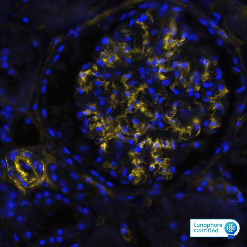
DR647MS) and counterstained with DAPI (blue; Lunaphore Catalog #
DR100). Specific staining was localized to the extracellular space. Protocol available in
COMET™ Panel Builder." alt="COL4A1 was detected in immersion fixed paraffin-embedded sections of human Kidney using Mouse Anti-Human COL4A1, Monoclonal Antibody (Catalog#MAB11758) at 15ug/mL at 37 ° Celsius for 8 minutes. Before incubation with the primary antibody, tissue underwent an all-in-one dewaxing and antigen retrieval preprocessing using PreTreatment Module (PT Module)and Dewax and HIER Buffer H (pH 9; Epredia Catalog #TA-999-DHBH).Tissue was stained using the Alexa Fluor™ 647 Goat anti-Mouse IgG Secondary Antibody at 1:200 at 37 ° Celsius for 2 minutes. (Yellow; Lunaphore Catalog #
DR647MS) and counterstained with DAPI (blue; Lunaphore Catalog #
DR100). Specific staining was localized to the extracellular space. Protocol available in
COMET™ Panel Builder." class="big_thumb" />
DR647MS) and counterstained with DAPI (blue; Lunaphore Catalog #
DR100). Specific staining was localized to the extracellular space. Protocol available in
COMET™ Panel Builder." class="big_lightbox">
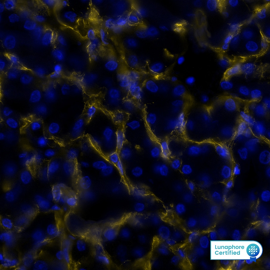
DR647MS) and counterstained with DAPI (blue; Lunaphore Catalog #
DR100). Specific staining was localized to the extracellular space. Protocol available in
COMET™ Panel Builder." class="big_thumb" />
DR647MS) and counterstained with DAPI (blue; Lunaphore Catalog #
DR100). Specific staining was localized to the extracellular space. Protocol available in
COMET™ Panel Builder." class="big_lightbox">
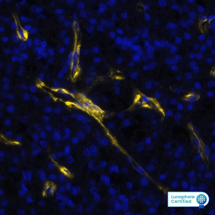
DR647MS) and counterstained with DAPI (blue; Lunaphore Catalog #
DR100). Specific staining was localized to the extracellular space. Protocol available in
COMET™ Panel Builder." class="big_thumb" />
 DR555RB) and counterstained with DAPI (blue; Lunaphore Catalog # DR100). Specific staining was localized to the cell membrane. Protocol available in COMET™ Panel Builder." alt="Collagen IV was detected in immersion fixed paraffin-embedded sections of human liver using Rabbit Anti-Human/Mouse/Rat Collagen IV alpha 1 Monoclonal Antibody (Novus Catalog # NBP3-25416) at 1µg/mL at 37° Celsius for 4 minutes. Before incubation with the primary antibody, tissue underwent an all-in-one dewaxing and antigen retrieval preprocessing using PreTreatment Module (PT Module) and Dewax and HIER Buffer H (pH 9). Tissue was stained using the Alexa Fluor™ Plus 555 Goat anti-Rabbit IgG Secondary Antibody at 1:100 at 37 ° Celsius for 2 minutes. (Yellow; Lunaphore Catalog # DR555RB) and counterstained with DAPI (blue; Lunaphore Catalog # DR100). Specific staining was localized to the cell membrane. Protocol available in COMET™ Panel Builder." class="big_thumb" />
 DR647MS) and counterstained with DAPI (blue; Lunaphore Catalog # DR100). Specific staining was localized to the extracellular space. Protocol available in COMET™ Panel Builder." alt="COL4A1 was detected in immersion fixed paraffin-embedded sections of human Kidney using Mouse Anti-Human COL4A1, Monoclonal Antibody (Catalog#MAB11758) at 15ug/mL at 37 ° Celsius for 8 minutes. Before incubation with the primary antibody, tissue underwent an all-in-one dewaxing and antigen retrieval preprocessing using PreTreatment Module (PT Module)and Dewax and HIER Buffer H (pH 9; Epredia Catalog #TA-999-DHBH).Tissue was stained using the Alexa Fluor™ 647 Goat anti-Mouse IgG Secondary Antibody at 1:200 at 37 ° Celsius for 2 minutes. (Yellow; Lunaphore Catalog # DR647MS) and counterstained with DAPI (blue; Lunaphore Catalog # DR100). Specific staining was localized to the extracellular space. Protocol available in COMET™ Panel Builder." class="big_thumb" />


![Immunohistochemistry-Paraffin: Collagen IV Antibody [NB120-6586] - IHC-P image submitted by a verified customer review.](http://images.novusbio.com/fullsize/Collagen-IV-Antibody-Immunohistochemistry-Paraffin-NB120-6586-img0013.jpg)
![Immunohistochemistry-Paraffin: Collagen IV Antibody [NB120-6586] - Analysis of Collagen IV in various paraffin embedded human tissues. Image courtesy of product review submitted by Daniel Pirici.](http://images.novusbio.com/fullsize/Collagen-IV-Antibody-Immunohistochemistry-Paraffin-NB120-6586-img0006.jpg)
![Immunohistochemistry-Paraffin: Collagen IV Antibody [NB120-6586] - Collagen IV antibody at 1:400, 45 min RT showed strong staining in FFPE sections of human kidney (left) with strong red staining observed in glomeruli and liver (right) with strong staining in sinusoids. Staining for both tissues was consistent with a basement membrane distribution. Slides were steamed in 0.01 M sodium citrate buffer, pH 6.0 at 100C for 20 minutes for antigen retrieval.](http://images.novusbio.com/fullsize/Collagen-IV-Antibody-Immunohistochemistry-Paraffin-NB120-6586-img0008.jpg)
![Immunohistochemistry-Paraffin: Collagen IV Antibody [NB120-6586] - Analysis of Collagen IV in paraffin embedded mouse cervix tissue using anti-Collagen IV antibody. IHC-P image submitted by a verified customer review.](http://images.novusbio.com/fullsize/Collagen-IV-Antibody-Immunohistochemistry-Paraffin-NB120-6586-img0010.jpg)
![Immunohistochemistry-Paraffin: Collagen IV Antibody [NB120-6586] - Analysis using the Biotin conjugate of NB120-6586. Staining of Collagen IV (red) and Insulin (green) in mouse pancreas. IHC-P image submitted by a verified customer review.](http://images.novusbio.com/fullsize/Collagen-IV-Antibody-Immunohistochemistry-Paraffin-NB120-6586-img0009.jpg)
![Immunohistochemistry-Frozen: Collagen IV Antibody [NB120-6586] - Mouse liver tissue. Collagen IV antibody at 1:200 dilution. Overnight incubation, no permeabilization. IHC-Fr image submitted by a verified customer review.](http://images.novusbio.com/fullsize/Collagen-IV-Antibody-Immunohistochemistry-Frozen-NB120-6586-img0011.jpg)
![Immunohistochemistry-Frozen: Collagen IV Antibody [NB120-6586] - Mouse brain tissue. Antibody at 1:500. IHC-Fr image submitted by a verified customer review.](http://images.novusbio.com/fullsize/Collagen-IV-Antibody-Immunohistochemistry-Frozen-NB120-6586-img0012.jpg)
![<b>Genetic Strategies Validation. </b>Immunocytochemistry/ Immunofluorescence: Collagen IV Antibody [NB120-6586] - Collagen IV was detected in immersion fixed THP-1 (positive) but not THP-1 KO (negative) cells using 10 ug/mL Rabbit Anti-Human/Bovine Polyclonal Antibody (Catalog # NB120-6586) for 3 hours at room temperature. Cells were stained with the NorthernLights(TM) 557-conjugated Donkey anti-Rabbit IgG Secondary Antibody [NL557] (red; Catalog # NL004) and counterstained with DAPI (blue).](http://images.novusbio.com/fullsize/Collagen-IV-Antibody-Knockout-Validated-NB120-6586-img0016.jpg)
![Immunohistochemistry-Frozen: Collagen IV Antibody [NBP1-26549] - Left panel: NBP1-26549 signal, identifying Collagen IV in extracellular basement membrane of brain endothelial cells. Right panel: counterstained image with DAPI. Mouse brain tissue (12um thickness) was incubated overnight with NBP1-26549 (1:400) in PBS 1x at 4C. The day after, the slide was washed 3 x PBS 1x and then incubated with secondary anti-goat antibody (Alex Fluor 568-conjugated), 1:1000 for 2 hours at r.t. After counterstaining with DAPI, image was acquired with confocal microscopy. Left panel: NBP1-26549 signal, identifying Collagen IV in extracellular basement membrane of brain endothelial cells. Right panel: counterstained image with DAPI. Image from verified customer review.](http://images.novusbio.com/fullsize/Collagen-IV-Antibody-Immunohistochemistry-Frozen-NBP1-26549-img0004.jpg "Immunohistochemistry-Frozen: Collagen IV Antibody [NBP1-26549] - Left panel: NBP1-26549 signal, identifying Collagen IV in extracellular basement membrane of brain endothelial cells. Right panel: counterstained image with DAPI. Mouse brain tissue (12um thickness) was incubated overnight with NBP1-26549 (1:400) in PBS 1x at 4C. The day after, the slide was washed 3 x PBS 1x and then incubated with secondary anti-goat antibody (Alex Fluor 568-conjugated), 1:1000 for 2 hours at r.t. After counterstaining with DAPI, image was acquired with confocal microscopy. Left panel: NBP1-26549 signal, identifying Collagen IV in extracellular basement membrane of brain endothelial cells. Right panel: counterstained image with DAPI. Image from verified customer review.")
![Immunohistochemistry-Paraffin: Collagen IV Antibody [NBP1-26549] - Human gastric cancer tissue was stained with Goat IgG-UNLB isotype control (left) and Goat Anti-Type IV Collagen-UNLB; (right) followed by Swine Anti-Goat IgG(H+L), Human/Rat/Mouse SP ads-HRP DAB, and hematoxylin.](http://images.novusbio.com/fullsize/Collagen-IV-alpha-1-Antibody-Immunohistochemistry-Paraffin-NBP1-26549-img0001.jpg)


![Immunohistochemistry: Collagen IV Antibody [NBP1-26549] - Photographs showing H&E staining & immunostaining for Col-I, Col-III, Col-IV & Col-V in the rhesus macaque & baboon intramural tube. Images of open fallopian tube (a, c, e, g, i, k, m, o, q & s) were obtained from control animals. Occluded tube (b, d, f, h, j, l, n, p, r & t) were from animals treated with 5% PF. Arrows indicate fibrotic response. All images were captured at original magnification ×200. Insets (frame r & s) show negative control with an irrelevant antibody. Treatment with PF increased collagen immunoreactivity in the lamina propria of the intramural tube. Image collected & cropped by CiteAb from the following publication (//linkinghub.elsevier.com/retrieve/pii/S0010782416301445), licensed under a CC-BY license. Not internally tested by Novus Biologicals.](http://images.novusbio.com/fullsize/nbp1-26549_goat-polyclonal-collagen-iv-antibody-310202415395955.jpg)
![Immunohistochemistry: Collagen IV Antibody [NBP1-26549] - Photographs showing H&E staining & immunostaining for Col-I, Col-III, Col-IV & Col-V in the rhesus macaque & baboon intramural tube. Images of open fallopian tube (a, c, e, g, i, k, m, o, q & s) were obtained from control animals. Occluded tube (b, d, f, h, j, l, n, p, r & t) were from animals treated with 5% PF. Arrows indicate fibrotic response. All images were captured at original magnification ×200. Insets (frame r & s) show negative control with an irrelevant antibody. Treatment with PF increased collagen immunoreactivity in the lamina propria of the intramural tube. Image collected & cropped by CiteAb from the following publication (//linkinghub.elsevier.com/retrieve/pii/S0010782416301445), licensed under a CC-BY license. Not internally tested by Novus Biologicals.](http://images.novusbio.com/fullsize/nbp1-26549_goat-polyclonal-collagen-iv-antibody-310202415334930.jpg)
![MiR-29a regulation of target gene expression in cochlea from miR-29a+/+ and miR-29a–/– mice. (A–G) With beta -actin as a control, COL4A1, COL4A2, COL4A3, COL4A4, COL4A5, LAMB2, and LAMC1 protein levels in cochlea were analyzed by Western blot. The independent t-test was used to compare data between two groups [COL4A1, t(4) = 9.920, P < 0.001; COL4A2, t(4) = 3.847, P = 0.018; COL4A3, t(4) = 3.389, P = 0.028; COL4A4, t(4) = 2.842, P = 0.047; COL4A5, t(4) = 3.782, P = 0.019; LAMB2, t(4) = 3.915, P = 0.017; LAMC1, t(4) = 3.088, P = 0.037]. (H) The mRNA expression levels of target genes as measured by qRT-PCR [independent t-test, Col4a1, t(4) = 6.050, P = 0.004; Col4a2, t(4) = 13.145, P < 0.001; Col4a3, t(4) = 3.735, P = 0.02; Col4a4, t(4) = 7.238, P = 0.002; Col4a5, t(4) = 13.141, P < 0.001; Lamb2, t(4) = 8.529, P = 0.001; Lamc1, t(4) = 3.126, P = 0.035]. All data are shown as the mean +/- SD, n = 3 biological replicates, *P < 0.05, **P < 0.01, and ***P < 0.001 between two groups. Image collected and cropped by CiteAb from the following open publication (//pubmed.ncbi.nlm.nih.gov/37275774), licensed under a CC-BY license. Not internally tested by Novus Biologicals.](http://images.novusbio.com/fullsize/nbp1-26549_goat-polyclonal-collagen-iv-antibody-western-blot-132202615104524.jpg)


![Immunohistochemistry-Frozen: Collagen IV Antibody (1043) [NBP1-97716] - Collagen type IV staining of basement membranes and connective tissue in a frozen section of human kidney.](http://images.novusbio.com/fullsize/Collagen-IV-alpha-1-Antibody-1043-Immunohistochemistry-Frozen-NBP1-97716-img0006.jpg)
![Immunohistochemistry-Paraffin: Collagen IV Antibody (1043) [NBP1-97716] - Human colon.](http://images.novusbio.com/fullsize/Collagen-IV-alpha-1-Antibody-1043-Immunohistochemistry-Paraffin-NBP1-97716-img0007.jpg)
![Immunohistochemistry-Frozen: Collagen IV Antibody (1043) [NBP1-97716] - Frozen section of Human colon](http://images.novusbio.com/fullsize/Collagen-IV-alpha-1-Antibody-1043-Immunohistochemistry-Frozen-NBP1-97716-img0005.jpg)

![Immunohistochemistry-Paraffin: Collagen IV Antibody (M3F7) [NBP3-07597] - Formalin-fixed, paraffin-embedded human kidney stained with Collagen IV Mouse Monoclonal Antibody (M3F7).](http://images.novusbio.com/fullsize/Collagen-IV-Antibody-M3F7-Immunohistochemistry-Paraffin-NBP3-07597-img0003.jpg "Immunohistochemistry-Paraffin: Collagen IV Antibody (M3F7) [NBP3-07597] - Formalin-fixed, paraffin-embedded human kidney stained with Collagen IV Mouse Monoclonal Antibody (M3F7).")
![Immunohistochemistry-Paraffin: Collagen IV Antibody (M3F7) [NBP3-07597] - Formalin-fixed, paraffin-embedded human kidney stained with Collagen IV Mouse Monoclonal Antibody (M3F7).](http://images.novusbio.com/fullsize/Collagen-IV-Antibody-M3F7-Immunohistochemistry-Paraffin-NBP3-07597-img0001.jpg)
![Immunohistochemistry-Paraffin: Collagen IV Antibody (M3F7) [NBP3-07597] - Formalin-fixed, paraffin-embedded human kidney stained with Collagen IV Mouse Monoclonal Antibody (M3F7).](http://images.novusbio.com/fullsize/Collagen-IV-Antibody-M3F7-Immunohistochemistry-Paraffin-NBP3-07597-img0002.jpg)
![ELISA: Human Collagen IV ELISA Kit (Colorimetric) [NBP2-75864] - Samples were spiked with high concentrations of Human Collagen IV and diluted with Reference Standard & Sample Diluent to produce samples with values within the range of the assay.](http://images.novusbio.com/fullsize/nbp2-75864_human-collagen-iv-elisa-kit-colorimetric-132202416232394.jpg "ELISA: Human Collagen IV ELISA Kit (Colorimetric) [NBP2-75864] - Samples were spiked with high concentrations of Human Collagen IV and diluted with Reference Standard & Sample Diluent to produce samples with values within the range of the assay.")
![ELISA: Human Collagen IV ELISA Kit (Colorimetric) [NBP2-75864] - Standard Curve Reference](http://images.novusbio.com/fullsize/nbp2-75864_human-collagen-iv-elisa-kit-colorimetric-4320249115622.jpg)
 DR555RB) and counterstained with DAPI (blue; Lunaphore Catalog #
  DR555RB) and counterstained with DAPI (blue; Lunaphore Catalog #
 ![COL4A1 antibody [HL1351] detects COL4A1 protein at vesicle by immunofluorescent analysis.</br>Sample: SK-N-SH cells were fixed in 4% paraformaldehyde at RT for 15 min.</br>Green: COL4A1 stained by COL4A1 antibody [HL1351] (NBP3-25416) diluted at 1:500.</br>Red: alpha Tubulin, a cytoskeleton marker, stained by alpha Tubulin antibody [GT114] diluted at 1:1000.</br>Blue: Fluoroshield with DAPI .](http://images.novusbio.com/fullsize/nbp3-25416_rabbit-collagen-iv-alpha-1-mab-hl1351-azide-and-bsa-free-immunocytochemistry-immunofluorescence-2611202518301211.jpg)
![COL4A1 antibody [HL1351] detects COL4A1 protein at cell membrane and secreted by immunohistochemical analysis.<br>Sample: Paraffin-embedded mouse placenta.<br>COL4A1 stained by COL4A1 antibody [HL1351] (NBP3-25416) diluted at 1:100.<br>Antigen Retrieval: Citrate buffer, pH 6.0, 15 min](http://images.novusbio.com/fullsize/nbp3-25416_rabbit-collagen-iv-alpha-1-mab-hl1351-azide-and-bsa-free-immunohistochemistry-paraffin-2611202518413915.jpg)
![COL4A1 antibody [HL1351] detects COL4A1 protein at cell membrane and secreted by immunohistochemical analysis.<br>Sample: Paraffin-embedded mouse placenta.<br>COL4A1 stained by COL4A1 antibody [HL1351] (NBP3-25416) diluted at 1:100.<br>Antigen Retrieval: Citrate buffer, pH 6.0, 15 min](http://images.novusbio.com/fullsize/nbp3-25416_rabbit-collagen-iv-alpha-1-mab-hl1351-azide-and-bsa-free-immunohistochemistry-paraffin-261120251823623.jpg)
![Various whole cell extracts (30 ug) were separated by 5% SDS-PAGE, and the membrane was blotted with COL4A1 antibody [HL1351] (NBP3-25416) diluted at 1:1000. The HRP-conjugated anti-rabbit IgG antibody was used to detect the primary antibody. Corresponding RNA expression data for the same cell lines are based on Human Protein Atlas program.](http://images.novusbio.com/fullsize/nbp3-25416_rabbit-collagen-iv-alpha-1-mab-hl1351-azide-and-bsa-free-western-blot-261120251834119.jpg)
![COL4A1 antibody [HL1351] detects COL4A1 protein at cell membrane by immunohistochemical analysis.<br>Sample: Paraffin-embedded human kidney.<br>COL4A1 stained by COL4A1 antibody [HL1351] (NBP3-25416) diluted at 1:50.<br>Antigen Retrieval: Citrate buffer, pH 6.0, 15 min](http://images.novusbio.com/fullsize/nbp3-25416_rabbit-collagen-iv-alpha-1-mab-hl1351-azide-and-bsa-free-immunohistochemistry-paraffin-261120251850422.jpg)
![SDS-Page: Collagen IV Native Protein [NBP1-97268] - Human Collagen Type IV. Lane 1: Human Collagen Type IV Reduced (5 ug). Lane 2: Prestained Molecular Weight Protein. Lane 3: Human Collagen Type IV Non-Reduced (5 ug). 4-20% Gel, Coomassie Stained. Observed MW: Type IV a1, a2, a3 bands.](http://images.novusbio.com/fullsize/Collagen-IV-Native-Protein-SDS-Page-NBP1-97268-img0001.jpg "SDS-Page: Collagen IV Native Protein [NBP1-97268] - Human Collagen Type IV. Lane 1: Human Collagen Type IV Reduced (5 ug). Lane 2: Prestained Molecular Weight Protein. Lane 3: Human Collagen Type IV Non-Reduced (5 ug). 4-20% Gel, Coomassie Stained. Observed MW: Type IV a1, a2, a3 bands.")
![Immunohistochemistry-Paraffin: Collagen IV Antibody (COL-94) [NBP1-22632] - Formalin fixed paraffin embedded human placenta stained with Collagen IV antibody. (NBP1-22632)](http://images.novusbio.com/fullsize/Collagen-IV-Antibody-COL-94-Immunohistochemistry-Paraffin-NBP1-22632-img0001.jpg "Immunohistochemistry-Paraffin: Collagen IV Antibody (COL-94) [NBP1-22632] - Formalin fixed paraffin embedded human placenta stained with Collagen IV antibody. (NBP1-22632)")
![ELISA: Mouse Collagen IV ELISA Kit (Colorimetric) [NBP2-75865] - Samples were spiked with high concentrations of Mouse Collagen IV and diluted with Reference Standard & Sample Diluent to produce samples with values within the range of the assay.](http://images.novusbio.com/fullsize/nbp2-75865_mouse-collagen-iv-elisa-kit-colorimetric-132202416254061.jpg "ELISA: Mouse Collagen IV ELISA Kit (Colorimetric) [NBP2-75865] - Samples were spiked with high concentrations of Mouse Collagen IV and diluted with Reference Standard & Sample Diluent to produce samples with values within the range of the assay.")
![ELISA: Mouse Collagen IV ELISA Kit (Colorimetric) [NBP2-75865] - Standard Curve Reference](http://images.novusbio.com/fullsize/nbp2-75865_mouse-collagen-iv-elisa-kit-colorimetric-43202495508.jpg)
![Western Blot: Collagen IV Antibody (3V3N3) [NBP3-33471] - Western blot analysis of lysates from HeLa cells, using Collagen IV Rabbit mAb at 1:1000 dilution.<br/>Secondary antibody: HRP-conjugated Goat anti-Rabbit IgG (H+L) at 1:10000 dilution.<br/>Lysates/proteins: 25ug per lane.<br/>Blocking buffer: 3% nonfat dry milk in TBST.<br/>Detection: ECL Basic Kit.<br/>Exposure time: 5s.](http://images.novusbio.com/fullsize/nbp3-33471_rabbit-collagen-iv-mab-3v3n3-912202415143633.jpg "Western Blot: Collagen IV Antibody (3V3N3) [NBP3-33471] - Western blot analysis of lysates from HeLa cells, using Collagen IV Rabbit mAb at 1:1000 dilution.<br/>Secondary antibody: HRP-conjugated Goat anti-Rabbit IgG (H+L) at 1:10000 dilution.<br/>Lysates/proteins: 25ug per lane.<br/>Blocking buffer: 3% nonfat dry milk in TBST.<br/>Detection: ECL Basic Kit.<br/>Exposure time: 5s.")
![Dot Blot: Collagen IV Antibody (3V3N3) [NBP3-33471] - Dot-blot analysis of all sorts of peptides using Collagen IV Rabbit mAb antibody at 1:1000 dilution.](http://images.novusbio.com/fullsize/nbp3-33471_rabbit-collagen-iv-mab-3v3n3-912202414163046.jpg)


![Immunohistochemistry-Paraffin: Collagen IV Antibody (COL4/4241R) [NBP3-07750] - Formalin-fixed, paraffin-embedded human tonsil stained with Collagen IV Recombinant Rabbit Monoclonal Antibody (COL4/4241R).](http://images.novusbio.com/fullsize/Collagen-IV-Antibody-COL4-4241R-Immunohistochemistry-Paraffin-NBP3-07750-img0002.jpg "Immunohistochemistry-Paraffin: Collagen IV Antibody (COL4/4241R) [NBP3-07750] - Formalin-fixed, paraffin-embedded human tonsil stained with Collagen IV Recombinant Rabbit Monoclonal Antibody (COL4/4241R).")
![Immunohistochemistry-Paraffin: Collagen IV Antibody (COL4/4241R) [NBP3-07750] - Formalin-fixed, paraffin-embedded human tonsil stained with Collagen IV Recombinant Rabbit Monoclonal Antibody (COL4/4241R).](http://images.novusbio.com/fullsize/Collagen-IV-Antibody-COL4-4241R-Immunohistochemistry-Paraffin-NBP3-07750-img0001.jpg)


![Immunohistochemistry-Paraffin: Collagen IV Antibody (COL4/4241R) [NBP3-07750] - Formalin-fixed, paraffin-embedded human tonsil stained with Collagen IV antibody (COL4/4241R). HIER: Tris/EDTA, pH9.0, 45min. Secondary: HRP-polymer, 30min. DAB, 5min.](http://images.novusbio.com/fullsize/nbp3-07750_rabbit-monoclonal-collagen-iv-antibody-col4-4241r-immunohistochemistry-paraffin-1772024101735..jpg)
 DR647MS) and counterstained with DAPI (blue; Lunaphore Catalog #
  DR647MS) and counterstained with DAPI (blue; Lunaphore Catalog #
  VCTS021). Tissue was stained using the HRP-conjugated Anti-Mouse IgG Secondary Antibody (Catalog #
  VCTS021). Tissue was stained using the HRP-conjugated Anti-Mouse IgG Secondary Antibody (Catalog #
  VCTS021). Tissue was stained using the HRP-conjugated Anti-Mouse IgG Secondary Antibody (Catalog #


