WBC023) and lysates of HeLa human cervical epithelial carcinoma cell line and human placenta tissue. PVDF membrane was probed with 1 µg/mL of Goat Anti-Human/Mouse TIMP-2 Antigen Affinity-purified Polyclonal Antibody (Catalog # AF971) followed by HRP-conjugated Anti-Goat IgG Secondary Antibody (Catalog #
HAF017). A specific band was detected for TIMP-2 at approximately 22 kDa (as indicated). This experiment was conducted under reducing conditions and using
Immunoblot Buffer Group 1." class="big_lightbox">
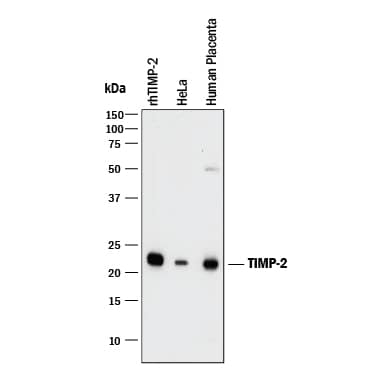
WBC023) and lysates of HeLa human cervical epithelial carcinoma cell line and human placenta tissue. PVDF membrane was probed with 1 µg/mL of Goat Anti-Human/Mouse TIMP-2 Antigen Affinity-purified Polyclonal Antibody (Catalog # AF971) followed by HRP-conjugated Anti-Goat IgG Secondary Antibody (Catalog #
HAF017). A specific band was detected for TIMP-2 at approximately 22 kDa (as indicated). This experiment was conducted under reducing conditions and using
Immunoblot Buffer Group 1." alt="Western blot shows Recombinant Human TIMP-2 Western Blot Standard Protein (Catalog #
WBC023) and lysates of HeLa human cervical epithelial carcinoma cell line and human placenta tissue. PVDF membrane was probed with 1 µg/mL of Goat Anti-Human/Mouse TIMP-2 Antigen Affinity-purified Polyclonal Antibody (Catalog # AF971) followed by HRP-conjugated Anti-Goat IgG Secondary Antibody (Catalog #
HAF017). A specific band was detected for TIMP-2 at approximately 22 kDa (as indicated). This experiment was conducted under reducing conditions and using
Immunoblot Buffer Group 1." class="big_thumb" />
HAF017). A specific band was detected for TIMP‑2 at approximately 22 kDa (as indicated) in the parental HeLa human cervical epithelial carcinoma cell line, but is not detectable in knockout HeLa human cervical epithelial carcinoma cell line. GAPDH (
AF5718) is shown as a loading control. This experiment was conducted under reducing conditions and using Western Blot Buffer Group 1." class="big_lightbox">
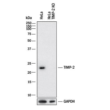
HAF017). A specific band was detected for TIMP‑2 at approximately 22 kDa (as indicated) in the parental HeLa human cervical epithelial carcinoma cell line, but is not detectable in knockout HeLa human cervical epithelial carcinoma cell line. GAPDH (
AF5718) is shown as a loading control. This experiment was conducted under reducing conditions and using Western Blot Buffer Group 1." class="big_thumb" />
902-MPNor
902-MP) activity is measured in the presence of Recombinant Human/Mouse TIMP-2 (0.143 µg/mL, Catalog #
971-TM) that has been preincubated with increasing concentrations of Goat Anti-Human TIMP-2 Antigen Affinity-purified Polyclonal Antibody (Catalog # AF971). The ND
50 is typically 2.6 µg/mL." class="big_lightbox">
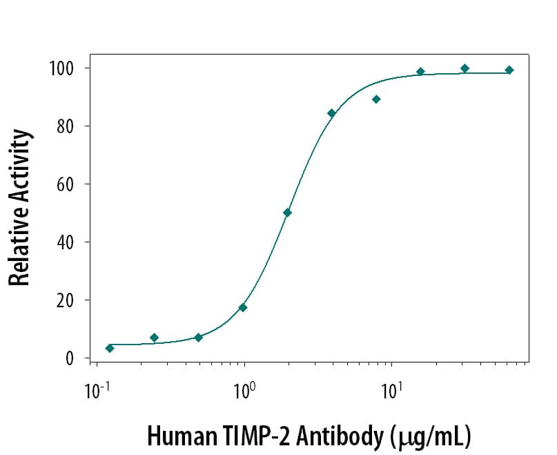
902-MPNor
902-MP) activity is measured in the presence of Recombinant Human/Mouse TIMP-2 (0.143 µg/mL, Catalog #
971-TM) that has been preincubated with increasing concentrations of Goat Anti-Human TIMP-2 Antigen Affinity-purified Polyclonal Antibody (Catalog # AF971). The ND
50 is typically 2.6 µg/mL." class="big_thumb" />
AF971) at 5ug/mL with overnight incubation at 4 degrees Celsius followed by incubation with anti-goat IgG VisUCyte HRP Polymer Antibody (Catalog #
VC004) and DAB chromogen (yellow-brown). Tissue was counterstained with hematoxylin (blue). Specific staining was localized to decidual cells. " class="big_lightbox">

AF971) at 5ug/mL with overnight incubation at 4 degrees Celsius followed by incubation with anti-goat IgG VisUCyte HRP Polymer Antibody (Catalog #
VC004) and DAB chromogen (yellow-brown). Tissue was counterstained with hematoxylin (blue). Specific staining was localized to decidual cells. " class="big_thumb" />
971-TM) was serially diluted and captured by Mouse Anti-Human TIMP‑2 Monoclonal Antibody (Catalog #
MAB9711) coated on a Clear Polystyrene Microplate (Catalog #
DY990). Goat Anti-Human/Mouse TIMP‑2 Antigen Affinity-purified Polyclonal Antibody (Catalog # AF971) was biotinylated and incubated with the protein captured on the plate. Detection of the standard curve was achieved by incubating Streptavidin-HRP (Catalog #
DY998)" class="big_lightbox">

971-TM) was serially diluted and captured by Mouse Anti-Human TIMP‑2 Monoclonal Antibody (Catalog #
MAB9711) coated on a Clear Polystyrene Microplate (Catalog #
DY990). Goat Anti-Human/Mouse TIMP‑2 Antigen Affinity-purified Polyclonal Antibody (Catalog # AF971) was biotinylated and incubated with the protein captured on the plate. Detection of the standard curve was achieved by incubating Streptavidin-HRP (Catalog #
DY998)" class="big_thumb" />
 WBC023) and lysates of HeLa human cervical epithelial carcinoma cell line and human placenta tissue. PVDF membrane was probed with 1 µg/mL of Goat Anti-Human/Mouse TIMP-2 Antigen Affinity-purified Polyclonal Antibody (Catalog # AF971) followed by HRP-conjugated Anti-Goat IgG Secondary Antibody (Catalog # HAF017). A specific band was detected for TIMP-2 at approximately 22 kDa (as indicated). This experiment was conducted under reducing conditions and using Immunoblot Buffer Group 1." alt="Western blot shows Recombinant Human TIMP-2 Western Blot Standard Protein (Catalog # WBC023) and lysates of HeLa human cervical epithelial carcinoma cell line and human placenta tissue. PVDF membrane was probed with 1 µg/mL of Goat Anti-Human/Mouse TIMP-2 Antigen Affinity-purified Polyclonal Antibody (Catalog # AF971) followed by HRP-conjugated Anti-Goat IgG Secondary Antibody (Catalog # HAF017). A specific band was detected for TIMP-2 at approximately 22 kDa (as indicated). This experiment was conducted under reducing conditions and using Immunoblot Buffer Group 1." class="big_thumb" />
 WBC023) and lysates of HeLa human cervical epithelial carcinoma cell line and human placenta tissue. PVDF membrane was probed with 2 µg/mL of Mouse Anti-Human TIMP-2 Monoclonal Antibody (Catalog # MAB971) followed by HRP-conjugated Anti-Mouse IgG Secondary Antibody (Catalog # HAF018). A specific band was detected for TIMP-2 at approximately 22 kDa (as indicated).This experiment was conducted under reducing conditions and using Immunoblot Buffer Group 1." alt="Western blot shows Recombinant Human TIMP-2 Western Blot Standard Protein (Catalog # WBC023) and lysates of HeLa human cervical epithelial carcinoma cell line and human placenta tissue. PVDF membrane was probed with 2 µg/mL of Mouse Anti-Human TIMP-2 Monoclonal Antibody (Catalog # MAB971) followed by HRP-conjugated Anti-Mouse IgG Secondary Antibody (Catalog # HAF018). A specific band was detected for TIMP-2 at approximately 22 kDa (as indicated).This experiment was conducted under reducing conditions and using Immunoblot Buffer Group 1." class="big_thumb" />
 HAF018). A specific band was detected for TIMP‑2 at approximately 22 kDa (as indicated) in the parental HeLa human cervical epithelial carcinoma cell line, but is not detectable in knockout HeLa human cervical epithelial carcinoma cell line. GAPDH (MAB5718) is shown as a loading control. This experiment was conducted under reducing conditions and using Western Blot Buffer Group 1." alt="Western blot shows lysates of HeLa human cervical epithelial carcinoma cell line and human TIMP-2 knockout HeLa human cervical epithelial carcinoma cell line (KO). PVDF membrane was probed with 4 µg/mL of Mouse Anti-Human TIMP‑2 Monoclonal Antibody (Catalog # MAB9711) followed by HRP-conjugated Anti-Mouse IgG Secondary Antibody (HAF018). A specific band was detected for TIMP‑2 at approximately 22 kDa (as indicated) in the parental HeLa human cervical epithelial carcinoma cell line, but is not detectable in knockout HeLa human cervical epithelial carcinoma cell line. GAPDH (MAB5718) is shown as a loading control. This experiment was conducted under reducing conditions and using Western Blot Buffer Group 1." class="big_thumb" />


![N/A TIMP-2 [HRP]](http://images.novusbio.com/fullsize/elisa/DATA_TIMP2_DTM200_ELISA_848.jpg "N/A TIMP-2 [HRP]")
![N/A TIMP-2 [HRP]](http://images.novusbio.com/fullsize/elisa/DATA_TIMP2_DTM200_ELISA_849.jpg)
![N/A TIMP-2 [HRP]](http://images.novusbio.com/fullsize/elisa/TIMP2_DTM200_ELISA_2722.jpg)
 CTS008) and counter-stained with hematoxylin (blue). View our protocol for
  HAF017). A specific band was detected for TIMP‑2 at approximately 22 kDa (as indicated) in the parental HeLa human cervical epithelial carcinoma cell line, but is not detectable in knockout HeLa human cervical epithelial carcinoma cell line. GAPDH (
 902-MPNor
  AF971) at 5ug/mL with overnight incubation at 4 degrees Celsius followed by incubation with anti-goat IgG VisUCyte HRP Polymer Antibody (Catalog #
  971-TM) was serially diluted and captured by Mouse Anti-Human TIMP‑2 Monoclonal Antibody (Catalog #
 ![N/A TIMP-2 [Biotin]](http://images.novusbio.com/fullsize/elisa/DATA_TIMP2_DY971_ELISA_2285.jpg "N/A TIMP-2 [Biotin]")
 CTS003) and counterstained with hematoxylin (blue). View our protocol for
 ![Western Blot: TIMP2 Antibody (1A6) [NBP2-01573] - HEK293T cells were transfected with the pCMV6-ENTRY control (Left lane) or pCMV6-ENTRY TIMP2 (Right lane) cDNA for 48 hrs and lysed. Equivalent amounts of cell lysates (5 ug per lane) were separated by SDS-PAGE and immunoblotted with anti-TIMP2.](http://images.novusbio.com/fullsize/TIMP2-Antibody-1A6-Western-Blot-NBP2-01573-img0003.jpg "Western Blot: TIMP2 Antibody (1A6) [NBP2-01573] - HEK293T cells were transfected with the pCMV6-ENTRY control (Left lane) or pCMV6-ENTRY TIMP2 (Right lane) cDNA for 48 hrs and lysed. Equivalent amounts of cell lysates (5 ug per lane) were separated by SDS-PAGE and immunoblotted with anti-TIMP2.")
![Immunohistochemistry-Paraffin: TIMP2 Antibody (1A6) [NBP2-01573] - Staining of paraffin-embedded Human pancreas tissue using anti-TIMP2 mouse monoclonal antibody.](http://images.novusbio.com/fullsize/TIMP2-Antibody-1A6-Immunohistochemistry-Paraffin-NBP2-01573-img0010.jpg)
![Flow Cytometry: TIMP2 Antibody (1A6) [NBP2-01573] - HEK293T cells transfected with either overexpression plasmid (Red) or empty vector control plasmid (Blue) were immunostained by anti-TIMP2 antibody, and then analyzed by flow cytometry.](http://images.novusbio.com/fullsize/TIMP2-Antibody-1A6-Flow-Cytometry-NBP2-01573-img0002.jpg)
![Immunohistochemistry-Paraffin: TIMP2 Antibody (1A6) [NBP2-01573] - Staining of paraffin-embedded Adenocarcinoma of Human breast tissue using anti-TIMP2 mouse monoclonal antibody.](http://images.novusbio.com/fullsize/TIMP2-Antibody-1A6-Immunohistochemistry-Paraffin-NBP2-01573-img0004.jpg)
![Immunohistochemistry-Paraffin: TIMP2 Antibody (1A6) [NBP2-01573] - Staining of paraffin-embedded Adenocarcinoma of Human endometrium tissue using anti-TIMP2 mouse monoclonal antibody.](http://images.novusbio.com/fullsize/TIMP2-Antibody-1A6-Immunohistochemistry-Paraffin-NBP2-01573-img0005.jpg)
![Immunohistochemistry-Paraffin: TIMP2 Antibody (1A6) [NBP2-01573] - Staining of paraffin-embedded Adenocarcinoma of Human ovary tissue using anti-TIMP2 mouse monoclonal antibody.](http://images.novusbio.com/fullsize/TIMP2-Antibody-1A6-Immunohistochemistry-Paraffin-NBP2-01573-img0006.jpg)
![Immunohistochemistry-Paraffin: TIMP2 Antibody (1A6) [NBP2-01573] - Staining of paraffin-embedded Carcinoma of Human lung tissue using anti-TIMP2 mouse monoclonal antibody.](http://images.novusbio.com/fullsize/TIMP2-Antibody-1A6-Immunohistochemistry-Paraffin-NBP2-01573-img0007.jpg)
![Immunohistochemistry-Paraffin: TIMP2 Antibody (1A6) [NBP2-01573] - Staining of paraffin-embedded Human breast tissue using anti-TIMP2 mouse monoclonal antibody.](http://images.novusbio.com/fullsize/TIMP2-Antibody-1A6-Immunohistochemistry-Paraffin-NBP2-01573-img0008.jpg)
![Immunohistochemistry-Paraffin: TIMP2 Antibody (1A6) [NBP2-01573] - Staining of paraffin-embedded Human endometrium tissue using anti-TIMP2 mouse monoclonal antibody.](http://images.novusbio.com/fullsize/TIMP2-Antibody-1A6-Immunohistochemistry-Paraffin-NBP2-01573-img0009.jpg)
![Flow Cytometry: TIMP2 Antibody (1A6) [NBP2-01573] - Analysis of Jurkat cells, using anti-TIMP2 antibody, (Red), compared to a nonspecific negative control antibody (Blue).](http://images.novusbio.com/fullsize/TIMP2-Antibody-1A6-Flow-Cytometry-NBP2-01573-img0001.jpg)
 971-TM) was serially diluted and captured by Mouse Anti-Human TIMP‑2 Monoclonal Antibody (Catalog # MAB9711) coated on a Clear Polystyrene Microplate (Catalog #
 ![Immunohistochemistry-Paraffin: TIMP-2 Antibody (3A4) [NBP2-53347] - Formalin-fixed, paraffin-embedded human Placenta stained with TIMP2 Monoclonal Antibody (3A4).](http://images.novusbio.com/fullsize/TIMP-2-Antibody-3A4-Immunohistochemistry-Paraffin-NBP2-53347-img0001.jpg "Immunohistochemistry-Paraffin: TIMP-2 Antibody (3A4) [NBP2-53347] - Formalin-fixed, paraffin-embedded human Placenta stained with TIMP2 Monoclonal Antibody (3A4).")
![Immunohistochemistry-Paraffin: TIMP-2 Antibody (SPM356) [NBP2-53348] - Formalin-fixed, paraffin-embedded human Placenta stained with TIMP2 Monoclonal Antibody (SPM356).](http://images.novusbio.com/fullsize/TIMP-2-Antibody-SPM356-Immunohistochemistry-Paraffin-NBP2-53348-img0001.jpg "Immunohistochemistry-Paraffin: TIMP-2 Antibody (SPM356) [NBP2-53348] - Formalin-fixed, paraffin-embedded human Placenta stained with TIMP2 Monoclonal Antibody (SPM356).")
![N/A TIMP-2 [Biotin]](http://images.novusbio.com/fullsize/elisa/DATA_TIMP2_DY630405_ELISA_2496.jpg "N/A TIMP-2 [Biotin]")
![Immunocytochemistry/Immunofluorescence: TIMP-2 Antibody (F27 P3 A4) [NB100-74552] - Cells were grown on chamber slides and fixed with formaldehyde prior to staining. Cells were probed without (control) or with a TIMP2 monoclonal antibody at a dilution of 1:200 overnight at 4 C, washed with PBS and incubated with a DyLight-488 conjugated secondary antibody and nuclei with DAPI (blue) is shown.](http://images.novusbio.com/fullsize/TIMP-2-Antibody-F27-P3-A4-Immunocytochemistry-Immunofluorescence-NB100-74552-img0006.jpg "Immunocytochemistry/Immunofluorescence: TIMP-2 Antibody (F27 P3 A4) [NB100-74552] - Cells were grown on chamber slides and fixed with formaldehyde prior to staining. Cells were probed without (control) or with a TIMP2 monoclonal antibody at a dilution of 1:200 overnight at 4 C, washed with PBS and incubated with a DyLight-488 conjugated secondary antibody and nuclei with DAPI (blue) is shown.")
![Immunohistochemistry-Paraffin: TIMP-2 Antibody (F27 P3 A4) [NB100-74552] - Normal biopsies of deparaffinized Mouse pancreas tissue at a dilution of 1:100.](http://images.novusbio.com/fullsize/TIMP-2-Antibody-F27-P3-A4-Immunohistochemistry-Paraffin-NB100-74552-img0007.jpg)
![Immunocytochemistry/Immunofluorescence: TIMP-2 Antibody (F27 P3 A4) [NB100-74552] - Cells were grown on chamber slides and fixed with formaldehyde prior to staining. Cells were probed without (control) or with a TIMP2 monoclonal antibody at a dilution of 1:200 overnight at 4 C, washed with PBS and incubated with a DyLight-488 conjugated secondary antibody and nuclei with DAPI (blue) is shown.](http://images.novusbio.com/fullsize/TIMP-2-Antibody-F27-P3-A4-Immunocytochemistry-Immunofluorescence-NB100-74552-img0004.jpg)
![Immunocytochemistry/Immunofluorescence: TIMP-2 Antibody (F27 P3 A4) [NB100-74552] - Cells were grown on chamber slides and fixed with formaldehyde prior to staining. Cells were probed without (control) or with a TIMP2 monoclonal antibody at a dilution of 1:200 overnight at 4 C, washed with PBS and incubated with a DyLight-488 conjugated secondary antibody and nuclei with DAPI (blue) is shown.](http://images.novusbio.com/fullsize/TIMP-2-Antibody-F27-P3-A4-Immunocytochemistry-Immunofluorescence-NB100-74552-img0005.jpg)
![Immunohistochemistry-Paraffin: TIMP-2 Antibody (F27 P3 A4) [NB100-74552] - Normal biopsies of deparaffinized Human pancreas tissue at a dilution of 1:500.](http://images.novusbio.com/fullsize/TIMP-2-Antibody-F27-P3-A4-Immunohistochemistry-Paraffin-NB100-74552-img0008.jpg)


