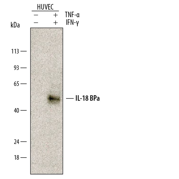
285-IF) and 40 ng/mL Recombinant Human TNF-alpha (
210-TA) for 48 hours. PVDF membrane was probed with 0.5 µg/mL of Goat Anti-Human IL-18 BPa Antigen Affinity-purified Polyclonal Antibody (Catalog # AF119) followed by HRP-conjugated Anti-Goat IgG Secondary Antibody (Catalog #
HAF109). A specific band was detected for IL-18 BPa at approximately 50 kDa (as indicated). This experiment was conducted under reducing conditions and using
Immunoblot Buffer Group 1." class="big_lightbox">
285-IF) and 40 ng/mL Recombinant Human TNF-alpha (
210-TA) for 48 hours. PVDF membrane was probed with 0.5 µg/mL of Goat Anti-Human IL-18 BPa Antigen Affinity-purified Polyclonal Antibody (Catalog # AF119) followed by HRP-conjugated Anti-Goat IgG Secondary Antibody (Catalog #
HAF109). A specific band was detected for IL-18 BPa at approximately 50 kDa (as indicated). This experiment was conducted under reducing conditions and using
Immunoblot Buffer Group 1." alt="Western blot shows conditioned media from HUVEC human umbilical vein endothelial cells untreated (-) or treated (+) with 50 ng/mL Recombinant Human IFN-gamma (Catalog #
285-IF) and 40 ng/mL Recombinant Human TNF-alpha (
210-TA) for 48 hours. PVDF membrane was probed with 0.5 µg/mL of Goat Anti-Human IL-18 BPa Antigen Affinity-purified Polyclonal Antibody (Catalog # AF119) followed by HRP-conjugated Anti-Goat IgG Secondary Antibody (Catalog #
HAF109). A specific band was detected for IL-18 BPa at approximately 50 kDa (as indicated). This experiment was conducted under reducing conditions and using
Immunoblot Buffer Group 1." class="big_thumb" />
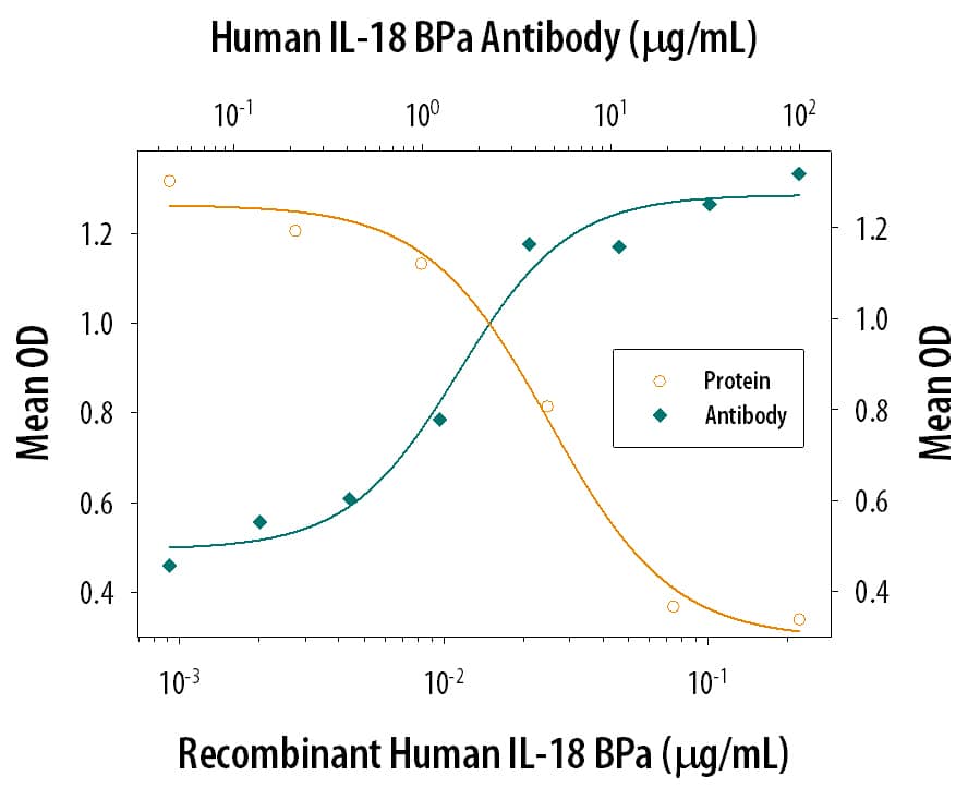
119-BP) inhibits Recombinant Human IL-18/IL-1F4 induced IFN-gamma secretion in the KG-1 human acute myelogenous leukemia cell line in a dose-dependent manner (orange line), as measured by the Human IFN-gamma Quantikine ELISA Kit (
DIF50C). Inhibition of Recombinant Human IL-18/IL-1F4 (40 ng/mL) activity elicited by Recombinant Human IL-18 BPa Fc Chimera (0.1 µg/mL) is neutralized (green line) by increasing concentrations of Goat Anti-Human IL-18 BPa Antigen Affinity-purified Polyclonal Antibody (Catalog # AF119). The ND
50 is typically 1-5 µg/mL in the presence of Recombinant Human TNF-alpha (20 ng/mL)." class="big_lightbox">
119-BP) inhibits Recombinant Human IL-18/IL-1F4 induced IFN-gamma secretion in the KG-1 human acute myelogenous leukemia cell line in a dose-dependent manner (orange line), as measured by the Human IFN-gamma Quantikine ELISA Kit (
DIF50C). Inhibition of Recombinant Human IL-18/IL-1F4 (40 ng/mL) activity elicited by Recombinant Human IL-18 BPa Fc Chimera (0.1 µg/mL) is neutralized (green line) by increasing concentrations of Goat Anti-Human IL-18 BPa Antigen Affinity-purified Polyclonal Antibody (Catalog # AF119). The ND
50 is typically 1-5 µg/mL in the presence of Recombinant Human TNF-alpha (20 ng/mL)." class="big_thumb" />
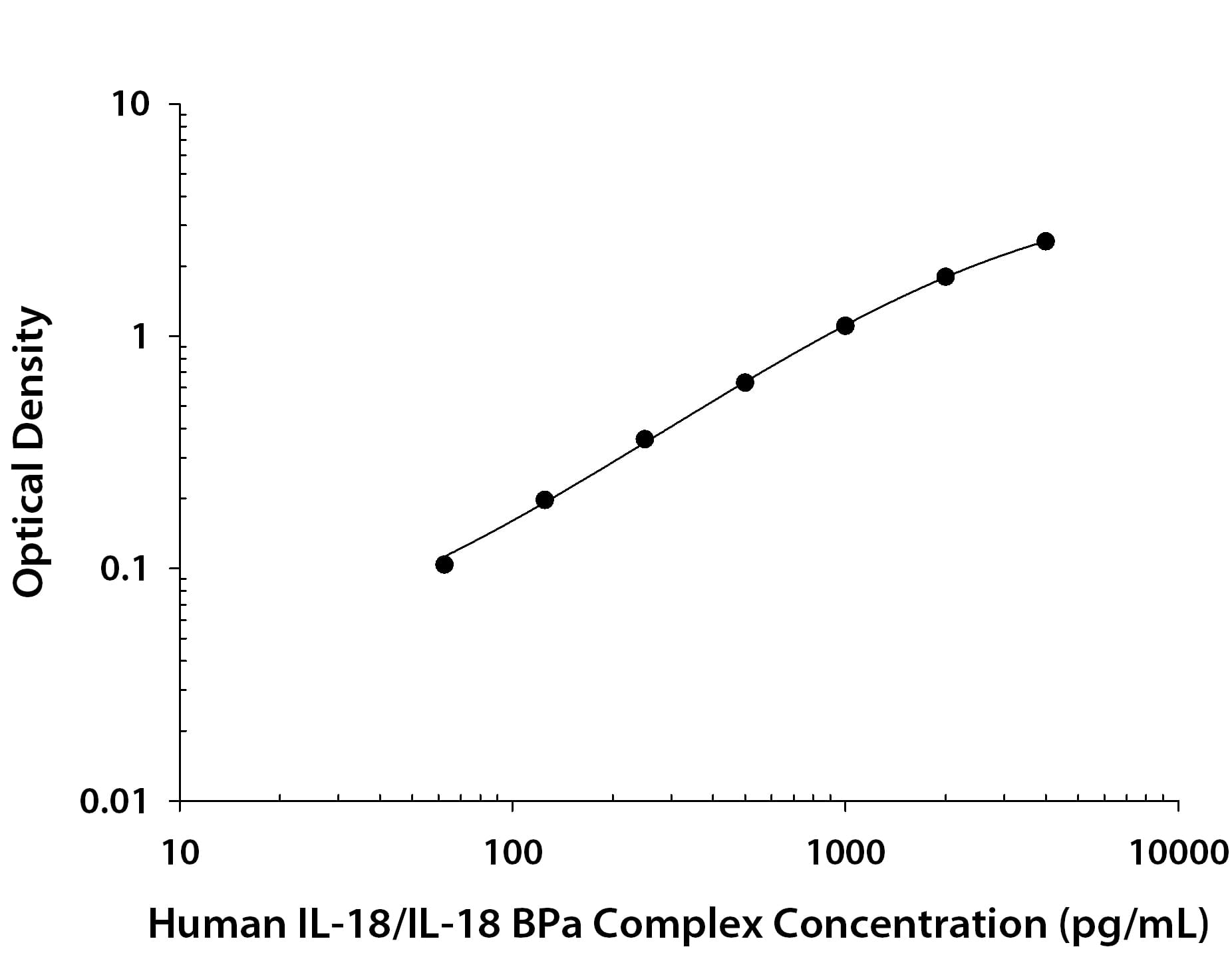
MAB91244) coated on a Clear Polystyrene Microplate (Catalog #
DY990). Goat Anti-Human IL-18 BPa Antigen Affinity-purified Polyclonal Antibody (Catalog # AF119) was biotinylated and incubated with the protein captured on the plate. Detection of the standard curve was achieved by incubating Streptavidin-HRP (Catalog #
DY998) followed by Substrate Solution (Catalog #
DY999) and stopping the enzymatic reaction with Stop Solution (Catalog #
DY994)." class="big_lightbox">
MAB91244) coated on a Clear Polystyrene Microplate (Catalog #
DY990). Goat Anti-Human IL-18 BPa Antigen Affinity-purified Polyclonal Antibody (Catalog # AF119) was biotinylated and incubated with the protein captured on the plate. Detection of the standard curve was achieved by incubating Streptavidin-HRP (Catalog #
DY998) followed by Substrate Solution (Catalog #
DY999) and stopping the enzymatic reaction with Stop Solution (Catalog #
DY994)." class="big_thumb" />
119-BP) was serially diluted and captured by Mouse Anti-Human IL‑18 BPa Monoclonal Antibody (Catalog #
MAB1192) coated on a Clear Polystyrene Microplate (Catalog #
DY990). Goat Anti-Human IL‑18 BPa Antigen Affinity-purified Polyclonal Antibody (Catalog # AF119) was biotinylated and incubated with the protein captured on the plate. Detection of the standard curve was achieved by incubating Streptavidin-HRP (Catalog #
DY998)" class="big_lightbox">

119-BP) was serially diluted and captured by Mouse Anti-Human IL‑18 BPa Monoclonal Antibody (Catalog #
MAB1192) coated on a Clear Polystyrene Microplate (Catalog #
DY990). Goat Anti-Human IL‑18 BPa Antigen Affinity-purified Polyclonal Antibody (Catalog # AF119) was biotinylated and incubated with the protein captured on the plate. Detection of the standard curve was achieved by incubating Streptavidin-HRP (Catalog #
DY998)" class="big_thumb" />
MAB91244) coated on a Clear Polystyrene Microplate (Catalog #
DY990). Goat Anti-Human IL‑18 BPa Antigen Affinity-purified Polyclonal Antibody (Catalog # AF119) was biotinylated and incubated with the protein captured on the plate. Detection of the standard curve was achieved by incubating Streptavidin-HRP (Catalog #
DY998)" class="big_lightbox">

MAB91244) coated on a Clear Polystyrene Microplate (Catalog #
DY990). Goat Anti-Human IL‑18 BPa Antigen Affinity-purified Polyclonal Antibody (Catalog # AF119) was biotinylated and incubated with the protein captured on the plate. Detection of the standard curve was achieved by incubating Streptavidin-HRP (Catalog #
DY998)" class="big_thumb" />
 285-IF) and 40 ng/mL Recombinant Human TNF-alpha (210-TA) for 48 hours. PVDF membrane was probed with 0.5 µg/mL of Goat Anti-Human IL-18 BPa Antigen Affinity-purified Polyclonal Antibody (Catalog # AF119) followed by HRP-conjugated Anti-Goat IgG Secondary Antibody (Catalog # HAF109). A specific band was detected for IL-18 BPa at approximately 50 kDa (as indicated). This experiment was conducted under reducing conditions and using Immunoblot Buffer Group 1." alt="Western blot shows conditioned media from HUVEC human umbilical vein endothelial cells untreated (-) or treated (+) with 50 ng/mL Recombinant Human IFN-gamma (Catalog # 285-IF) and 40 ng/mL Recombinant Human TNF-alpha (210-TA) for 48 hours. PVDF membrane was probed with 0.5 µg/mL of Goat Anti-Human IL-18 BPa Antigen Affinity-purified Polyclonal Antibody (Catalog # AF119) followed by HRP-conjugated Anti-Goat IgG Secondary Antibody (Catalog # HAF109). A specific band was detected for IL-18 BPa at approximately 50 kDa (as indicated). This experiment was conducted under reducing conditions and using Immunoblot Buffer Group 1." class="big_thumb" />
 122-BP) inhibits Recombinant Mouse IL-18/IL-1F4 induced IFN-gamma secretion in activated mouse T cells in a dose-dependent manner (orange line), as measured by the Mouse IFN-gamma Quantikine ELISA Kit (Catalog # MIF00). Inhibition of Recombinant Mouse IL-18/ IL-1F4 (0.5 ng/mL) activity elicited by Recombinant Mouse IL-18 BPd Fc Chimera (2 µg/mL) is neutral-ized (green line) by increasing concentrations of Goat Anti-Mouse IL-18 BPd Antigen Affinity-purified Polyclonal Antibody (Catalog # AF122). The ND50 is typically 10-30 µg/mL in the presence of Recombinant Mouse IL-12 (0.1 ng/mL)." alt="Recombinant Mouse IL-18 BPd Fc Chimera (Catalog # 122-BP) inhibits Recombinant Mouse IL-18/IL-1F4 induced IFN-gamma secretion in activated mouse T cells in a dose-dependent manner (orange line), as measured by the Mouse IFN-gamma Quantikine ELISA Kit (Catalog # MIF00). Inhibition of Recombinant Mouse IL-18/ IL-1F4 (0.5 ng/mL) activity elicited by Recombinant Mouse IL-18 BPd Fc Chimera (2 µg/mL) is neutral-ized (green line) by increasing concentrations of Goat Anti-Mouse IL-18 BPd Antigen Affinity-purified Polyclonal Antibody (Catalog # AF122). The ND50 is typically 10-30 µg/mL in the presence of Recombinant Mouse IL-12 (0.1 ng/mL)." class="big_thumb" />
 119-BP) was serially diluted and captured by Mouse Anti-Human IL‑18 BPa Monoclonal Antibody (Catalog # MAB1192) coated on a Clear Polystyrene Microplate (Catalog # DY990). Goat Anti-Human IL‑18 BPa Antigen Affinity-purified Polyclonal Antibody (Catalog # AF119) was biotinylated and incubated with the protein captured on the plate. Detection of the standard curve was achieved by incubating Streptavidin-HRP (Catalog # DY998)" alt="Recombinant Human IL‑18 BPa Fc Chimera (Catalog # 119-BP) was serially diluted and captured by Mouse Anti-Human IL‑18 BPa Monoclonal Antibody (Catalog # MAB1192) coated on a Clear Polystyrene Microplate (Catalog # DY990). Goat Anti-Human IL‑18 BPa Antigen Affinity-purified Polyclonal Antibody (Catalog # AF119) was biotinylated and incubated with the protein captured on the plate. Detection of the standard curve was achieved by incubating Streptavidin-HRP (Catalog # DY998)" class="big_thumb" />
 119-BP) inhibition of Recombinant Human IL-18/IL-1F4 (9124-IL) induced IFN gamma secretion in the KG-1 human acute myelogenous leukemia cell line as measured by the Human IFN-gamma Quantikine ELISA Kit (DIF50C). The ND50 for this effect is typically 0.800-12.0 µg/mL." alt="Mouse Anti-Human IL-18 BPa Monoclonal Antibody (Catalog # MAB1191) neutralizes Recombinant Human IL-18 BPa Fc Chimera (119-BP) inhibition of Recombinant Human IL-18/IL-1F4 (9124-IL) induced IFN gamma secretion in the KG-1 human acute myelogenous leukemia cell line as measured by the Human IFN-gamma Quantikine ELISA Kit (DIF50C). The ND50 for this effect is typically 0.800-12.0 µg/mL." class="big_thumb" />


![N/A IL-18 BPa/IL18BP [Biotin]](http://images.novusbio.com/fullsize/elisa/DATA_IL18_BPa_DY119_ELISA_1598.jpg "N/A IL-18 BPa/IL18BP [Biotin]")
 119-BP) inhibits Recombinant Human IL-18/IL-1F4 induced IFN-gamma secretion in the KG-1 human acute myelogenous leukemia cell line in a dose-dependent manner (orange line), as measured by the Human IFN-gamma Quantikine ELISA Kit (
 MAB91244) coated on a Clear Polystyrene Microplate (Catalog #
  119-BP) was serially diluted and captured by Mouse Anti-Human IL‑18 BPa Monoclonal Antibody (Catalog #
  MAB91244) coated on a Clear Polystyrene Microplate (Catalog #
 ![N/A IL-18 BPd [Biotin]](http://images.novusbio.com/fullsize/elisa/DATA_IL18_BPd_DY12205_ELISA_2345.jpg "N/A IL-18 BPd [Biotin]")
 MAB122) coated on a Clear Polystyrene Microplate (


![Western Blot: IL-18 BPa/IL18BP Antibody (MM0379-10G36) [NBP2-11678]](http://images.novusbio.com/fullsize/IL-18-BPa-IL18BP-Antibody-MM0379-10G36-Western-Blot-NBP2-11678-img0001.jpg "Western Blot: IL-18 BPa/IL18BP Antibody (MM0379-10G36) [NBP2-11678]")


