285-IF) using Mouse Anti-Human Indoleamine 2,3-dioxygenase/IDO Monoclonal Antibody (Catalog # MAB6030) at 10 µg/mL for 3 hours at room temperature. Cells were stained using the NorthernLights™ 557-conjugated Anti-Mouse IgG Secondary Antibody (red; Catalog #
NL007) and counter-stained with DAPI (blue). Specific staining was localized to cytoplasm. View our protocol for
Fluorescent ICC Staining of Cells on Coverslips." class="big_lightbox">
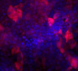
285-IF) using Mouse Anti-Human Indoleamine 2,3-dioxygenase/IDO Monoclonal Antibody (Catalog # MAB6030) at 10 µg/mL for 3 hours at room temperature. Cells were stained using the NorthernLights™ 557-conjugated Anti-Mouse IgG Secondary Antibody (red; Catalog #
NL007) and counter-stained with DAPI (blue). Specific staining was localized to cytoplasm. View our protocol for
Fluorescent ICC Staining of Cells on Coverslips." alt="Indoleamine 2,3-dioxygenase/IDO was detected in immersion fixed A431 human epithelial carcinoma cells stimulated with 0.5 ng/mL of Recombinant Human IFN-gamma (Catalog #
285-IF) using Mouse Anti-Human Indoleamine 2,3-dioxygenase/IDO Monoclonal Antibody (Catalog # MAB6030) at 10 µg/mL for 3 hours at room temperature. Cells were stained using the NorthernLights™ 557-conjugated Anti-Mouse IgG Secondary Antibody (red; Catalog #
NL007) and counter-stained with DAPI (blue). Specific staining was localized to cytoplasm. View our protocol for
Fluorescent ICC Staining of Cells on Coverslips." class="big_thumb" />
206-IL) and 10 ng/mL Recombinant Human GM-CSF (Catalog #
215-GM) for 7 days were stained with Mouse Anti-Human Siglec-3/CD33 APC-conjugated Monoclonal Antibody (Catalog #
FAB1137A) and either (A) Mouse Anti-Human Indoleamine 2,3-dioxygenase/IDO Monoclonal Antibody (Catalog # MAB6030) or (B) Mouse IgG
1Isotype Control (Catalog #
MAB002) followed by Phycoerythrin-conjugated Anti-Mouse IgG Secondary Antibody (Catalog #
F0102B). To facilitate intracellular staining, cells were fixed with paraformaldehyde and permeabilized with saponin. View our protocol for
Staining Intracellular Molecules." class="big_lightbox">
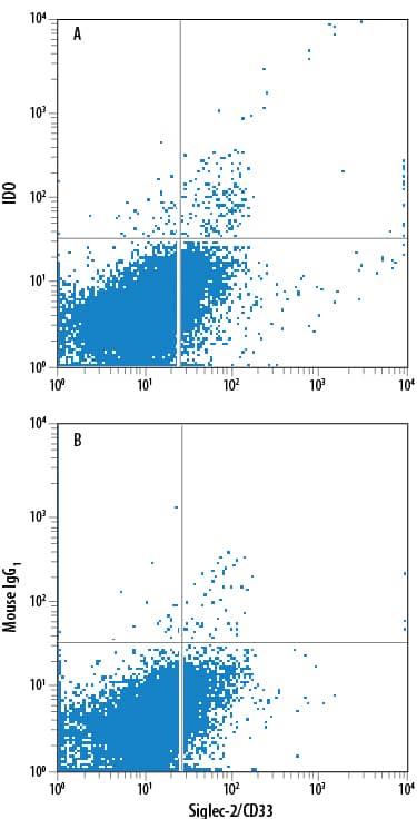
206-IL) and 10 ng/mL Recombinant Human GM-CSF (Catalog #
215-GM) for 7 days were stained with Mouse Anti-Human Siglec-3/CD33 APC-conjugated Monoclonal Antibody (Catalog #
FAB1137A) and either (A) Mouse Anti-Human Indoleamine 2,3-dioxygenase/IDO Monoclonal Antibody (Catalog # MAB6030) or (B) Mouse IgG
1Isotype Control (Catalog #
MAB002) followed by Phycoerythrin-conjugated Anti-Mouse IgG Secondary Antibody (Catalog #
F0102B). To facilitate intracellular staining, cells were fixed with paraformaldehyde and permeabilized with saponin. View our protocol for
Staining Intracellular Molecules." class="big_thumb" />
MAGH105) and cultured overnight with (A) 50 ng/mL Recombinant Human MCSF (Catalog #
216-MC), 50 ng/mL Recombinant Human IFNg (Catalog #
285-IF) and 50 ng/mL LPS, or (B) Recombinant Human MCSF alone. Cells were stained with Mouse Anti-Human Indoleamine 2,3-dioxygenase/IDO Monoclonal Antibody (Catalog # MAB6030) followed by APC-conjugated Anti-Mouse IgG Secondary Antibody (Catalog #
F0101B) and Mouse Anti-Human CD14 PE-conjugated Monoclonal Antibody (Catalog #
FAB3832P). Quadrant markers were set based on Mouse IgG1 Isotype Control (Catalog #
MAB002). To facilitate intracellular staining, cells were fixed with 1% paraformaldehyde and permeabilized with saponin. View our protocol for
Staining Intracellular Molecules." class="big_lightbox">
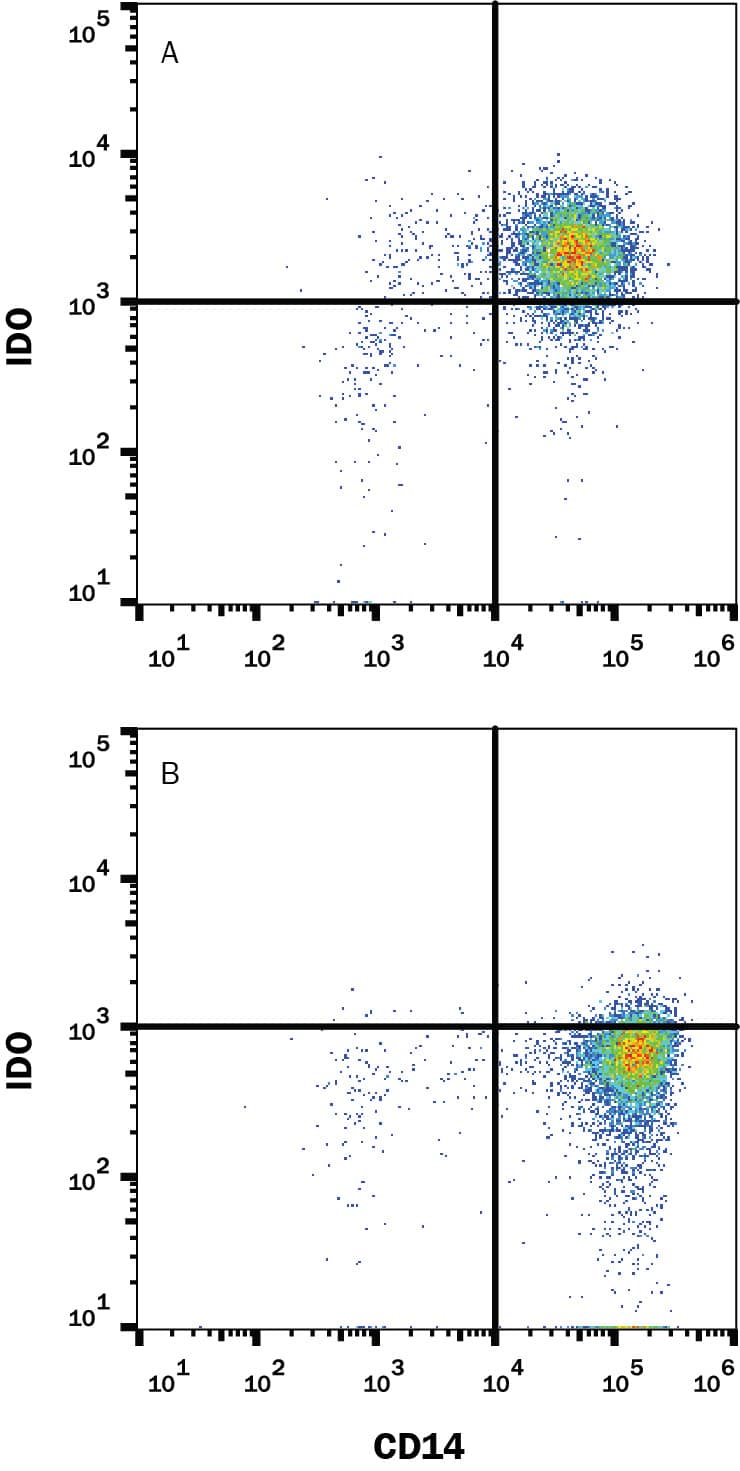
MAGH105) and cultured overnight with (A) 50 ng/mL Recombinant Human MCSF (Catalog #
216-MC), 50 ng/mL Recombinant Human IFNg (Catalog #
285-IF) and 50 ng/mL LPS, or (B) Recombinant Human MCSF alone. Cells were stained with Mouse Anti-Human Indoleamine 2,3-dioxygenase/IDO Monoclonal Antibody (Catalog # MAB6030) followed by APC-conjugated Anti-Mouse IgG Secondary Antibody (Catalog #
F0101B) and Mouse Anti-Human CD14 PE-conjugated Monoclonal Antibody (Catalog #
FAB3832P). Quadrant markers were set based on Mouse IgG1 Isotype Control (Catalog #
MAB002). To facilitate intracellular staining, cells were fixed with 1% paraformaldehyde and permeabilized with saponin. View our protocol for
Staining Intracellular Molecules." class="big_thumb" />
 285-IF) using Mouse Anti-Human Indoleamine 2,3-dioxygenase/IDO Monoclonal Antibody (Catalog # MAB6030) at 10 µg/mL for 3 hours at room temperature. Cells were stained using the NorthernLights™ 557-conjugated Anti-Mouse IgG Secondary Antibody (red; Catalog # NL007) and counter-stained with DAPI (blue). Specific staining was localized to cytoplasm. View our protocol for Fluorescent ICC Staining of Cells on Coverslips." alt="Indoleamine 2,3-dioxygenase/IDO was detected in immersion fixed A431 human epithelial carcinoma cells stimulated with 0.5 ng/mL of Recombinant Human IFN-gamma (Catalog # 285-IF) using Mouse Anti-Human Indoleamine 2,3-dioxygenase/IDO Monoclonal Antibody (Catalog # MAB6030) at 10 µg/mL for 3 hours at room temperature. Cells were stained using the NorthernLights™ 557-conjugated Anti-Mouse IgG Secondary Antibody (red; Catalog # NL007) and counter-stained with DAPI (blue). Specific staining was localized to cytoplasm. View our protocol for Fluorescent ICC Staining of Cells on Coverslips." class="big_thumb" />


![Immunohistochemistry-Paraffin: Indoleamine 2,3-dioxygenase/IDO Antibody [NBP1-87702] - Staining in human placenta and cerebral cortex tissues . Corresponding IDO1 RNA-seq data are presented for the same tissues.](http://images.novusbio.com/fullsize/Indoleamine-2-3-dioxygenase-IDO-Antibody-Immunohistochemistry-Paraffin-NBP1-87702-img0016.jpg "<b>Orthogonal Strategies Validation. </b>Immunohistochemistry-Paraffin: Indoleamine 2,3-dioxygenase/IDO Antibody [NBP1-87702] - Staining in human placenta and cerebral cortex tissues . Corresponding IDO1 RNA-seq data are presented for the same tissues.")
![<b>Orthogonal Strategies Validation. </b>Western Blot: Indoleamine 2,3-dioxygenase/IDO Antibody [NBP1-87702] - Analysis in human cell line EFO-21.](http://images.novusbio.com/fullsize/Indoleamine-2-3-dioxygenase-IDO-Antibody-Western-Blot-NBP1-87702-img0013.jpg)
![Immunohistochemistry-Paraffin: Indoleamine 2,3-dioxygenase/IDO Antibody [NBP1-87702] - Staining of human lymph node shows strong cytoplasmic and nuclear positivity in non-germinal center cells.](http://images.novusbio.com/fullsize/Indoleamine-2-3-dioxygenase-IDO-Antibody-Immunohistochemistry-Paraffin-NBP1-87702-img0018.jpg)
![Immunohistochemistry-Paraffin: Indoleamine 2,3-dioxygenase/IDO Antibody [NBP1-87702] - Immunohistochemical staining of human placenta shows strong cytoplasmic and nuclear positivity in endothelial cells.](http://images.novusbio.com/fullsize/Indoleamine-2-3-dioxygenase-IDO-Antibody-Immunohistochemistry-Paraffin-NBP1-87702-img0009.jpg)
![Immunohistochemistry-Paraffin: Indoleamine 2,3-dioxygenase/IDO Antibody [NBP1-87702] - Immunohistochemical staining of human cerebral cortex shows no positivity as expected.](http://images.novusbio.com/fullsize/Indoleamine-2-3-dioxygenase-IDO-Antibody-Immunohistochemistry-Paraffin-NBP1-87702-img0010.jpg)
![Immunohistochemistry-Paraffin: Indoleamine 2,3-dioxygenase/IDO Antibody [NBP1-87702] - Immunohistochemical staining of human small intestine shows strong cytoplasmic and nuclear positivity in a subset of leukocytes.](http://images.novusbio.com/fullsize/Indoleamine-2-3-dioxygenase-IDO-Antibody-Immunohistochemistry-Paraffin-NBP1-87702-img0011.jpg)
![Simple Western: Indoleamine 2,3-dioxygenase/IDO Antibody [NBP1-87702] - Simple Western lane view shows a specific band for IDO1 in 0.2 mg/ml of OVCAR-3 (left), h. Tonsil, raji, daudi lysate. This experiment was performed under reducing conditions using the 12-230 kDa separation system.](http://images.novusbio.com/fullsize/Indoleamine-2-3-dioxygenase-IDO-Antibody-Simple-Western-NBP1-87702-img0005.jpg)
![Simple Western: Indoleamine 2,3-dioxygenase/IDO Antibody [NBP1-87702] - Electropherogram image(s) of corresponding Simple Western lane view. Indoleamine 2,3-dioxygenase/IDO antibody was used at 1:20 dilution on OVCAR-3, h. Tonsil, Raji, and Daudi lysate(s).](http://images.novusbio.com/fullsize/Indoleamine-2-3-dioxygenase-IDO-Antibody-Simple-Western-NBP1-87702-img0008.jpg)
![ELISA: Human Indoleamine 2,3-dioxygenase/IDO ELISA Kit (Colorimetric) [NBP2-62765] - Samples were spiked with high concentrations of Human Indoleamine 2,3-dioxygenase/IDO and diluted with Reference Standard & Sample Diluent to produce samples with values within the range of the assay.](http://images.novusbio.com/fullsize/nbp2-62765_human-indoleamine-2-3-dioxygenase-ido-elisa-kit-color-132202416143052.jpg "ELISA: Human Indoleamine 2,3-dioxygenase/IDO ELISA Kit (Colorimetric) [NBP2-62765] - Samples were spiked with high concentrations of Human Indoleamine 2,3-dioxygenase/IDO and diluted with Reference Standard & Sample Diluent to produce samples with values within the range of the assay.")
![ELISA: Human Indoleamine 2,3-dioxygenase/IDO ELISA Kit (Colorimetric) [NBP2-62765] - Standard Curve Reference](http://images.novusbio.com/fullsize/nbp2-62765_human-indoleamine-2-3-dioxygenase-ido-elisa-kit-color-432024983550.jpg)
![Western Blot: Indoleamine 2,3-dioxygenase/IDO Antibody [NBP1-87703] - Analysis in EFO-21 cells transfected with control siRNA, target specific siRNA probe #1 and #2, using anti-IDO1 antibody. Remaining relative intensity is presented. Loading control: anti-PPIB.](http://images.novusbio.com/fullsize/Indoleamine-2-3-dioxygenase-IDO-Antibody-Western-Blot-NBP1-87703-img0014.jpg "<b>Genetic Strategies Validation. </b>Western Blot: Indoleamine 2,3-dioxygenase/IDO Antibody [NBP1-87703] - Analysis in EFO-21 cells transfected with control siRNA, target specific siRNA probe #1 and #2, using anti-IDO1 antibody. Remaining relative intensity is presented. Loading control: anti-PPIB.")
![<b>Genetic Strategies Validation and Orthogonal Strategies Validation.</b>Immunohistochemistry-Paraffin: Indoleamine 2,3-dioxygenase/IDO Antibody [NBP1-87703] - Staining in human placenta and cerebral cortex tissues . Corresponding IDO1 RNA-seq data are presented for the same tissues.](http://images.novusbio.com/fullsize/Indoleamine-2-3-dioxygenase-IDO-Antibody-Immunohistochemistry-Paraffin-NBP1-87703-img0013.jpg)
![Western Blot: Indoleamine 2,3-dioxygenase/IDO Antibody [NBP1-87703] - Analysis in control (vector only transfected HEK293T lysate) and IDO1 over-expression lysate (Co-expressed with a C-terminal myc-DDK tag (3.1 kDa) in mammalian HEK293T cells).](http://images.novusbio.com/fullsize/Indoleamine-2-3-dioxygenase-IDO-Antibody-Western-Blot-NBP1-87703-img0015.jpg)
![Immunohistochemistry-Paraffin: Indoleamine 2,3-dioxygenase/IDO Antibody [NBP1-87703] - Staining of human placenta shows moderate to strong cytoplasmic positivity in endothelial cells.](http://images.novusbio.com/fullsize/Indoleamine-2-3-dioxygenase-IDO-Antibody-Immunohistochemistry-Paraffin-NBP1-87703-img0006.jpg)
![Immunohistochemistry-Paraffin: Indoleamine 2,3-dioxygenase/IDO Antibody [NBP1-87703] - Staining of human cerebral cortex shows no cytoplasmic positivity in neuronal cells as expected.](http://images.novusbio.com/fullsize/Indoleamine-2-3-dioxygenase-IDO-Antibody-Immunohistochemistry-Paraffin-NBP1-87703-img0010.jpg)
![Immunohistochemistry-Paraffin: Indoleamine 2,3-dioxygenase/IDO Antibody [NBP1-87703] - Staining of human lymph node shows strong positivity in non - germinal center cells.](http://images.novusbio.com/fullsize/Indoleamine-2-3-dioxygenase-IDO-Antibody-Immunohistochemistry-Paraffin-NBP1-87703-img0011.jpg)
![Immunohistochemistry-Paraffin: Indoleamine 2,3-dioxygenase/IDO Antibody [NBP1-87703] - Staining of human small intestine shows moderate to strong positivity in lymphoid cells.](http://images.novusbio.com/fullsize/Indoleamine-2-3-dioxygenase-IDO-Antibody-Immunohistochemistry-Paraffin-NBP1-87703-img0012.jpg)
 206-IL) and 10 ng/mL Recombinant Human GM-CSF (Catalog #
  MAGH105) and cultured overnight with (A) 50 ng/mL Recombinant Human MCSF (Catalog #
 ![Western Blot: Indoleamine 2,3-dioxygenase/IDO Antibody (BLR031F) [NBP2-76404] - Detection of human IDO1 by WB of HeLa treated with IFN-gamma (+) or mock treated (-), HEK293T, HDLM-2, Hep-G2, IDO2 over-expressing HEK, untransfected HEK, and IDO1 over-expresssing HEK lysate. Antibody: Rabbit anti-IDO1 recombinant monoclonal [BLR031F] (NBP2-76404). Secondary: HRP-conjugated goat anti-rabbit IgG. Lower Panel: Rabbit anti-Vinculin.](http://images.novusbio.com/fullsize/Indoleamine-2-3-dioxygenase-IDO-Antibody-BLR031F-Western-Blot-NBP2-76404-img0005.jpg "<b>Biological Strategies Validation. </b>Western Blot: Indoleamine 2,3-dioxygenase/IDO Antibody (BLR031F) [NBP2-76404] - Detection of human IDO1 by WB of HeLa treated with IFN-gamma (+) or mock treated (-), HEK293T, HDLM-2, Hep-G2, IDO2 over-expressing HEK, untransfected HEK, and IDO1 over-expresssing HEK lysate. Antibody: Rabbit anti-IDO1 recombinant monoclonal [BLR031F] (NBP2-76404). Secondary: HRP-conjugated goat anti-rabbit IgG. Lower Panel: Rabbit anti-Vinculin.")
![<b>Biological Strategies Validation. </b>Immunocytochemistry/Immunofluorescence: Indoleamine 2,3-dioxygenase/IDO Antibody (BLR031F) [NBP2-76404] - Detection of human IDO1 (red) in FFPE skin carcinoma by IHC-IF. Antibody: Rabbit anti-IDO1 recombinant monoclonal [BLR031F] NBP2-76404. Secondary: HRP-conjugated goat anti-rabbit IgG. Substrate: Opal™. Counterstain: DAPI (blue).](http://images.novusbio.com/fullsize/Indoleamine-2-3-dioxygenase-IDO-Antibody-BLR031F-Immunocytochemistry-Immunofluorescence-NBP2-76404-img0006.jpg)
![Immunohistochemistry-Paraffin: Indoleamine 2,3-dioxygenase/IDO Antibody (BLR031F) [NBP2-76404] - Detection of human IDO1 in FFPE tonsil by IHC. Antibody: Rabbit anti-IDO1 recombinant monoclonal [BLR031F] (NBP2-76404). Secondary: HRP-conjugated goat anti-rabbit IgG. Substrate: DAB.](http://images.novusbio.com/fullsize/Indoleamine-2-3-dioxygenase-IDO-Antibody-BLR031F-Immunohistochemistry-Paraffin-NBP2-76404-img0003.jpg)
![<b>Biological Strategies Validation. </b>Immunocytochemistry/Immunofluorescence: Indoleamine 2,3-dioxygenase/IDO Antibody (BLR031F) [NBP2-76404] - Detection of human IDO1 in FFPE A-549 cells untreated (left) or treated with hIFN-gamma (right) by ICC. Antibody: Rabbit anti-IDO1 recombinant monoclonal [BLR031F] (NBP2-76404). Secondary: HRP-conjugated goat anti-rabbit IgG. Substrate: DAB.](http://images.novusbio.com/fullsize/Indoleamine-2-3-dioxygenase-IDO-Antibody-BLR031F-Immunocytochemistry-NBP2-76404-img0004.jpg)
![Immunohistochemistry-Paraffin: Indoleamine 2,3-dioxygenase/IDO Antibody (BLR031F) [NBP2-76404] - Detection of human IDO1 in FFPE colon carcinoma by IHC. Antibody: Rabbit anti-IDO1 recombinant monoclonal [BLR031F] (NBP2-76404). Secondary: HRP-conjugated goat anti-rabbit IgG. Substrate: DAB.](http://images.novusbio.com/fullsize/Indoleamine-2-3-dioxygenase-IDO-Antibody-BLR031F-Immunohistochemistry-Paraffin-NBP2-76404-img0001.jpg)
![Immunohistochemistry-Paraffin: Indoleamine 2,3-dioxygenase/IDO Antibody (BLR031F) [NBP2-76404] - Detection of human IDO1 in FFPE ovarian carcinoma by IHC. Antibody: Rabbit anti-IDO1 recombinant monoclonal [BLR031F] (NBP2-76404). Secondary: HRP-conjugated goat anti-rabbit IgG. Substrate: DAB.](http://images.novusbio.com/fullsize/Indoleamine-2-3-dioxygenase-IDO-Antibody-BLR031F-Immunohistochemistry-Paraffin-NBP2-76404-img0002.jpg)
![Western Blot: Indoleamine 2,3-dioxygenase/IDO Antibody (2B5) [NBP2-46020] - Analysis of extracts (10ug) from 2 different cell lines by using anti-IDO1 monoclonal antibody at 1:200.](http://images.novusbio.com/fullsize/Indoleamine-2-3-dioxygenase-IDO-Antibody-2B5-Western-Blot-NBP2-46020-img0010.jpg "Western Blot: Indoleamine 2,3-dioxygenase/IDO Antibody (2B5) [NBP2-46020] - Analysis of extracts (10ug) from 2 different cell lines by using anti-IDO1 monoclonal antibody at 1:200.")
![Immunocytochemistry/Immunofluorescence: Indoleamine 2,3-dioxygenase/IDO Antibody (2B5) [NBP2-46020] - Analysis of COS7 cells transiently transfected by pCMV6-ENTRY IDO1.](http://images.novusbio.com/fullsize/Indoleamine-2-3-dioxygenase-IDO-Antibody-2B5-Immunocytochemistry-Immunofluorescence-NBP2-46020-img0006.jpg)
![Immunohistochemistry-Paraffin: Indoleamine 2,3-dioxygenase/IDO Antibody (2B5) [NBP2-46020] - Staining of paraffin-embedded Human lymphoma tissue using anti-IDO1 mouse monoclonal antibody. (Heat-induced epitope retrieval by 10mM citric buffer, pH6.0, 120C for 3min.](http://images.novusbio.com/fullsize/Indoleamine-2-3-dioxygenase-IDO-Antibody-2B5-Immunohistochemistry-Paraffin-NBP2-46020-img0008.jpg)
![Western Blot: Indoleamine 2,3-dioxygenase/IDO Antibody (2B5) [NBP2-46020] - Analysis of extracts (10ug) from 2 different cell lines.](http://images.novusbio.com/fullsize/Indoleamine-2-3-dioxygenase-IDO-Antibody-2B5-Western-Blot-NBP2-46020-img0005.jpg)
![Western Blot: Indoleamine 2,3-dioxygenase/IDO Antibody (2B5) [NBP2-46020] - Analysis of HEK293T cells were transfected with the pCMV6-ENTRY control (Left lane) or pCMV6-ENTRY IDO.](http://images.novusbio.com/fullsize/Indoleamine-2-3-dioxygenase-IDO-Antibody-2B5-Western-Blot-NBP2-46020-img0004.jpg)
![Immunohistochemistry-Paraffin: Indoleamine 2,3-dioxygenase/IDO Antibody (2B5) [NBP2-46020] - Staining of paraffin-embedded Human pancreas tissue within the normal limits using anti-IDO1 mouse monoclonal antibody. (Heat-induced epitope retrieval by 10mM citric buffer, pH6.0, 120C for 3min.](http://images.novusbio.com/fullsize/Indoleamine-2-3-dioxygenase-IDO-Antibody-2B5-Immunohistochemistry-Paraffin-NBP2-46020-img0009.jpg)
![Western Blot: Indoleamine 2,3-dioxygenase/IDO Antibody [NB100-2459] - (0.5ug/ml) staining of Human Lymph Node (A), Spleen (B) and Placenta (C) lysates (35ug protein in RIPA buffer). Detected by chemiluminescence.](http://images.novusbio.com/fullsize/Indoleamine-2-3-dioxygenase-IDO-Antibody-Western-Blot-NB100-2459-img0007.jpg "Western Blot: Indoleamine 2,3-dioxygenase/IDO Antibody [NB100-2459] - (0.5ug/ml) staining of Human Lymph Node (A), Spleen (B) and Placenta (C) lysates (35ug protein in RIPA buffer). Detected by chemiluminescence.")
![Immunohistochemistry-Paraffin: Indoleamine 2,3-dioxygenase/IDO Antibody [NB100-2459] - Negative Control showing staining of paraffin embedded Human Placenta, with no primary antibody.](http://images.novusbio.com/fullsize/NB100-2459_Immunohistochemistry-Paraffin-Indoleamine-2-3-dioxygenase-IDO-Antibody-NB100-2459-Negative-Control-showing-staining-of-paraffin-embedded-Human-Placenta-with-no-primary-antibody--29320239254017.jpg)
![Immunohistochemistry-Paraffin: Indoleamine 2,3-dioxygenase/IDO Antibody [NB100-2459] - (6ug/ml) staining of paraffin embedded Human Placenta. Heat induced antigen retrieval with citrate buffer pH 6, HRP-staining.](http://images.novusbio.com/fullsize/NB100-2459_Immunohistochemistry-Paraffin-Indoleamine-2-3-dioxygenase-IDO-Antibody-NB100-2459-6ug-ml-staining-of-paraffin-embedded-Human-Placenta-Heat-induced-antigen-retrieval-with-citrate-buffer-pH-6-HRP-staining--29320239254031.jpg)
![Western Blot: Indoleamine 2,3-dioxygenase/IDO Antibody [NBP1-87701] - Analysis in control (vector only transfected HEK293T lysate) and IDO1 over-expression lysate (Co-expressed with a C-terminal myc-DDK tag (3.1 kDa) in mammalian HEK293T cells).](http://images.novusbio.com/fullsize/Indoleamine-2-3-dioxygenase-IDO-Antibody-Western-Blot-NBP1-87701-img0005.jpg "Western Blot: Indoleamine 2,3-dioxygenase/IDO Antibody [NBP1-87701] - Analysis in control (vector only transfected HEK293T lysate) and IDO1 over-expression lysate (Co-expressed with a C-terminal myc-DDK tag (3.1 kDa) in mammalian HEK293T cells).")


![Western Blot: Indoleamine 2,3-dioxygenase/IDO Antibody [NBP2-80106] - analysis of mouse IDO using anti-IDO (mouse), pAb at 1:2,000 dilution. 1. Recombinant mouse IDO (His-tagged). 2. Con-A treated mouse peripheral blood lymphocyte lysate.](http://images.novusbio.com/fullsize/Indoleamine-2-3-dioxygenase-IDO-Antibody-Western-Blot-NBP2-80106-img0002.jpg "<b>Biological Strategies Validation. </b>Western Blot: Indoleamine 2,3-dioxygenase/IDO Antibody [NBP2-80106] - analysis of mouse IDO using anti-IDO (mouse), pAb at 1:2,000 dilution. 1. Recombinant mouse IDO (His-tagged). 2. Con-A treated mouse peripheral blood lymphocyte lysate.")
![<b>Biological Strategies Validation. </b>Immunohistochemistry-Paraffin: Indoleamine 2,3-dioxygenase/IDO Antibody [NBP2-80106] - Staining of mouse gut using anti-IDO (mouse), pAb. Colorization by ABS Reagent (Vectastain) and counterstaining with hematoxylin.](http://images.novusbio.com/fullsize/Indoleamine-2-3-dioxygenase-IDO-Antibody-Immunohistochemistry-Paraffin-NBP2-80106-img0001.jpg)
![Immunohistochemistry: Indoleamine 2,3-dioxygenase/IDO Antibody (PD00-62) [NBP3-32457] - Immunohistochemical analysis of paraffin-embedded human endometrial carcinoma tissue with Rabbit anti-Indoleamine 2,3-dioxygenase/IDO antibody (NBP3-32457) at 1/200 dilution.<br/><br/> The section was pre-treated using heat mediated antigen retrieval with Tris-EDTA buffer (pH 9.0) for 20 minutes. The tissues were blocked in 1% BSA for 20 minutes at room temperature, washed with ddH2O and PBS, and then probed with the primary antibody (NBP3-32457) at 1/200 dilution for 1 hour at room temperature. The detection was performed using an HRP conjugated compact polymer system. DAB was used as the chromogen. Tissues were counterstained with hematoxylin and mounted with DPX.](http://images.novusbio.com/fullsize/nbp3-32457_rabbit-indoleamine-2-3-dioxygenase-ido-mab-pd00-62-298202413585591.jpg "Immunohistochemistry: Indoleamine 2,3-dioxygenase/IDO Antibody (PD00-62) [NBP3-32457] - Immunohistochemical analysis of paraffin-embedded human endometrial carcinoma tissue with Rabbit anti-Indoleamine 2,3-dioxygenase/IDO antibody (NBP3-32457) at 1/200 dilution.<br/><br/> The section was pre-treated using heat mediated antigen retrieval with Tris-EDTA buffer (pH 9.0) for 20 minutes. The tissues were blocked in 1% BSA for 20 minutes at room temperature, washed with ddH2O and PBS, and then probed with the primary antibody (NBP3-32457) at 1/200 dilution for 1 hour at room temperature. The detection was performed using an HRP conjugated compact polymer system. DAB was used as the chromogen. Tissues were counterstained with hematoxylin and mounted with DPX.")
![Immunocytochemistry/ Immunofluorescence: Indoleamine 2,3-dioxygenase/IDO Antibody (PD00-62) [NBP3-32457] - Immunocytochemistry analysis of HeLa cells treated with or without 50 ng/mL IFN-gamma for 16 hours labeling Indoleamine 2,3-dioxygenase/IDO with Rabbit anti-Indoleamine 2,3-dioxygenase/IDO antibody (NBP3-32457) at 1/100 dilution.<br/><br/> Cells were fixed in 4% paraformaldehyde for 20 minutes at room temperature, permeabilized with 0.1% Triton X-100 in PBS for 5 minutes at room temperature, then blocked with 1% BSA in 10% negative goat serum for 1 hour at room temperature. Cells were then incubated with Rabbit anti-Indoleamine 2,3-dioxygenase/IDO antibody (NBP3-32457) at 1/100 dilution in 1% BSA in PBST overnight at 4 ℃. Goat Anti-Rabbit IgG H&L (iFluor™ 488) was used as the secondary antibody at 1/1,000 dilution. PBS instead of the primary antibody was used as the secondary antibody only control. Nuclear DNA was labelled in blue with DAPI.<br/><br/> Beta tubulin (red) was stained at 1/100 dilution overnight at +4℃. Goat Anti-Mouse IgG H&L (iFluor™ 594) was used as the secondary antibody at 1/1,000 dilution.](http://images.novusbio.com/fullsize/nbp3-32457_rabbit-indoleamine-2-3-dioxygenase-ido-mab-pd00-62-2920241192334.jpg)
![Western Blot: Indoleamine 2,3-dioxygenase/IDO Antibody (BLR040F) [NBP2-76408] - Detection of human IDO1 by WB of HeLa treated with IFN-gamma (+) or mock treated (-). Antibody: Rabbit anti-IDO1 recombinant monoclonal [BLR040F] (NBP2-76408). Secondary: HRP-conjugated goat anti-rabbit IgG. Lower Panel: Rabbit anti-Vinculin.](http://images.novusbio.com/fullsize/Indoleamine-2-3-dioxygenase-IDO-Antibody-BLR040F-Western-Blot-NBP2-76408-img0002.jpg "<b>Biological Strategies Validation. </b>Western Blot: Indoleamine 2,3-dioxygenase/IDO Antibody (BLR040F) [NBP2-76408] - Detection of human IDO1 by WB of HeLa treated with IFN-gamma (+) or mock treated (-). Antibody: Rabbit anti-IDO1 recombinant monoclonal [BLR040F] (NBP2-76408). Secondary: HRP-conjugated goat anti-rabbit IgG. Lower Panel: Rabbit anti-Vinculin.")
![<b>Biological Strategies Validation. </b>Western Blot: Indoleamine 2,3-dioxygenase/IDO Antibody (BLR040F) [NBP2-76408] - Detection of human IDO1 by WB of immunoprecipitates from HeLa lysate treated with IFN-gamma. Antibodies: Rabbit anti-IDO1 recombinant monoclonal [BLR040F] (NBP2-76408) and rabbit anti-IDO1 recombinant monoclonal [BLR031F] (NBP2-76404).](http://images.novusbio.com/fullsize/Indoleamine-2-3-dioxygenase-IDO-Antibody-BLR040F-Western-Blot-NBP2-76408-img0001.jpg)


