DR647RT) and counterstained with DAPI (blue; Lunaphore Catalog #
DR100). Specific staining was localized to the cytoplasm and cytoskeleton. Protocol available in
COMET™ Panel Builder." class="big_lightbox">
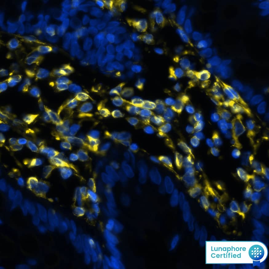
DR647RT) and counterstained with DAPI (blue; Lunaphore Catalog #
DR100). Specific staining was localized to the cytoplasm and cytoskeleton. Protocol available in
COMET™ Panel Builder." alt="Vimentin was detected in immersion fixed paraffin-embedded sections of human colon using Rat Anti-Human/Mouse/Rat Vimentin Monoclonal Antibody (Catalog # MAB2105) at 1 µg/mL at 37 ° Celsius for 4 minutes. Before incubation with the primary antibody, tissue underwent an all-in-one dewaxing and antigen retrieval preprocessing using PreTreatment Module (PT Module) and Dewax and HIER Buffer H (pH 9). Tissue was stained using the Alexa Fluor™ Plus 647 Goat anti-Rat IgG Secondary Antibody at 1:200 at 37 ° Celsius for 2 minutes. (Yellow; Lunaphore Catalog #
DR647RT) and counterstained with DAPI (blue; Lunaphore Catalog #
DR100). Specific staining was localized to the cytoplasm and cytoskeleton. Protocol available in
COMET™ Panel Builder." class="big_thumb" />
MAB2105) at 1 µg/mL at 37 ° Celsius for 4 minutes. Before incubation with the primary antibody, tissue underwent an all-in-one dewaxing and antigen retrieval preprocessing using PreTreatment Module (PT Module) and Dewax and HIER Buffer H (pH 9; Epredia Catalog # TA-999-DHBH). Tissue was stained using the Alexa Fluor™ 647 Goat anti-Rat IgG Secondary Antibody at 1:200 at 37 ° Celsius for 2 minutes. (Yellow; Lunaphore Catalog #
DR647RT) and counterstained with DAPI (blue; Lunaphore Catalog #
DR100). Specific staining was localized to the cytoplasm and cytoskeleton. Protocol available in
COMET™ Panel Builder." class="big_lightbox">
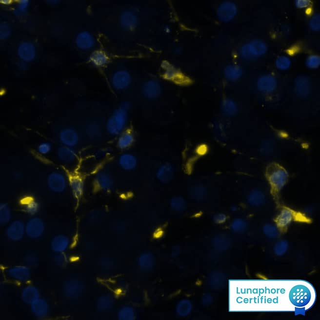
MAB2105) at 1 µg/mL at 37 ° Celsius for 4 minutes. Before incubation with the primary antibody, tissue underwent an all-in-one dewaxing and antigen retrieval preprocessing using PreTreatment Module (PT Module) and Dewax and HIER Buffer H (pH 9; Epredia Catalog # TA-999-DHBH). Tissue was stained using the Alexa Fluor™ 647 Goat anti-Rat IgG Secondary Antibody at 1:200 at 37 ° Celsius for 2 minutes. (Yellow; Lunaphore Catalog #
DR647RT) and counterstained with DAPI (blue; Lunaphore Catalog #
DR100). Specific staining was localized to the cytoplasm and cytoskeleton. Protocol available in
COMET™ Panel Builder." class="big_thumb" />
DR647RT) and counterstained with DAPI (blue; Lunaphore Catalog #
DR100). Specific staining was localized to the cytoplasm and cytoskeleton. Protocol available in
COMET™ Panel Builder." class="big_lightbox">
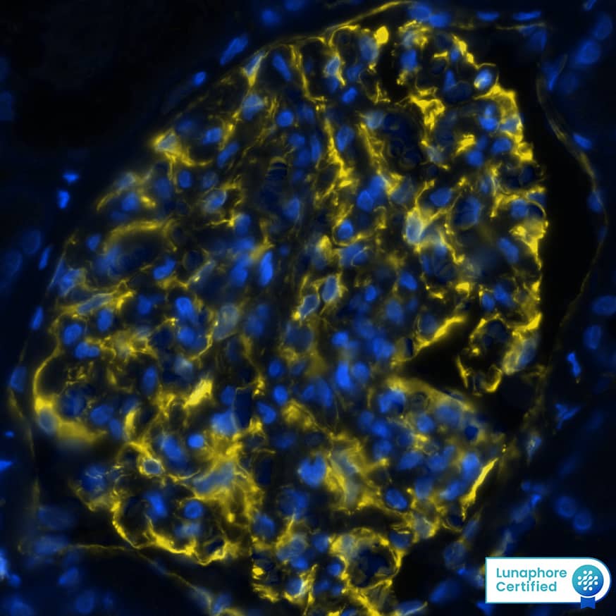
DR647RT) and counterstained with DAPI (blue; Lunaphore Catalog #
DR100). Specific staining was localized to the cytoplasm and cytoskeleton. Protocol available in
COMET™ Panel Builder." class="big_thumb" />
MAB2105) at 10ug/mL at 37 ° Celsius for 4 minutes. Before incubation with the primary antibody, tissue underwent an all-in-one dewaxing and antigen retrieval preprocessing using PreTreatment Module (PT Module) and Dewax and HIER Buffer H (pH 9; Epredia Catalog # TA-999-DHBH). Tissue was stained using the Alexa Fluor™ 647 Goat anti-Rat IgG Secondary Antibody at 1:200 at 37 ° Celsius for 2 minutes. (Yellow; Lunaphore Catalog #
DR647RT) and counterstained with DAPI (blue; Lunaphore Catalog #
DR100). Specific staining was localized to the membrane. Protocol available in
COMET™ Panel Builder." class="big_lightbox">
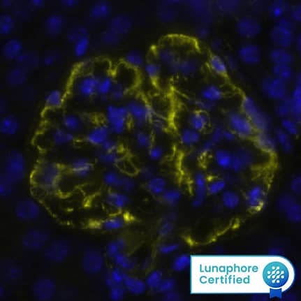
MAB2105) at 10ug/mL at 37 ° Celsius for 4 minutes. Before incubation with the primary antibody, tissue underwent an all-in-one dewaxing and antigen retrieval preprocessing using PreTreatment Module (PT Module) and Dewax and HIER Buffer H (pH 9; Epredia Catalog # TA-999-DHBH). Tissue was stained using the Alexa Fluor™ 647 Goat anti-Rat IgG Secondary Antibody at 1:200 at 37 ° Celsius for 2 minutes. (Yellow; Lunaphore Catalog #
DR647RT) and counterstained with DAPI (blue; Lunaphore Catalog #
DR100). Specific staining was localized to the membrane. Protocol available in
COMET™ Panel Builder." class="big_thumb" />
HAF005). A specific band was detected for Vimentin at approximately 55 kDa (as indicated). This experiment was conducted under reducing conditions and using
Immunoblot Buffer Group 1." class="big_lightbox">
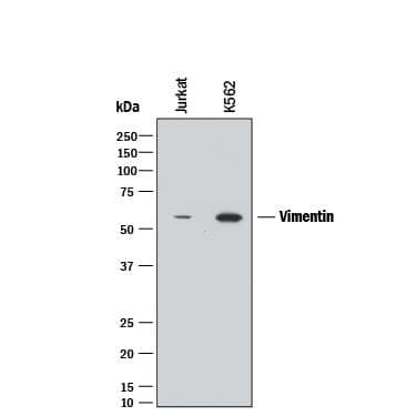
HAF005). A specific band was detected for Vimentin at approximately 55 kDa (as indicated). This experiment was conducted under reducing conditions and using
Immunoblot Buffer Group 1." class="big_thumb" />
HAF005). A specific band was detected for Vimentin at approximately 55 kDa (as indicated). This experiment was conducted under reducing conditions and using
Immunoblot Buffer Group 1." class="big_lightbox">
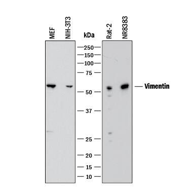
HAF005). A specific band was detected for Vimentin at approximately 55 kDa (as indicated). This experiment was conducted under reducing conditions and using
Immunoblot Buffer Group 1." class="big_thumb" />
MAB006, open histogram) followed by anti-Rat IgG PE-conjugated secondary antibody (Catalog #
F0105B). To facilitate intracellular staining, cells were fixed with Flow Cytometry Fixation Buffer (Catalog #
FC004) and permeabilized with Flow Cytometry Permeabilization/Wash Buffer I (Catalog #
FC005). View our protocol for
Staining Intracellular Molecules." class="big_lightbox">
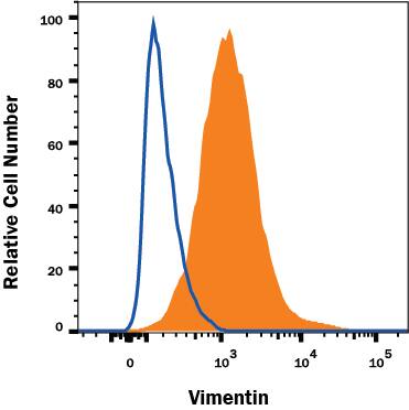
MAB006, open histogram) followed by anti-Rat IgG PE-conjugated secondary antibody (Catalog #
F0105B). To facilitate intracellular staining, cells were fixed with Flow Cytometry Fixation Buffer (Catalog #
FC004) and permeabilized with Flow Cytometry Permeabilization/Wash Buffer I (Catalog #
FC005). View our protocol for
Staining Intracellular Molecules." class="big_thumb" />
HAF005). This experiment was conducted under reducing conditions and using the 12-230 kDa separation system." class="big_lightbox">
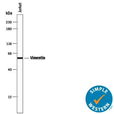
HAF005). This experiment was conducted under reducing conditions and using the 12-230 kDa separation system." class="big_thumb" />
HAF017). A specific band was detected for Vimentin at approximately 55 kDa (as indicated) in the parental K562 cell line, but is not detectable in knockout K562 cell line. GAPDH (
MAB5718) is shown as a loading control. This experiment was conducted under reducing conditions and using Western Blot Buffer Group 1." class="big_lightbox">
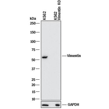
HAF017). A specific band was detected for Vimentin at approximately 55 kDa (as indicated) in the parental K562 cell line, but is not detectable in knockout K562 cell line. GAPDH (
MAB5718) is shown as a loading control. This experiment was conducted under reducing conditions and using Western Blot Buffer Group 1." class="big_thumb" />

![Widespread GLI expression in human donor and cirrhotic liver.(A) Frozen (4 μm) human donor (n = 5), and cirrhotic liver sections [ALD (n = 6), NASH (n = 3), PBC (n = 1)] were screened for GLI2 (red) expression by immunofluorescence. Representative images taken at 5x or 40x (insets) objective shown. DAPI, blue. (B) qRT-PCR for GLI1 and GLI3 transcript in human donor or ALD samples. Mean±S.E.M. Significant (*) difference between means (One-sided student t-test, **p<0.005). Western blot for full-length GLI1 protein (>150 kDa) in donor (Don) or ALD patient samples. Densitometry analysis with GLI1 normalised to GAPDH (Image J). Mean±S.E.M; **p = 0.0093 (Two-sided student t-test). (C) Nuclear GLI2 (green) expression in EpCAM+ (red) LPCs in donor, ALD, PBC and NASH liver. (D) Nuclear GLI2 (green) expression demonstrated within CD31+ (red) ECs, CK18+ (red) hepatocytes, CD45+ (red) leukocytes and vimentin+ (red) HSCs/myofibroblasts, in ALD. 63x objective. (E) Maximum intensity projection illustrating close physical association between EpCAM+ LPCs (green) and vimentin+ HSCs/myofibroblasts (red), both of which express GLI2 (grey), in ALD tissue. Arrows indicate myofibroblasts directly contacting LPCs. Confocal microscopy, 63x objective. Quantitation (%) of EpCAM+ GLI2+ cells and vimentin+ GLI2+ cells within the same FOV (n = 3 ALD samples, 8 FOV/sample). Image collected and cropped by CiteAb from the following publication (//dx.plos.org/10.1371/journal.pone.0171480), licensed under a CC-BY license. Not internally tested by R&D Systems.](//images.novusbio.com/images/mab2105_human-mouse-rat-vimentin-mab-clone-280618-41202410331991.jpg)
human intestinal organoid culture protocol. Human intestinal organoids were stained using a Rat Anti-Human/Mouse/Rat Vimentin Monoclonal Antibody (Catalog # MAB2105; green) and a Goat Anti-Human/Mouse Desmin Antigen Affinity-purified Polyclonal Antibody (Catalog # AF3844; red) to visualize myofibroblast cells and counterstained with DAPI (Catalog # 5748; blue). " class="big_lightbox">

human intestinal organoid culture protocol. Human intestinal organoids were stained using a Rat Anti-Human/Mouse/Rat Vimentin Monoclonal Antibody (Catalog # MAB2105; green) and a Goat Anti-Human/Mouse Desmin Antigen Affinity-purified Polyclonal Antibody (Catalog # AF3844; red) to visualize myofibroblast cells and counterstained with DAPI (Catalog # 5748; blue). " class="big_thumb" />
 DR647RT) and counterstained with DAPI (blue; Lunaphore Catalog # DR100). Specific staining was localized to the cytoplasm and cytoskeleton. Protocol available in COMET™ Panel Builder." alt="Vimentin was detected in immersion fixed paraffin-embedded sections of human colon using Rat Anti-Human/Mouse/Rat Vimentin Monoclonal Antibody (Catalog # MAB2105) at 1 µg/mL at 37 ° Celsius for 4 minutes. Before incubation with the primary antibody, tissue underwent an all-in-one dewaxing and antigen retrieval preprocessing using PreTreatment Module (PT Module) and Dewax and HIER Buffer H (pH 9). Tissue was stained using the Alexa Fluor™ Plus 647 Goat anti-Rat IgG Secondary Antibody at 1:200 at 37 ° Celsius for 2 minutes. (Yellow; Lunaphore Catalog # DR647RT) and counterstained with DAPI (blue; Lunaphore Catalog # DR100). Specific staining was localized to the cytoplasm and cytoskeleton. Protocol available in COMET™ Panel Builder." class="big_thumb" />
 HAF017). A specific band was detected for Vimentin at approximately 55 kDa (as indicated). This experiment was conducted under reducing conditions and using Western Blot Buffer Group 1." alt="Western blot shows lysates of K562 human chronic myelogenous leukemia cell line, MEF mouse embryonic feeder cells, RAW 264.7 mouse monocyte/macrophage cell line, NR8383 rat alveolar macrophage cell line, and Rat‑2 rat embryonic fibroblast cell line. PVDF membrane was probed with 2 µg/mL of Goat Anti-Human/Mouse/Rat Vimentin Antigen Affinity-purified Polyclonal Antibody (Catalog # AF2105) followed by HRP-conjugated Anti-Goat IgG Secondary Antibody (HAF017). A specific band was detected for Vimentin at approximately 55 kDa (as indicated). This experiment was conducted under reducing conditions and using Western Blot Buffer Group 1." class="big_thumb" />
 DR647MS) and counterstained with DAPI (blue; Lunaphore Catalog # DR100). Specific staining was localized to the cytoplasm and cytoskeleton. Protocol available in COMET™ Panel Builder." alt="Vimentin was detected in immersion fixed paraffin-embedded sections of human kidney using Mouse Anti-Human/Mouse/Rat Vimentin Monoclonal Antibody (Catalog # MAB21052) at 0.05 µg/mL at 37 ° Celsius for 4 minutes. Before incubation with the primary antibody, tissue underwent an all-in-one dewaxing and antigen retrieval preprocessing using PreTreatment Module (PT Module) and Dewax and HIER Buffer H (pH 9; Epredia Catalog # TA-999-DHBH).Tissue was stained using the Alexa Fluor™ 647 Goat anti-Mouse IgG Secondary Antibody at 1:200 at 37 ° Celsius for 2 minutes. (Yellow; Lunaphore Catalog # DR647MS) and counterstained with DAPI (blue; Lunaphore Catalog # DR100). Specific staining was localized to the cytoplasm and cytoskeleton. Protocol available in COMET™ Panel Builder." class="big_thumb" />


![Western Blot: Vimentin Antibody [NB300-223] - Analysis of tissue and cell lysates. Antibody at 1:5000 in red. [1] protein standard (red), [2] rat whole brain lysate, [3] HeLa, [4] SH-SY5Y, [5] HEK293, and [6] NIH-3T3 cell lysates. NB300-223 binds to the vimentin protein showing a single band at ~50 kDa. The blot was simultaneously probed with mouse mAb to MAP2C/D, dilution 1:5000 in green, revealing multiple bands around 280 kDa that correspond to full length MAP2A/2B isotypes, and ~70 kDa bands which are MAP2C/D isotypes. MAP2 isotypes are seen only in extracts containing neuronal lineage cells.](http://images.novusbio.com/fullsize/Vimentin-Antibody-Western-Blot-NB300-223-img0002.jpg "Western Blot: Vimentin Antibody [NB300-223] - Analysis of tissue and cell lysates. Antibody at 1:5000 in red. [1] protein standard (red), [2] rat whole brain lysate, [3] HeLa, [4] SH-SY5Y, [5] HEK293, and [6] NIH-3T3 cell lysates. NB300-223 binds to the vimentin protein showing a single band at ~50 kDa. The blot was simultaneously probed with mouse mAb to MAP2C/D, dilution 1:5000 in green, revealing multiple bands around 280 kDa that correspond to full length MAP2A/2B isotypes, and ~70 kDa bands which are MAP2C/D isotypes. MAP2 isotypes are seen only in extracts containing neuronal lineage cells.")

![Immunocytochemistry/Immunofluorescence: Vimentin Antibody [NB300-223] - Human A7 cells. Cells were fixed with 10% formalin and permeabilized with 0.1% TritonX-100. After blocking, cells were incubated with primary antibody (1:600) at 4C O/N followed with Alexa Fluor 488 labeled secondary antibody and counterstained with DAPI and Phalloidin for nuclei and F-actin respectively. Imaged with Leica TCS SP5 confocal microscope. ICC/IF image submitted by a verified customer review.](http://images.novusbio.com/fullsize/Vimentin-Antibody-Immunocytochemistry-Immunofluorescence-NB300-223-img0004.jpg)


![Immunocytochemistry/Immunofluorescence: Vimentin Antibody [NB300-223] - Analysis of HeLa cell culture. Antibody at 1:10,000 in green, and costained with mouse mAb to actin, dilution 1:500 in red. The blue is DAPI staining of nuclear DNA. The vimentin antibody stains the intermediate filament network while the actin antibody labels the submembranous cytoskeleton, stress fibers, and bundles of actin associated with cell adhesion sites.](http://images.novusbio.com/fullsize/Vimentin-Antibody-Immunocytochemistry-Immunofluorescence-NB300-223-img0001.jpg)
![Immunohistochemistry-Frozen: Vimentin Antibody [NB300-223] - 10 um Cryosections from PFA fixed murine brain acute slices stained with Vimentin antibody (1:600) and Alexa Fluor 488 anti chicken IgG. Antibody showed specific staining, also of blood vessels. IHC-Fr image submitted by a verified customer review.](http://images.novusbio.com/fullsize/Vimentin-Antibody-Immunohistochemistry-Frozen-NB300-223-img0003.jpg)
![Immunohistochemistry-Frozen: Vimentin Antibody [NB300-223] - Infarcted mouse heart frozen sections stained with vimentin antibody at 1:200. Secondary antibody at 1:500. Fixation with 4% PFA. IHC-Fr image submitted by a verified customer review.](http://images.novusbio.com/fullsize/Vimentin-Antibody-Immunohistochemistry-Frozen-NB300-223-img0005.jpg)
![Immunocytochemistry/ Immunofluorescence: Vimentin Antibody [NB300-223] - Increased numbers of differentiating NSPCs in SVZ of G93A-SOD1 brain. (A) Confocal microscopy images of dorsal & ventral regions of SVZ in G93A-SOD1 & Wt-SOD1 brain at postnatal week 18, stained for nestin (green), GFAP (red), & vimentin (blue). Scale bar = 50 μm. (B) Quantification of nestin-, GFAP-, & vimentin-positive cells in SVZ of G93A-SOD1 & Wt-SOD1 mice. Data are means ± SD of 3 mice per group. **p < 0.001; limma moderated t-test. (C) Hematoxylin & eosin staining of SVZ sections adjacent to those analyzed by confocal microscopy. Scale bar = 50 μm. D: dorsal. V: ventral. (D) Confocal microscopy images showing Dlx2-stained cells in ventral SVZ in G93A-SOD1 & Wt-SOD1 brain. Scale bar = 50 μm. (E) Quantification of Dlx2-positive cells in G93A-SOD1 & Wt-SOD1 SVZ. Data are means ± SD of 3 mice per group. **p < 0.001; limma moderated t-test. Image collected & cropped by CiteAb from the following publication (//molecularbrain.biomedcentral.com/articles/10.1186/s13041-015-0095-0), licensed under a CC-BY license. Not internally tested by Novus Biologicals.](http://images.novusbio.com/fullsize/nb300-223_chicken-polyclonal-vimentin-antibody-310202416205154.jpg)
![Immunocytochemistry/ Immunofluorescence: Vimentin Antibody [NB300-223] - Attenuated reactive astrocytosis after stroke in Smad1 cKO mice.(A) Images of IHC for reactive astrocyte marker GFAP on ipsilateral hemisphere at 7 days (top two panels) or 3 months (bottom panels) post-stroke. The border between the stroke core & peri-infarct area is outlined by dotted lines. Arrows point to striatal infarct core. (B) Enlarged IHC images of boxed areas in (A) at the cortical peri-infarct area (blue box in D) with the indicated reactive astrocyte markers GFAP, Nestin, & Vimentin. Enlarged images of GFAP IHC highlight the hypertrophic morphology of GFAP+ astrocytes in mutants. (C) Quantification of the number of GFAP+ astrocytes & the intensity of GFAP immunoreactivity (IR) at the peri-infarct area shown in (B). n = 4, one-way ANOVA for the number of astrocytes, unpaired Student’s t-test for GFAP intensity, ***p<0.001, Ipsi, ipsilateral; Ctra, contralateral cortex. (D) Diagram of infarct territory affected by tMCAO (blue) & peri-infarct cortical area (blue box). (E) Reactive astrocytosis was similarly attenuated in the ipsilateral hippocampus of Smad1 cKO mice. Scale, 500 μm (A), 100 μm (B), & 200 μm (E). Image collected & cropped by CiteAb from the following publication (//dx.plos.org/10.1371/journal.pone.0136967), licensed under a CC-BY license. Not internally tested by Novus Biologicals.](http://images.novusbio.com/fullsize/nb300-223_chicken-polyclonal-vimentin-antibody-310202416205162.jpg)
![Immunocytochemistry/ Immunofluorescence: Vimentin Antibody [NB300-223] - Tumor formation & metastasis by PyMT-1099 cells. (A) PyMT-1099 cells untreated or treated with TGF beta for >20 days (PyMT-1099 LT) were injected orthotopically into mammary fat pads of NSG mice. The graph represents tumor growth in PyMT-1099 & PyMT-1099 LT group of mice. (B) Histological tumor sections from tumors of PyMT-1099 or PyMT-1099 LT cells described in (A) were stained with H&E to assess the morphology of primary tumors. Representative microphotographs are shown from tumors of 2 out of the 6 mice used in the experiment. (C) Immunofluorescence analysis was performed to assess the expression of the EMT markers FN1, VIM, E-CAD & N-CAD in tumors formed by PyMT-1099 or PyMT-1099 LT cells in the experiment described in (A). DAPI was used as a nuclear counterstain. Representative pictures are shown from tumors of 2 out of the 6 mice used in the experiment. Scale bar, 100 μm. (D) The graph represents the number of lung metastases formed in NSG mice orthotopically transplanted with PyMT-1099 or PyMT-1099 LT cells; n = 6. (E) The graph represents the number of lung metastases formed in NSG mice injected with PyMT-1099 or PyMT-1099 LT cells through the tail vein; n = 6. The mice were sacrificed 8 weeks post-injection, & lungs were resected for the analysis of cancer cell colonization/ metastases formation. Image collected & cropped by CiteAb from the following publication (//pubmed.ncbi.nlm.nih.gov/30108334), licensed under a CC-BY license. Not internally tested by Novus Biologicals.](http://images.novusbio.com/fullsize/nb300-223_chicken-polyclonal-vimentin-antibody-31020241621828.jpg)


 MAB2105) at 1 µg/mL at 37 ° Celsius for 4 minutes. Before incubation with the primary antibody, tissue underwent an all-in-one dewaxing and antigen retrieval preprocessing using PreTreatment Module (PT Module) and Dewax and HIER Buffer H (pH 9; Epredia Catalog # TA-999-DHBH). Tissue was stained using the Alexa Fluor™ 647 Goat anti-Rat IgG Secondary Antibody at 1:200 at 37 ° Celsius for 2 minutes. (Yellow; Lunaphore Catalog #
  DR647RT) and counterstained with DAPI (blue; Lunaphore Catalog #
  MAB2105) at 10ug/mL at 37 ° Celsius for 4 minutes. Before incubation with the primary antibody, tissue underwent an all-in-one dewaxing and antigen retrieval preprocessing using PreTreatment Module (PT Module) and Dewax and HIER Buffer H (pH 9; Epredia Catalog # TA-999-DHBH). Tissue was stained using the Alexa Fluor™ 647 Goat anti-Rat IgG Secondary Antibody at 1:200 at 37 ° Celsius for 2 minutes. (Yellow; Lunaphore Catalog #
  HAF005). A specific band was detected for Vimentin at approximately 55 kDa (as indicated). This experiment was conducted under reducing conditions and using
  HAF005). A specific band was detected for Vimentin at approximately 55 kDa (as indicated). This experiment was conducted under reducing conditions and using
  NL013) and counterstained with DAPI (blue). View our protocol for
  NL015) and counterstained with DAPI (blue). View our protocol for
  NL013) and counterstained with DAPI (blue). Specific staining was localized to cytoskeleton. View our protocol for
  NL013) and counterstained with DAPI (blue). Specific staining was localized to cytoskeleton. View our protocol for
  VC005). Tissue was stained using DAB (brown) and counterstained with hematoxylin (blue). Specific staining was localized to cytoplasm. View our protocol for
  MAB006, open histogram) followed by anti-Rat IgG PE-conjugated secondary antibody (Catalog #
  HAF005). This experiment was conducted under reducing conditions and using the 12-230 kDa separation system." class="big_thumb" />
 HAF017). A specific band was detected for Vimentin at approximately 55 kDa (as indicated) in the parental K562 cell line, but is not detectable in knockout K562 cell line. GAPDH (
![Widespread GLI expression in human donor and cirrhotic liver.(A) Frozen (4 μm) human donor (n = 5), and cirrhotic liver sections [ALD (n = 6), NASH (n = 3), PBC (n = 1)] were screened for GLI2 (red) expression by immunofluorescence. Representative images taken at 5x or 40x (insets) objective shown. DAPI, blue. (B) qRT-PCR for GLI1 and GLI3 transcript in human donor or ALD samples. Mean±S.E.M. Significant (*) difference between means (One-sided student t-test, **p<0.005). Western blot for full-length GLI1 protein (>150 kDa) in donor (Don) or ALD patient samples. Densitometry analysis with GLI1 normalised to GAPDH (Image J). Mean±S.E.M; **p = 0.0093 (Two-sided student t-test). (C) Nuclear GLI2 (green) expression in EpCAM+ (red) LPCs in donor, ALD, PBC and NASH liver. (D) Nuclear GLI2 (green) expression demonstrated within CD31+ (red) ECs, CK18+ (red) hepatocytes, CD45+ (red) leukocytes and vimentin+ (red) HSCs/myofibroblasts, in ALD. 63x objective. (E) Maximum intensity projection illustrating close physical association between EpCAM+ LPCs (green) and vimentin+ HSCs/myofibroblasts (red), both of which express GLI2 (grey), in ALD tissue. Arrows indicate myofibroblasts directly contacting LPCs. Confocal microscopy, 63x objective. Quantitation (%) of EpCAM+ GLI2+ cells and vimentin+ GLI2+ cells within the same FOV (n = 3 ALD samples, 8 FOV/sample). Image collected and cropped by CiteAb from the following publication (//dx.plos.org/10.1371/journal.pone.0171480), licensed under a CC-BY license. Not internally tested by R&D Systems.](http://images.novusbio.com/fullsize/mab2105_human-mouse-rat-vimentin-mab-clone-280618-41202410331991.jpg)
 VCTS021). Tissue was stained using the HRP-conjugated Anti-Rat IgG Secondary Antibody (Catalog #
  human intestinal organoid culture protocol. Human intestinal organoids were stained using a Rat Anti-Human/Mouse/Rat Vimentin Monoclonal Antibody (Catalog # MAB2105; green) and a Goat Anti-Human/Mouse Desmin Antigen Affinity-purified Polyclonal Antibody (Catalog # AF3844; red) to visualize myofibroblast cells and counterstained with DAPI (Catalog # 5748; blue). " class="big_thumb" />
 CTS008) and counterstained with hematoxylin (blue). View our protocol for
  NL001) and counterstained with DAPI (blue). Specific staining was localized to intermediate filaments in cytoplasm. View our protocol for
  HAF109). This experiment was conducted under reducing conditions and using the 12-230 kDa separation system. Non-specific interaction with the 230 kDa Simple Western standard may be seen with this antibody." class="big_thumb" />
 NL003) and counterstained with DAPI (blue). Specific staining was localized to cytoplasm. Staining was performed using our Fluorescent ICC Staining of Non-adherent Cells protocol." class="big_thumb" />
 HAF017). A specific band was detected for Vimentin at approximately 55 kDa (as indicated) in the parental K562 cell line, but is not detectable in knockout K562 cell line. GAPDH (

![<b>Biological Strategies Validation. </b>NRF1 and/or E2 re-programming contributed to the overexpression of pluripotency and epithelial-mesenchymal transition (EMT) markers and differentiation of tumor initiating breast cancer cells to other-types of cells. Experimental conditions were the same as Figure 1. (A) Immunofluorescent and flow cytometry detections of MCF10A (T) tumor initiating pluripotent stem cells markers (SOX2, Oct4, and Nanog) in transformed breast epithelial cells of MCF10A [MCF-10A (T)] overexpressing vector (control) or NRF1 treated with DMSO and E2. (B) Immunofluorescent and flow cytometry detections of pluripotent stem cells markers (SOX2 and Oct4) in transformed breast epithelial cells of MCF10A [MCF-10A (T)] overexpressing vector (control) or NRF1 treated with DMSO and E2. (C) Immunofluorescent and Western detections of the expression of EMT markers E-cadherin, N-cadherin, and vimentin in transformed breast epithelial cells of MCF10A [MCF-10A (T)] overexpressing vector (control) or NRF1 treated with DMSO and E2. (D) Differentiation of NRF1 tumor initiating stem cells to chondrocytes, neurons, and smooth muscle cells compared to the vector. The vector did not show any differentiation to other cells. Image collected and cropped by CiteAb from the following publication (//pubmed.ncbi.nlm.nih.gov/30486409), licensed under a CC-BY license. Not internally tested by R&D Systems.](http://images.novusbio.com/fullsize/af2105_human-mouse-rat-vimentin-affinity-purified-polyclonal-ab-41202410485417.jpg)

![Western Blot: Vimentin Antibody [NBP1-31327] - Non-transfected (-) and transfected (+) 293T whole cell extracts (30 ug) were separated by 10% SDS-PAGE, and the membrane was blotted with Vimentin antibody.](http://images.novusbio.com/fullsize/Vimentin-Antibody-Western-Blot-NBP1-31327-img0031.jpg "<b>Genetic Strategies Validation. </b>Western Blot: Vimentin Antibody [NBP1-31327] - Non-transfected (-) and transfected (+) 293T whole cell extracts (30 ug) were separated by 10% SDS-PAGE, and the membrane was blotted with Vimentin antibody.")
![<b>Genetic Strategies Validation. </b>Immunocytochemistry/Immunofluorescence: Vimentin Antibody [NBP1-31327] - Cellular aggregates composed of equine endometrial fibroblast. Co-staining using the vimentin antibody and a cytokeratin antibody (mouse). Image submitted by a verified customer review.](http://images.novusbio.com/fullsize/Vimentin-Antibody-Immunocytochemistry-NBP1-31327-img0037.jpg)
![Immunocytochemistry/Immunofluorescence: Vimentin Antibody [NBP1-31327] - Human primary cardiac fibroblasts stained with Vimentin (green) and smooth muscle actin (red) antibodies. Image from verified customer review.](http://images.novusbio.com/fullsize/Vimentin-Antibody-Immunocytochemistry-Immunofluorescence-NBP1-31327-img0024.jpg)
![Immunohistochemistry-Paraffin: Vimentin Antibody [NBP1-31327] - Paraffin-embedded rat ovary. Vimentin antibody at 1:500.](http://images.novusbio.com/fullsize/Vimentin-Antibody-Immunohistochemistry-Paraffin-NBP1-31327-img0036.jpg)
![Western Blot: Vimentin Antibody [NBP1-31327] - 293T cell lysate: A. 20 ug. B. 10 ug. C. 5 ug. D. 1 ug.](http://images.novusbio.com/fullsize/Vimentin-Antibody-Western-Blot-NBP1-31327-img0014.jpg)
![Western Blot: Vimentin Antibody [NBP1-31327] - Various whole cell extracts (30 ug) were separated by 10% SDS-PAGE, membrane was blotted with Vimentin antibody at 1:50000.](http://images.novusbio.com/fullsize/Vimentin-Antibody-Western-Blot-NBP1-31327-img0023.jpg)
![Immunohistochemistry-Paraffin: Vimentin Antibody [NBP1-31327] - Paraffin-embedded human lung adenocarcinoma. Vimentin antibody diluted at 1:500.](http://images.novusbio.com/fullsize/Vimentin-Antibody-Immunohistochemistry-Paraffin-NBP1-31327-img0022.jpg)
![Immunohistochemistry-Paraffin: Vimentin Antibody [NBP1-31327] - Paraffin-embedded rat testis. Vimentin antibody at 1:500.](http://images.novusbio.com/fullsize/Vimentin-Antibody-Immunohistochemistry-Paraffin-NBP1-31327-img0034.jpg)
![Immunoprecipitation: Vimentin Antibody [NBP1-31327] - Vimentin protein from HeLa whole cell extracts using 5 ug of Vimentin antibody. Western blot analysis was performed using Vimentin antibody. EasyBlot anti-Rabbit IgG was used as a secondary reagent.](http://images.novusbio.com/fullsize/Vimentin-Antibody-Immunoprecipitation-NBP1-31327-img0019.jpg)
![Sandwich ELISA: Vimentin Antibody [NBP1-31327] - Detection of recombinant full-length Vimentin protein using as capture antibody at concentration of 5 ug/mL as detection antibody at concentration of 1 ug/mL. Rabbit IgG antibody (HRP) (NBP2-19301) was diluted at 1:10000 and used to detect the primary antibody.](http://images.novusbio.com/fullsize/Vimentin-Antibody-Sandwich-ELISA-NBP1-31327-img0039.jpg)
![Western Blot: Vimentin Antibody [NBP1-31327] - Various whole cell extracts (30 ug) were separated by 10% SDS-PAGE, and the membrane was blotted with Vimentin antibody (NBP1-31327) diluted at 1:5000. The HRP-conjugated anti-rabbit IgG antibody was used to detect the primary antibody.](http://images.novusbio.com/fullsize/nbp1-31327_rabbit-polyclonal-vimentin-antibody-9102024204128.jpg)
![Western Blot: Vimentin Antibody [NBP1-31327] - Non-transfected (-) and transfected (+) 293T whole cell extracts (10 ug) were separated by 10% SDS-PAGE, and the membrane was blotted with Vimentin antibody (NBP1-31327) diluted at 1:10000. The HRP-conjugated anti-rabbit IgG antibody was used to detect the primary antibody.](http://images.novusbio.com/fullsize/nbp1-31327_rabbit-polyclonal-vimentin-antibody-91020242010448.jpg)
![ELISA: Vimentin Antibody [NBP1-31327] - Sandwich ELISA detection of recombinant full-length Vimentin protein using the capture antibody at concentration of 5 ug/mL and NBP1-31327 as detection antibody at concentration of 1 ug/mL. Rabbit IgG antibody (HRP) was diluted at 1:10000 and used to detect the primary antibody.](http://images.novusbio.com/fullsize/nbp1-31327_rabbit-polyclonal-vimentin-antibody-91020242063381.jpg)
![Immunohistochemistry-Paraffin: Vimentin Antibody [NBP1-31327] - Vimentin antibody detects Vimentin protein at cytoplasm by immunohistochemical analysis.</br>Sample: Paraffin-embedded mouse intestine.</br>Vimentin stained by Vimentin antibody (NBP1-31327) diluted at 1:500.</br>Antigen Retrieval: Citrate buffer, pH 6.0, 15 min](http://images.novusbio.com/fullsize/nbp1-31327_rabbit-polyclonal-vimentin-antibody-910202419593530.jpg)
![Western Blot: Vimentin Antibody [NBP1-31327] - Various whole cell extracts (30 ug) were separated by 10% SDS-PAGE, and the membrane was blotted with Vimentin antibody (NBP1-31327) diluted at 1:5000. The HRP-conjugated anti-rabbit IgG antibody was used to detect the primary antibody.](http://images.novusbio.com/fullsize/nbp1-31327_rabbit-polyclonal-vimentin-antibody-910202420131228.jpg)
![Immunohistochemistry-Paraffin: Vimentin Antibody [NBP1-31327] - Vimentin antibody detects Vimentin protein expression by immunohistochemical analysis.<br>Sample:Paraffin-Embedded adult mouse retina. <br>Green: Vimentin protein stained by Vimentin antibody (NBP1-31327) diluted at 1:250.<br>Red: beta Tubulin 3/ TUJ1, stained by beta Tubulin 3/ TUJ1 antibody [GT11710] diluted at 1:500.<br>Blue: Fluoroshield with DAPI .<br>](http://images.novusbio.com/fullsize/nbp1-31327_rabbit-polyclonal-vimentin-antibody-910202420131227.jpg)
![Western Blot: Vimentin Antibody [NBP1-31327] - Various whole cell extracts (30 ug) were separated by 10% SDS-PAGE, and the membrane was blotted with Vimentin antibody (NBP1-31327) diluted at 1:20000. The HRP-conjugated anti-rabbit IgG antibody was used to detect the primary antibody.](http://images.novusbio.com/fullsize/nbp1-31327_rabbit-polyclonal-vimentin-antibody-9102024201149.jpg)
![Immunocytochemistry/ Immunofluorescence: Vimentin Antibody [NBP1-31327] - Vimentin antibody detects Vimentin protein at cytoskeleton and nucleus by immunofluorescent analysis.</br>Sample: HeLa cells were fixed in 4% paraformaldehyde at RT for 15 min.</br>Green: Vimentin stained by Vimentin antibody (NBP1-31327) diluted at 1:500.</br>Red: alpha Tubulin, a cytoskeleton marker, stained by alpha Tubulin antibody [GT114] diluted at 1:1000.</br>Blue: Fluoroshield with DAPI .](http://images.novusbio.com/fullsize/nbp1-31327_rabbit-polyclonal-vimentin-antibody-91020242011456.jpg)
![Immunohistochemistry-Paraffin: Vimentin Antibody [NBP1-31327] - Vimentin antibody detects Vimentin protein at cytoplasm by immunohistochemical analysis.</br>Sample: Paraffin-embedded mouse colon.</br>Vimentin stained by Vimentin antibody (NBP1-31327) diluted at 1:500.</br>Antigen Retrieval: Citrate buffer, pH 6.0, 15 min](http://images.novusbio.com/fullsize/nbp1-31327_rabbit-polyclonal-vimentin-antibody-91020242021974.jpg)
![Immunohistochemistry-Paraffin: Vimentin Antibody [NBP1-31327] - Vimentin antibody detects Vimentin protein at cell membrane and cytoplasm by immunohistochemical analysis.</br>Sample: Paraffin-embedded rat ovary.</br>Vimentin stained by Vimentin antibody (NBP1-31327) diluted at 1:500.</br>Antigen Retrieval: Citrate buffer, pH 6.0, 15 min](http://images.novusbio.com/fullsize/nbp1-31327_rabbit-polyclonal-vimentin-antibody-910202420134066.jpg)
![Immunohistochemistry-Paraffin: Vimentin Antibody [NBP1-31327] - Vimentin antibody detects Vimentin protein at cytoplasm by immunohistochemical analysis.</br>Sample: Paraffin-embedded mouse testis.</br>Vimentin stained by Vimentin antibody (NBP1-31327) diluted at 1:500.</br>Antigen Retrieval: Citrate buffer, pH 6.0, 15](http://images.novusbio.com/fullsize/nbp1-31327_rabbit-polyclonal-vimentin-antibody-910202420134020.jpg)
![Immunohistochemistry-Paraffin: Vimentin Antibody [NBP1-31327] - Vimentin antibody detects Vimentin protein by immunohistochemical analysis.</br>Sample: Paraffin-embedded dog pancreas.</br>Vimentin stained by Vimentin antibody (NBP1-31327) diluted at 1:500.</br>Antigen Retrieval: Citrate buffer, pH 6.0, 15 min](http://images.novusbio.com/fullsize/nbp1-31327_rabbit-polyclonal-vimentin-antibody-910202420115052.jpg)
![Immunocytochemistry/ Immunofluorescence: Vimentin Antibody [NBP1-31327] - Vimentin antibody detects Vimentin protein at cytoskeleton by immunofluorescent analysis.</br>Sample: MDCK cells were fixed in 4% paraformaldehyde at RT for 15 min.</br>Green: Vimentin stained by Vimentin antibody (NBP1-31327) diluted at 1:500.</br>Blue: Hoechst 33342 staining.</br>Scale bar= 10 um.](http://images.novusbio.com/fullsize/nbp1-31327_rabbit-polyclonal-vimentin-antibody-910202420134098.jpg)
![<b>Genetic Strategies Validation. </b>Western Blot: Vimentin Antibody [NBP1-31327] - Wild-type (WT) and Vimentin knockout (KO) 293T cell extracts (30 ug) were separated by 10% SDS-PAGE, and the membrane was blotted with Vimentin antibody diluted at 1:50000. The HRP-conjugated anti-rabbit IgG antibody was used to detect the primary antibody.](http://images.novusbio.com/fullsize/nbp1-31327_rabbit-polyclonal-vimentin-antibody-1610202419414514.jpg)


![Western Blot: Vimentin Antibody (2D1) [NBP1-92687] - Western blot shows lysates of K562 human Chronic Myelogenous Leukemia parental cell line and Vimentin knockout (KO) K562 cell line. PVDF membrane was probed with 1:10,000 of Mouse Anti-Human Vimentin Monoclonal Antibody (Catalog # NBP1-92687) followed by HRP-conjugated Anti-Mouse IgG Secondary Antibody (Catalog #HAF018). Specific band was detected for Vimentin at approximately 55 kDa (as indicated) in the parental K562 cell line, but is not detectable in the knockout K562 cell line. This experiment was conducted under reducing conditions.](http://images.novusbio.com/fullsize/Vimentin-Antibody-2D1-Knockdown-Validated-NBP1-92687-img0011.jpg "<b>Genetic Strategies Validation. </b>Western Blot: Vimentin Antibody (2D1) [NBP1-92687] - Western blot shows lysates of K562 human Chronic Myelogenous Leukemia parental cell line and Vimentin knockout (KO) K562 cell line. PVDF membrane was probed with 1:10,000 of Mouse Anti-Human Vimentin Monoclonal Antibody (Catalog # NBP1-92687) followed by HRP-conjugated Anti-Mouse IgG Secondary Antibody (Catalog #HAF018). Specific band was detected for Vimentin at approximately 55 kDa (as indicated) in the parental K562 cell line, but is not detectable in the knockout K562 cell line. This experiment was conducted under reducing conditions.")
![<b>Genetic Strategies Validation. </b>Immunocytochemistry/Immunofluorescence: Vimentin Antibody (2D1) [NBP1-92687] - View of mixed neuron/glial cultures stained with NBP1-92687 (green) and the GFAP rabbit polyclonal (NB300-141, red). Vimentin is expressed alone in fibroblastic and endothelial cells, which are the flattened cells in the middle of the image which appear green. Astrocytes may express primarily GFAP, or GFAP and Vimentin, and so appear red (GFAP only) or golden yellow (GFAP and Vimentin). In cells which express both GFAP and Vimentin, the two proteins assemble to produce heteropolymer filaments.](http://images.novusbio.com/fullsize/Vimentin-Antibody-2D1-Immunocytochemistry-Immunofluorescence-NBP1-92687-img0003.jpg)
![Western Blot: Vimentin Antibody (2D1) [NBP1-92687] - Analysis of different cell lysates using mouse mAb to vimentin, NBP1-92687, dilution 1:10,000 in green: [1] protein standard (red), [2] HEK293, [3] HeLa, [4] SH-SY5Y, [5] COS-1, and [6] C6 cells. The band at about 50kDa mark corresponds to the vimentin protein.](http://images.novusbio.com/fullsize/Vimentin-Antibody-2D1-Western-Blot-NBP1-92687-img0009.jpg)
![Immunocytochemistry/Immunofluorescence: Vimentin Antibody (2D1) [NBP1-92687] - Analysis of cortical neuron-glial cell cultures from E20 rat stained with mouse mAb to vimentin, NBP1-92687, dilution 1:2,000 in red, and costained with chicken pAb to glial fibrillary acidic protein (GFAP), dilution 1:5,000, in green. The blue is DAPI staining of nuclear DNA. Fibroblastic and other developing cells express only vimentin and appear red. Astrocytes that express GFAP only are green while those that express both GFAP and vimentin appear golden yellow.](http://images.novusbio.com/fullsize/Vimentin-Antibody-2D1-Immunocytochemistry-Immunofluorescence-NBP1-92687-img0010.jpg)
![Immunohistochemistry-Paraffin: Vimentin Antibody (2D1) [NBP1-92687] - Human breast cancer tissue stained for Vimentin (white) and countersatined with DAPI (blue). Alexa Fluor 750 version of antibody used (NBP1-92687AF750). Image from verified customer review.](http://images.novusbio.com/fullsize/Vimentin-Antibody-2D1-Immunohistochemistry-Paraffin-NBP1-92687-img0012.jpg)
![Immunohistochemistry: Vimentin Antibody (2D1) [NBP1-92687] - Mouse cortex microslice section (from postnatal day 3) was immunostained with (1:500 dil.). This image was submitted via customer Review.](http://images.novusbio.com/fullsize/Vimentin-Antibody-2D1-Immunohistochemistry-NBP1-92687-img0005.jpg)
![Simple Western: Vimentin Antibody (2D1) [NBP1-92687] - Simple Western lane view shows a specific band for Vimentin in 0.5 mg/ml of HeLa lysate. This experiment was performed under reducing conditions using the 12-230 kDa separation system.](http://images.novusbio.com/fullsize/Vimentin-Antibody-2D1-Simple-Western-NBP1-92687-img0004.jpg)
![Western Blot: Vimentin Antibody (2D1) [NBP1-92687] - High glucose (HG) concentrations induced epithelial-to-mesenchymal transition protein expression & enhanced migration activity in colorectal cancer (CRC) cells. SW480 (low metastatic potential) & SW620 (high metastatic potential) cells were cultured in different concentrations of glucose (normal: NG; HG; & osmotic control: NG + l-glucose). (A) Morphological change occurred from epithelial to mesenchymal type in the HG-concentration group. (B) HG concentration caused downregulation of E-cadherin & upregulation of N-cadherin, beta CTN, & vimentin, but c-myc was unchanged, as detected using Western blotting. beta -actin was evaluated as an internal control. (C,D) Wound healing assay showed that HG concentration promoted cell motility in SW480 & SW620 CRC cells after 48 & 72 h of culture, compared with the NG & NG + l-glucose groups. (E) In a Transwell migration assay, 3.5 × 10⁵ SW480 & SW620 CRC cells were plated onto a 24-well plate & cultured in NG & HG-concentration medium for 96 h. HG concentration promoted cell motility in SW480 & SW620 cells. NG + l-glucose cells were evaluated as ostomic controls. (F) These data show that HG concentration caused upregulation of p-IGF1R in CRC. In addition, HG concentration promoted IGF1R downstream signaling, including p-Src & p-ERK; these proteins were increased when CRC cells were cultured in HG-concentration medium. Levels of beta -actin were evaluated as loading controls. Statistically significant differences between the two groups were judged using Student’s t-tests; * p < 0.05, ** p < 0.005, *** p < 0.001; n.s. = nonsignificant. Image collected & cropped by CiteAb from the following publication (//pubmed.ncbi.nlm.nih.gov/30965609), licensed under a CC-BY license. Not internally tested by Novus Biologicals.](http://images.novusbio.com/fullsize/nbp1-92687_mouse-monoclonal-vimentin-antibody-2d1-310202416113533.jpg)
![Immunocytochemistry/Immunofluorescence: Vimentin Antibody (RV202) [NBP1-97672] - Staining of a 1 month old zebrafish embryo](http://images.novusbio.com/fullsize/Vimentin-Antibody-RV202-Immunocytochemistry-Immunofluorescence-NBP1-97672-img0006.jpg "Immunocytochemistry/Immunofluorescence: Vimentin Antibody (RV202) [NBP1-97672] - Staining of a 1 month old zebrafish embryo")
![Immunohistochemistry-Frozen: Vimentin Antibody (RV202) [NBP1-97672] - Immunohistochemistry on frozen section of swine colon showing positive staining in connective tissue cells and no reactivity in epithelial cells.](http://images.novusbio.com/fullsize/Vimentin-Antibody-RV202-Immunohistochemistry-Frozen-NBP1-97672-img0007.jpg)
![Immunocytochemistry/Immunofluorescence: Vimentin Antibody (RV202) [NBP1-97672] - Staining of the developing neural tube in a 2 days old zebrafish embryo. Left panel: DAPI-staining of cell nuclei, providing an overview of the tissue section used for immunostaining](http://images.novusbio.com/fullsize/Vimentin-Antibody-RV202-Immunocytochemistry-Immunofluorescence-NBP1-97672-img0005.jpg)
![Immunohistochemistry-Frozen: Vimentin Antibody (RV202) [NBP1-97672] - MUB1900L1: Frozen section of swine colon immunostained with RV202-FITC (1:200)](http://images.novusbio.com/fullsize/Vimentin-Antibody-RV202-Immunohistochemistry-Frozen-NBP1-97672-img0002.jpg)
![Immunohistochemistry-Frozen: Vimentin Antibody (RV202) [NBP1-97672] - Frozen section of swine colon showing positive staining in connective tissue cells and no reactivity in epithelial cells](http://images.novusbio.com/fullsize/Vimentin-Antibody-RV202-Immunohistochemistry-Frozen-NBP1-97672-img0001.jpg)
![Immunohistochemistry-Frozen: Vimentin Antibody (RV202) [NBP1-97672] - Frozen section of swine colon immunostained with RV202-HRP (1:200)](http://images.novusbio.com/fullsize/Vimentin-Antibody-RV202-Immunohistochemistry-Frozen-NBP1-97672-img0003.jpg)
![Immunohistochemistry-Paraffin: Vimentin Antibody (RV202) [NBP1-97672] - Paraffin section of human colon](http://images.novusbio.com/fullsize/Vimentin-Antibody-RV202-Immunohistochemistry-Paraffin-NBP1-97672-img0004.jpg)

![<b>Genetic Strategies Validation. </b>Immunohistochemistry-Frozen: Vimentin Antibody (V9) [NBP1-97670] - Strong specific staining of connective tissue cells in a frozen tissue section of human colon. No reactivity in epithelial cells.](http://images.novusbio.com/fullsize/Vimentin-Antibody-V9-Immunohistochemistry-Frozen-NBP1-97670-img0005.jpg)
![Flow Cytometry: Vimentin Antibody (V9) [NBP1-97670] - An intracellular stain was performed on Jurkat cells with Vimentin antibody (V9) NBP1-97670PE (blue) and a matched isotype control NBP2-27287PE (orange). Cells were fixed with 4% PFA and then permeablized with 0.1% saponin. Cells were incubated in an antibody dilution of 1 ug/mL for 30 minutes at room temperature. Both antibodies were conjugated to phycoerythrin.](http://images.novusbio.com/fullsize/Vimentin-Antibody-V9-Flow-Cytometry-NBP1-97670-img0002.jpg)
![Immunocytochemistry/Immunofluorescence: Vimentin Antibody (V9) [NBP1-97670] - Strong specific staining of glomeruli and connetive tissue in human kidney with in a 1:200 dilution. No reactivity in epithelial cells.](http://images.novusbio.com/fullsize/Vimentin-Antibody-V9-Immunocytochemistry-Immunofluorescence-NBP1-97670-img0003.jpg)
![Immunocytochemistry/Immunofluorescence: Vimentin Antibody (V9) [NBP1-97670] - Strong specific staining of connective tissue cells in swine colon with in a 1:200 dilution. No reactivity in epithelial cells.](http://images.novusbio.com/fullsize/Vimentin-Antibody-V9-Immunocytochemistry-Immunofluorescence-NBP1-97670-img0004.jpg)
![Flow Cytometry: Vimentin Antibody (V9) [NBP1-97670] - Analysis of PE conjugate of NBP1-97670. An intracellular stain was performed on THP-1 cells with Vimentin antibody (V9) NBP1-97670PE (blue) and a matched isotype control NBP2-27287PE (orange). Cells were fixed with 4% PFA and then permeablized with 0.1% s](http://images.novusbio.com/fullsize/Vimentin-Antibody-V9-Flow-Cytometry-NBP1-97670-img0001.jpg)
![Immunohistochemistry-Paraffin: Vimentin Antibody (V9) [NBP1-97670] - Vimentin immunoreactivity in human tonsil. NBP1-97670 was diluted to 0.5ug/mL and was left on sections for 30m at room temperature. Secondary antibody was Horse-Anti Mouse HRP Veterinary Reagent. Image from verified customer review.](http://images.novusbio.com/fullsize/antibody/nbp1-97670_mouse-monoclonal-vimentin-antibody-v9-immunohistochemistry-paraffin-1462023111714..jpg)
![Immunohistochemistry-Paraffin: Vimentin Antibody (V9) [NBP1-97670] - Vimentin immunoreactivity in horse intestine. NBP1-97670 was used at a concentration of 0.5ug/mL and was left on slides for 30m at room temperature. Secondary antibody was Horse Anti-Mouse HRP Veterinary Reagent. Image from verified customer review.](http://images.novusbio.com/fullsize/antibody/nbp1-97670_mouse-monoclonal-vimentin-antibody-v9-immunohistochemistry-paraffin-1462023112025..jpg)
![Immunohistochemistry-Paraffin: Vimentin Antibody (V9) [NBP1-97670] - Vimentin immunoreactivity in feline intestine. NBP1-97670 was used at a concentration of 0.5ug/mL. Secondary antibody was Horse Anti-Mouse HRP Veterinary Reagent. Image from verified customer review.](http://images.novusbio.com/fullsize/antibody/nbp1-97670_mouse-monoclonal-vimentin-antibody-v9-immunohistochemistry-paraffin-1462023112626.jpg)
![Immunohistochemistry-Paraffin: Vimentin Antibody (V9) [NBP1-97670] - Vimentin immunoreactivity in canine intestine. NBP1-97670 was used at a concentration of 0.5ug/mL and was left on tissue sections for 30m at room temperature. Secondary was Horse Anti-Mouse Veterinary Reagent. Image from verified customer review.](http://images.novusbio.com/fullsize/antibody/nbp1-97670_mouse-monoclonal-vimentin-antibody-v9-immunohistochemistry-paraffin-1462023115136..jpg)
![Immunohistochemistry-Paraffin: Mouse Monoclonal Vimentin Antibody (V9) [NBP1-97670] - Vimentin was stained in human FFPE tonsil tissue. HIER antigen retrieval at pH 9 for 20min was performed. Concentration used: 1:100. AF750 conjugated version of the antibody was used (Catalog # NBP1-97670AF750). Image from a verified customer review.](http://images.novusbio.com/fullsize/antibody/nbp1-97670_mouse-monoclonal-vimentin-antibody-v9-immunohistochemistry-paraffin-1122023155957..jpg)


![Immunohistochemistry-Paraffin: Vimentin Antibody [NBP1-85814] - Analysis in human ovary and skeletal muscle tissues. Corresponding VIM RNA-seq data are presented for the same tissues.](http://images.novusbio.com/fullsize/Vimentin-Antibody-Immunohistochemistry-Paraffin-NBP1-85814-img0023.jpg "<b>Orthogonal Strategies Validation. </b>Immunohistochemistry-Paraffin: Vimentin Antibody [NBP1-85814] - Analysis in human ovary and skeletal muscle tissues. Corresponding VIM RNA-seq data are presented for the same tissues.")
![<b>Orthogonal Strategies Validation. </b>Simple Western: Vimentin Antibody [NBP1-85814] - Simple Western lane view shows a specific band for VIM in 0.2 mg/ml of NIH-3T3 lysate. This experiment was performed under reducing conditions using the 12-230 kDa separation system.](http://images.novusbio.com/fullsize/Vimentin-Antibody-Simple-Western-NBP1-85814-img0006.jpg)
![Immunohistochemistry-Paraffin: Vimentin Antibody [NBP1-85814] - Staining of human cervix, uterine shows moderate cytoplasmic positivity in stromal cells.](http://images.novusbio.com/fullsize/Vimentin-Antibody-Immunohistochemistry-Paraffin-NBP1-85814-img0027.jpg)
![Immunohistochemistry-Paraffin: Vimentin Antibody [NBP1-85814] - Staining of human testis shows strong membranous positivity in cells in seminiferous ducts.](http://images.novusbio.com/fullsize/Vimentin-Antibody-Immunohistochemistry-Paraffin-NBP1-85814-img0024.jpg)
![Immunohistochemistry-Paraffin: Vimentin Antibody [NBP1-85814] - Staining of human skeletal muscle shows no positivity in myocytes.](http://images.novusbio.com/fullsize/Vimentin-Antibody-Immunohistochemistry-Paraffin-NBP1-85814-img0025.jpg)
![Immunohistochemistry-Paraffin: Vimentin Antibody [NBP1-85814] - Staining of human ovary shows moderate cytoplasmic positivity in stromal cells.](http://images.novusbio.com/fullsize/Vimentin-Antibody-Immunohistochemistry-Paraffin-NBP1-85814-img0026.jpg)
![Immunohistochemistry-Paraffin: Vimentin Antibody [NBP1-85814] - Staining of human fallopian tube shows moderate membranous positivity in glandular cells.](http://images.novusbio.com/fullsize/Vimentin-Antibody-Immunohistochemistry-Paraffin-NBP1-85814-img0028.jpg)
![Simple Western: Vimentin Antibody [NBP1-85814] - Simple Western lane view shows a specific band for VIM in 0.2 mg/ml of U-251MG lysate. This experiment was performed under reducing conditions using the 12-230 kDa separation system.](http://images.novusbio.com/fullsize/Vimentin-Antibody-Simple-Western-NBP1-85814-img0007.jpg)
![Simple Western: Vimentin Antibody [NBP1-85814] - Electropherogram image(s) of corresponding Simple Western lane view. Vimentin antibody was used at 1:20 dilution on NIH-3T3 lysates(s).](http://images.novusbio.com/fullsize/Vimentin-Antibody-Simple-Western-NBP1-85814-img0008.jpg)


![Western Blot: Vimentin Antibody (VIM/1937R) [NBP3-07191] - Western Blot Analysis of human U-87 cell lysate using Vimentin Rabbit Recombinant Monoclonal Antibody (VIM/1937R).](http://images.novusbio.com/fullsize/Vimentin-Antibody-VIM-1937R-Western-Blot-NBP3-07191-img0005.jpg "Western Blot: Vimentin Antibody (VIM/1937R) [NBP3-07191] - Western Blot Analysis of human U-87 cell lysate using Vimentin Rabbit Recombinant Monoclonal Antibody (VIM/1937R).")
![Immunohistochemistry-Paraffin: Vimentin Antibody (VIM/1937R) [NBP3-07191] - Formalin-fixed, paraffin-embedded human tonsil stained with Vimentin Rabbit Recombinant Monoclonal Antibody (VIM/1937R).](http://images.novusbio.com/fullsize/Vimentin-Antibody-VIM-1937R-Immunohistochemistry-Paraffin-NBP3-07191-img0003.jpg)
![Western Blot: Vimentin Antibody (VIM/1937R) [NBP3-07191] - Western Blot Analysis of human A375 cell lysate using Vimentin Rabbit Recombinant Monoclonal Antibody (VIM/1937R).](http://images.novusbio.com/fullsize/Vimentin-Antibody-VIM-1937R-Western-Blot-NBP3-07191-img0004.jpg)
![Immunohistochemistry-Paraffin: Vimentin Antibody (VIM/1937R) [NBP3-07191] - Formalin-fixed, paraffin-embedded human melanoma stained with Vimentin Rabbit Recombinant Monoclonal Antibody (VIM/1937R).](http://images.novusbio.com/fullsize/Vimentin-Antibody-VIM-1937R-Immunohistochemistry-Paraffin-NBP3-07191-img0001.jpg)
![Immunohistochemistry-Paraffin: Vimentin Antibody (VIM/1937R) [NBP3-07191] - Formalin-fixed, paraffin-embedded human colon carcinoma stained with Vimentin Rabbit Recombinant Monoclonal Antibody (VIM/1937R).](http://images.novusbio.com/fullsize/Vimentin-Antibody-VIM-1937R-Immunohistochemistry-Paraffin-NBP3-07191-img0002.jpg)
![Western Blot: Vimentin Antibody (812) [NBP2-43560] - Wild-type (WT) and Vimentin knockout (KO) 293T cell extracts (30 ug) were separated by 10% SDS-PAGE, and the membrane was blotted with Vimentin antibody (812) diluted at 1:5000. HRP-conjugated anti-mouse IgG antibody was used to detect the primary antibody.](http://images.novusbio.com/fullsize/Vimentin-Antibody-812-Western-Blot-NBP2-43560-img0012.jpg "<b>Genetic Strategies Validation. </b>Western Blot: Vimentin Antibody (812) [NBP2-43560] - Wild-type (WT) and Vimentin knockout (KO) 293T cell extracts (30 ug) were separated by 10% SDS-PAGE, and the membrane was blotted with Vimentin antibody (812) diluted at 1:5000. HRP-conjugated anti-mouse IgG antibody was used to detect the primary antibody.")
![<b>Genetic Strategies Validation. </b>Immunohistochemistry-Frozen: Vimentin Antibody (812) [NBP2-43560] - Frozen sectioned E13.5 Rat brain. Green: Vimentin protein stained by Vimentin antibody [812] diluted at 1:250.Red: beta Tubulin 3/ TUJ1, a mature neuron marker, stained by beta Tubulin 3/ TUJ1 antibody diluted at 1:250. Blue: Fluoroshield with DAPI.](http://images.novusbio.com/fullsize/Vimentin-Antibody-812-Immunohistochemistry-Frozen-NBP2-43560-img0006.jpg)
![Immunohistochemistry-Frozen: Vimentin Antibody (812) [NBP2-43560] - Analysis of Frozen section of embryonic mouse brain (mE18.5). Red: Vimentin antibody [812] diluted at 1:250. Blue: DAPI.](http://images.novusbio.com/fullsize/Vimentin-Antibody-812-Immunohistochemistry-Frozen-NBP2-43560-img0001.jpg)
![Western Blot: Vimentin Antibody (812) [NBP2-43560] - Analysis of A. 30 ug 293T whole cell lysate/extract B. 30 ug HeLa whole cell lysate/extract 10 % SDS-PAGE Vimentin antibody [812] dilution: 1:1000](http://images.novusbio.com/fullsize/Vimentin-Antibody-812-Western-Blot-NBP2-43560-img0002.jpg)
![Sandwich ELISA: Vimentin Antibody (812) [NBP2-43560] - Sandwich ELISA detection of recombinant full-length Vimentin protein using Vimentin Antibody (812) as capture antibody at concentration of 5 ug/mL. HRP-conjugated rabbit IgG antibody was diluted at 1:10000 and used to detect the primary antibody.](http://images.novusbio.com/fullsize/Vimentin-Antibody-812-Sandwich-ELISA-NBP2-43560-img0011.jpg)
![Western Blot: Vimentin Antibody (812) [NBP2-43560] - Non-transfected (-) and transfected (+) 293T whole cell extracts (10 ug) were separated by 10% SDS-PAGE, and the membrane was blotted with Vimentin antibody [GT812] (NBP2-43560) diluted at 1:10000. The HRP-conjugated anti-mouse IgG antibody was used to detect the primary antibody.](http://images.novusbio.com/fullsize/nbp2-43560_mouse-monoclonal-vimentin-antibody-812-910202420131230.jpg)
![ELISA: Vimentin Antibody (812) [NBP2-43560] - Sandwich ELISA detection of recombinant full-length Vimentin protein using NBP2-43560 as capture antibody at concentration of 5 ug/mL and detection antibody at concentration of 1 ug/mL. Rabbit IgG antibody (HRP) was diluted at 1:10000 and used to detect the primary antibody.](http://images.novusbio.com/fullsize/nbp2-43560_mouse-monoclonal-vimentin-antibody-812-91020242041256.jpg)
![Western Blot: Vimentin Antibody (812) [NBP2-43560] - Whole cell extract (30 ug) was separated by 10% SDS-PAGE, and the membrane was blotted with Vimentin antibody (NBP2-43560) diluted at 1:500000. The HRP-conjugated anti-mouse IgG antibody was used to detect the primary antibody, and the signal was developed with Trident ECL plus-Enhanced.](http://images.novusbio.com/fullsize/nbp2-43560_mouse-monoclonal-vimentin-antibody-812-910202420131251.jpg)
![Various whole cell extracts (30 ug) were separated by 10% SDS-PAGE, and the membrane was blotted with Vimentin antibody [GT812] (NBP2-43560) diluted at 1:3000.](http://images.novusbio.com/fullsize/nbp2-43560_mouse-monoclonal-vimentin-antibody-812-western-blot-2611202519369.jpg)
![<b>Genetic Strategies Validation. </b>Non-transfected (-) and transfected (+) 293T whole cell extracts (30 ug) were separated by 10% SDS-PAGE, and the membrane was blotted with Vimentin antibody [GT812] (NBP2-43560) diluted at 1:5000.](http://images.novusbio.com/fullsize/nbp2-43560_mouse-monoclonal-vimentin-antibody-812-western-blot-2611202518434512.jpg)
![Various whole cell extracts (30 ug) were separated by 10% SDS-PAGE, and the membrane was blotted with Vimentin antibody [GT812] (NBP2-43560) diluted at 1:5000.](http://images.novusbio.com/fullsize/nbp2-43560_mouse-monoclonal-vimentin-antibody-812-western-blot-261120251820010.jpg)
![Whole cell extract (30 ug) was separated by 10% SDS-PAGE, and the membrane was blotted with Vimentin antibody [GT812] (NBP2-43560) diluted at 1:75000.](http://images.novusbio.com/fullsize/nbp2-43560_mouse-monoclonal-vimentin-antibody-812-western-blot-261120251837248.jpg)
 DR647MS) and counterstained with DAPI (blue; Lunaphore Catalog #
  DR647MS) and counterstained with DAPI (blue; Lunaphore Catalog #
  HAF018). A specific band was detected for Vimentin at approximately 55 kDa (as indicated). This experiment was conducted under reducing conditions and using
 NL007) and counterstained with DAPI (blue). Specific staining was localized to cytoplasm and cytoskeleton. View our protocol for
  VC001). Tissue was stained using DAB (brown) and counterstained with hematoxylin (blue). Specific staining was localized to cytoplasm in lymphocytes. View our protocol for
  NL009) and counterstained with DAPI (blue). Specific staining was localized to cytoplasm. View our protocol for
  VC001). Tissue was stained using ACD RNAscope®2.5 HD Duplex Detection Reagents (Catalog # 322500)." class="big_thumb" />
![Immunocytochemistry/Immunofluorescence: Vimentin Antibody (J144) [NB100-74564] - Vimentin staining (green), F-Actin staining with Phalloidin (red) and nuclei with DAPI (blue) is shown. Cells were grown on chamber slides and fixed with formaldehyde prior to staining. Cells were probed without (control) or with or an antibody recognizing Vimentin at a dilution of 1:100-1:200 over night at 4C, washed with PBS and incubated with a DyLight-488 conjugated.](http://images.novusbio.com/fullsize/Vimentin-Antibody-J144-Immunocytochemistry-Immunofluorescence-NB100-74564-img0005.jpg "Immunocytochemistry/Immunofluorescence: Vimentin Antibody (J144) [NB100-74564] - Vimentin staining (green), F-Actin staining with Phalloidin (red) and nuclei with DAPI (blue) is shown. Cells were grown on chamber slides and fixed with formaldehyde prior to staining. Cells were probed without (control) or with or an antibody recognizing Vimentin at a dilution of 1:100-1:200 over night at 4C, washed with PBS and incubated with a DyLight-488 conjugated.")
![Immunohistochemistry-Paraffin: Vimentin Antibody (J144) [NB100-74564] - Biopsies of deparaffinized Human melanoma tissue. Cells were probed without (control) or with or an antibody recognizing Vimentin](http://images.novusbio.com/fullsize/Vimentin-Antibody-J144-Immunohistochemistry-Paraffin-NB100-74564-img0003.jpg)
![Western Blot: Vimentin Antibody (J144) [NB100-74564] - Analysis of rat brain cells.](http://images.novusbio.com/fullsize/Vimentin-Antibody-J144-Western-Blot-NB100-74564-img0002.jpg)
![Immunocytochemistry/Immunofluorescence: Vimentin Antibody (J144) [NB100-74564] - Vimentin staining (green), F-Actin staining with Phalloidin (red) and nuclei with DAPI (blue) is shown. Cells were grown on chamber slides and fixed with formaldehyde prior to staining. Cells were probed without (control) or with or an antibody recognizing Vimentin at a dilution of 1:100-1:200 over night at 4C, washed with PBS and incubated with a DyLight-488 conjugated.](http://images.novusbio.com/fullsize/Vimentin-Antibody-J144-Immunocytochemistry-Immunofluorescence-NB100-74564-img0007.jpg)
![Immunohistochemistry-Paraffin: Vimentin Antibody (J144) [NB100-74564] - Both normal and cancer biopsies of deparaffinized Human tonsil tissue.](http://images.novusbio.com/fullsize/Vimentin-Antibody-J144-Immunohistochemistry-Paraffin-NB100-74564-img0004.jpg)
![Immunocytochemistry/Immunofluorescence: Vimentin Antibody (J144) [NB100-74564] - Vimentin staining (green), F-Actin staining with Phalloidin (red) and nuclei with DAPI (blue) is shown. Cells were grown on chamber slides and fixed with formaldehyde prior to staining. Cells were probed without (control) or with or an antibody recognizing Vimentin at a dilution of 1:100-1:200 over night at 4C, washed with PBS and incubated with a DyLight-488 conjugated.](http://images.novusbio.com/fullsize/Vimentin-Antibody-J144-Immunocytochemistry-Immunofluorescence-NB100-74564-img0006.jpg)


