DR647MS) and counterstained with DAPI (blue; Lunaphore Catalog #
DR100). Specific staining was localized to the nucleus. Protocol available in
COMET™ Panel Builder." class="big_lightbox">
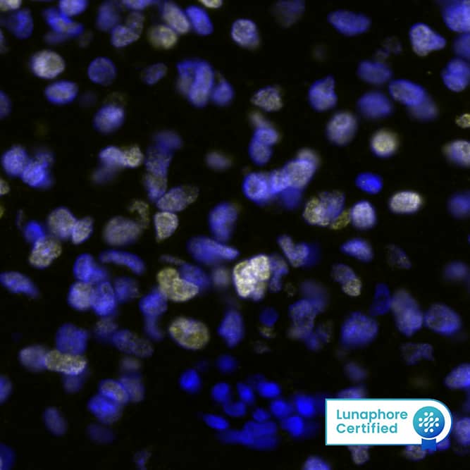
DR647MS) and counterstained with DAPI (blue; Lunaphore Catalog #
DR100). Specific staining was localized to the nucleus. Protocol available in
COMET™ Panel Builder." alt="SOX2 Antibody was detected in immersion fixed paraffin-embedded sections of human Glioblastoma using Mouse Anti-Human SOX2, Monoclonal Antibody (Catalog # MAB2018) at 5ug/mL at 37 ° Celsius for 4 minutes. Before incubation with the primary antibody, tissue underwent an all-in-one dewaxing and antigen retrieval preprocessing using PreTreatment Module (PT Module) and Dewax and HIER Buffer H (pH 9; Epredia Catalog # TA-999-DHBH). Tissue was stained using the Alexa Fluor™ 647 Goat anti-Mouse IgG Secondary Antibody at 1:200 at 37 ° Celsius for 2 minutes. (Yellow; Lunaphore Catalog #
DR647MS) and counterstained with DAPI (blue; Lunaphore Catalog #
DR100). Specific staining was localized to the nucleus. Protocol available in
COMET™ Panel Builder." class="big_thumb" />
AF648) and 10 µg/mL Mouse Anti-Human/Mouse/Rat SOX2 Monoclonal Antibody (Catalog # MAB2018). Cells were incubated with primary antibodies for 3 hours at room temperature. Cells were stained for E-Cadherin using the NorthernLights™ 557-conjugated Anti-Goat IgG Secondary Antibody (red;
NL001) and for SOX2 using the NorthernLights 493-conjugated Anti-Mouse Secondary Antibody (green;
NL009). Cells were counterstained with DAPI (blue). View our protocol for
Fluorescent ICC Staining of Cells on Coverslips." class="big_lightbox">
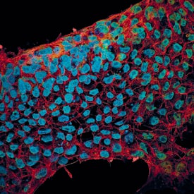
AF648) and 10 µg/mL Mouse Anti-Human/Mouse/Rat SOX2 Monoclonal Antibody (Catalog # MAB2018). Cells were incubated with primary antibodies for 3 hours at room temperature. Cells were stained for E-Cadherin using the NorthernLights™ 557-conjugated Anti-Goat IgG Secondary Antibody (red;
NL001) and for SOX2 using the NorthernLights 493-conjugated Anti-Mouse Secondary Antibody (green;
NL009). Cells were counterstained with DAPI (blue). View our protocol for
Fluorescent ICC Staining of Cells on Coverslips." class="big_thumb" />
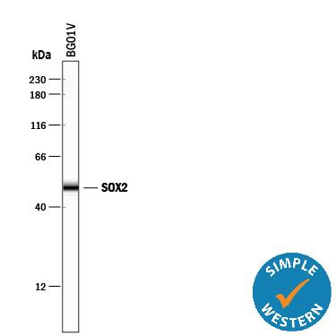
HAF018). A specific band was detected for SOX2 at approximately 36 kDa (as indicated). This experiment was conducted under reducing conditions and using
Immunoblot Buffer Group 1." class="big_lightbox">
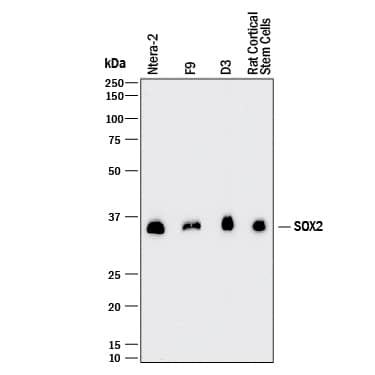
HAF018). A specific band was detected for SOX2 at approximately 36 kDa (as indicated). This experiment was conducted under reducing conditions and using
Immunoblot Buffer Group 1." class="big_thumb" />
MAB003, open histogram), followed by Phycoerythrin-conjugated Anti-Mouse IgG Secondary Antibody (
F0102B). To facilitate intracellular staining, cells were fixed with Flow Cytometry Fixation Buffer (
FC004) and permeabilized with Flow Cytometry Permeabilization/Wash Buffer I (
FC005). View our protocol for
Staining Intracellular Molecules." class="big_lightbox">
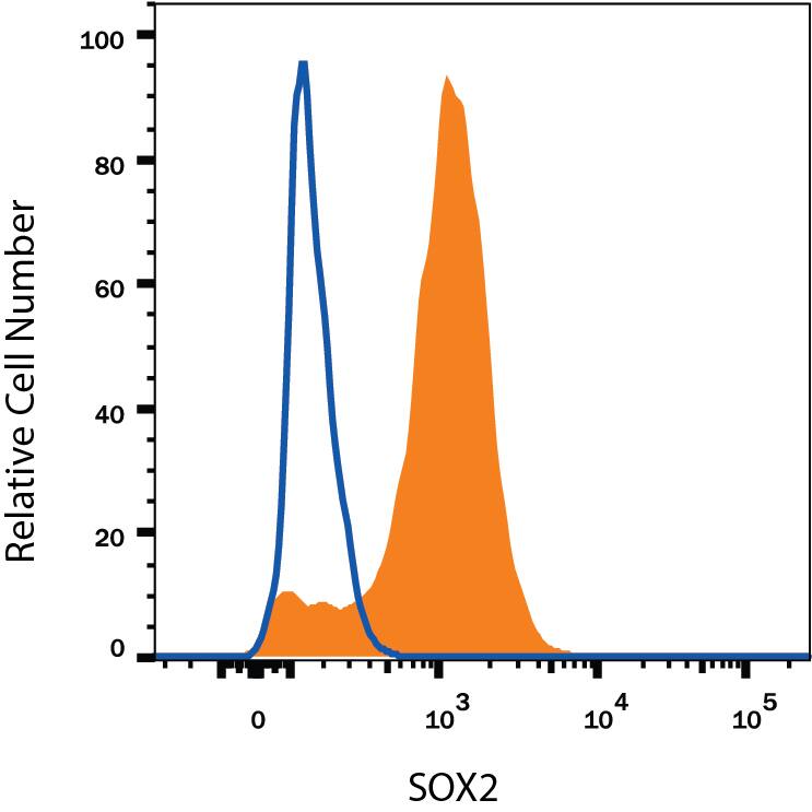
MAB003, open histogram), followed by Phycoerythrin-conjugated Anti-Mouse IgG Secondary Antibody (
F0102B). To facilitate intracellular staining, cells were fixed with Flow Cytometry Fixation Buffer (
FC004) and permeabilized with Flow Cytometry Permeabilization/Wash Buffer I (
FC005). View our protocol for
Staining Intracellular Molecules." class="big_thumb" />
VC001). Before incubation with the primary antibody, tissue was subjected to heat-induced epitope retrieval using VisUCyte Antigen Retrieval Reagent-Basic (Catalog #
VCTS021). Tissue was stained using DAB (brown) and counterstained with hematoxylin (blue). Specific staining was localized to nuclei in cancer cells. View our protocol for
IHC Staining with VisUCyte HRP Polymer Detection Reagents." class="big_lightbox">

VC001). Before incubation with the primary antibody, tissue was subjected to heat-induced epitope retrieval using VisUCyte Antigen Retrieval Reagent-Basic (Catalog #
VCTS021). Tissue was stained using DAB (brown) and counterstained with hematoxylin (blue). Specific staining was localized to nuclei in cancer cells. View our protocol for
IHC Staining with VisUCyte HRP Polymer Detection Reagents." class="big_thumb" />


![<b>Biological Strategies Validation. </b>Soluble factors increase the numbers of Hes3+ cells in the adult rat hippocampus.(a) Treatment with the soluble factors induced a powerful increase in the number of Sox2+ cells in the hilus [Quantifications are from images taken from the tip of the hilus to approximately 1,300 micrometers towards the CA3 region – representative images are shown. Staining for the young neuron marker doublecortin (DCX) helps to visualize the subgranular zone]. (b) The same treatment also induced an increase in the numbers of Hes3+ cells in the hilus. (c) Sox2+ cells inside the hilus co-expressed Hes3; in contrast, Sox2+ cells in the subgranular zone did not co-express Hes3. (d,e) Quantification graphs of the increases in Sox2+ and Hes3+ cell numbers in the hilus (p<0.05). For the graph, the number of cells in the sham operated control animals was set to 100%. [Width of images: a, 650 micrometers; b, 650 micrometers]. Image collected and cropped by CiteAb from the following publication (//dx.plos.org/10.1371/journal.pone.0051630), licensed under a CC-BY license. Not internally tested by R&D Systems.](//images.novusbio.com/images/mab2018_human-mouse-rat-sox2-mab-clone-245610-41202410325570.jpg)
![<b>Biological Strategies Validation. </b>Soluble factors increase the numbers of Hes3+ cells in the adult rat hippocampus.(a) Treatment with the soluble factors induced a powerful increase in the number of Sox2+ cells in the hilus [Quantifications are from images taken from the tip of the hilus to approximately 1,300 micrometers towards the CA3 region – representative images are shown. Staining for the young neuron marker doublecortin (DCX) helps to visualize the subgranular zone]. (b) The same treatment also induced an increase in the numbers of Hes3+ cells in the hilus. (c) Sox2+ cells inside the hilus co-expressed Hes3; in contrast, Sox2+ cells in the subgranular zone did not co-express Hes3. (d,e) Quantification graphs of the increases in Sox2+ and Hes3+ cell numbers in the hilus (p<0.05). For the graph, the number of cells in the sham operated control animals was set to 100%. [Width of images: a, 650 micrometers; b, 650 micrometers]. Image collected and cropped by CiteAb from the following publication (//dx.plos.org/10.1371/journal.pone.0051630), licensed under a CC-BY license. Not internally tested by R&D Systems.](//images.novusbio.com/images/mab2018_human-mouse-rat-sox2-mab-clone-245610-4120241033305.jpg)



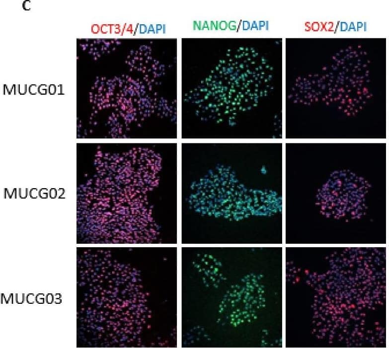
human lung organoid culture protocol. Lung organoids were stained with a (A) Goat Anti-Human p63/TP73L Polyclonal Antibody (Catalog # AF1916; red) and a rabbit anti-human cytokeratin 5 (KRT5) monoclonal antibody (green) to visualize basal cells; a (B) Hamster Anti-Mouse Podoplanin (PDPN) Monoclonal Antibody (Novus Biologicals, Catalog # NB600-1015; green) to visualize alveolar type I cells and a Goat Anti-Human p63/TP73L Polyclonal Antibody (Catalog # AF1916; red) to visualize basal cells; and a (C, D) Mouse Anti-MUC5AC Monoclonal Antibody (Novus Biologicals, Catalog # NBP2-15196; green) to visualize goblet cells and a Mouse Anti-Human/Mouse/Rat SOX2 Monoclonal Antibody (Catalog # MAB2018; red). All samples were counterstained with DAPI (Catalog # 5748; blue). " class="big_lightbox">

human lung organoid culture protocol. Lung organoids were stained with a (A) Goat Anti-Human p63/TP73L Polyclonal Antibody (Catalog # AF1916; red) and a rabbit anti-human cytokeratin 5 (KRT5) monoclonal antibody (green) to visualize basal cells; a (B) Hamster Anti-Mouse Podoplanin (PDPN) Monoclonal Antibody (Novus Biologicals, Catalog # NB600-1015; green) to visualize alveolar type I cells and a Goat Anti-Human p63/TP73L Polyclonal Antibody (Catalog # AF1916; red) to visualize basal cells; and a (C, D) Mouse Anti-MUC5AC Monoclonal Antibody (Novus Biologicals, Catalog # NBP2-15196; green) to visualize goblet cells and a Mouse Anti-Human/Mouse/Rat SOX2 Monoclonal Antibody (Catalog # MAB2018; red). All samples were counterstained with DAPI (Catalog # 5748; blue). " class="big_thumb" />
![Characterization of cell lines.(A) H&E staining of tumor 5653, the arrow indicates the location of the tumor. The right panel shows a higher magnification. (B) Genotyping PCR on genomic wildtype mouse, genomic NPcis mouse (heterozygous for Nf1 and Tp53) DNA and DNA extracted from cell lines 17, 5653 and 5746. The glioma cell lines show complete loss of the wildtype allele for Nf1 and Tp53. (C) Western blot shows the expression of markers consistent with OPC lineage origin of our cell lines. SOX2 is expressed in Neural Progenitor cells (NPC) and Oligodendrocyte precursor cells (OPC); NG2 is expressed in OPCs. Markers (CPNase and MAG) expressed in pre-oligodendrocytes (pre-OL) or mature oligodendrocytes (OL) are not present, highlighting the progenitor status of our lines [22]. (D) The ATRX mutation (E2281*) present in TM31 was confirmed using sequencing. Top shows the reference genome DNA and protein sequence, bottom shows the TM31 sequence with STOP mutation. (E) The homozygous 4bp NF1 deletion (p.LF1247fs*18) present in TM31 was confirmed using sequencing. Top shows the reference genome DNA and protein sequence, bottom shows the TM31 sequence with the homozygous 4bp deletion inducing a frameshift. (F) Western blot for TP53 and CDKN2A, showing that both genes are absent in TM31, but not in glioma line LN319. Image collected and cropped by CiteAb from the following open publication (//pubmed.ncbi.nlm.nih.gov/36730269), licensed under a CC-BY license. Not internally tested by R&D Systems.](//images.novusbio.com/images/mab2018_human-mouse-rat-sox2-mab-clone-245610-western-blot-1212202596399.jpg)
![Characterization of cell lines.(A) H&E staining of tumor 5653, the arrow indicates the location of the tumor. The right panel shows a higher magnification. (B) Genotyping PCR on genomic wildtype mouse, genomic NPcis mouse (heterozygous for Nf1 and Tp53) DNA and DNA extracted from cell lines 17, 5653 and 5746. The glioma cell lines show complete loss of the wildtype allele for Nf1 and Tp53. (C) Western blot shows the expression of markers consistent with OPC lineage origin of our cell lines. SOX2 is expressed in Neural Progenitor cells (NPC) and Oligodendrocyte precursor cells (OPC); NG2 is expressed in OPCs. Markers (CPNase and MAG) expressed in pre-oligodendrocytes (pre-OL) or mature oligodendrocytes (OL) are not present, highlighting the progenitor status of our lines [22]. (D) The ATRX mutation (E2281*) present in TM31 was confirmed using sequencing. Top shows the reference genome DNA and protein sequence, bottom shows the TM31 sequence with STOP mutation. (E) The homozygous 4bp NF1 deletion (p.LF1247fs*18) present in TM31 was confirmed using sequencing. Top shows the reference genome DNA and protein sequence, bottom shows the TM31 sequence with the homozygous 4bp deletion inducing a frameshift. (F) Western blot for TP53 and CDKN2A, showing that both genes are absent in TM31, but not in glioma line LN319. Image collected and cropped by CiteAb from the following open publication (//pubmed.ncbi.nlm.nih.gov/36730269), licensed under a CC-BY license. Not internally tested by R&D Systems.](//images.novusbio.com/images/mab2018_human-mouse-rat-sox2-mab-clone-245610-western-blot-121220259161717.jpg)
 HAF017). A specific band was detected for SOX2 at approximately 36 kDa (as indicated). This experiment was conducted under reducing conditions and using Immunoblot Buffer Group 1." alt="Western blot shows lysates of D3 mouse embryonic stem cell line, NTera-2 human testicular embryonic carcinoma cell line, F9 mouse teratocarcinoma stem cells, and rat cortical stem cells. PVDF membrane was probed with 1 µg/mL of Goat Anti-Human/Mouse/Rat SOX2 Antigen Affinity-purified Polyclonal Antibody (Catalog # AF2018) followed by HRP-conjugated Anti-Goat IgG Secondary Antibody (HAF017). A specific band was detected for SOX2 at approximately 36 kDa (as indicated). This experiment was conducted under reducing conditions and using Immunoblot Buffer Group 1." class="big_thumb" />
 DR647MS) and counterstained with DAPI (blue; Lunaphore Catalog # DR100). Specific staining was localized to the nucleus. Protocol available in COMET™ Panel Builder." alt="SOX2 Antibody was detected in immersion fixed paraffin-embedded sections of human Glioblastoma using Mouse Anti-Human SOX2, Monoclonal Antibody (Catalog # MAB2018) at 5ug/mL at 37 ° Celsius for 4 minutes. Before incubation with the primary antibody, tissue underwent an all-in-one dewaxing and antigen retrieval preprocessing using PreTreatment Module (PT Module) and Dewax and HIER Buffer H (pH 9; Epredia Catalog # TA-999-DHBH). Tissue was stained using the Alexa Fluor™ 647 Goat anti-Mouse IgG Secondary Antibody at 1:200 at 37 ° Celsius for 2 minutes. (Yellow; Lunaphore Catalog # DR647MS) and counterstained with DAPI (blue; Lunaphore Catalog # DR100). Specific staining was localized to the nucleus. Protocol available in COMET™ Panel Builder." class="big_thumb" />
 DR647RB) and counterstained with DAPI (blue; Lunaphore Catalog # DR100). Specific staining was localized to the nucleus and cytoplasm. Protocol available in COMET™ Panel Builder." alt="SOX2 Antibody was detected in immersion fixed paraffin-embedded sections of mouse Brain Cortex using Rabbit Anti-Mouse SOX2, Monoclonal Antibody (Catalog # NB110-37235) at 1:125 dilution at 37 ° Celsius for 4 minutes. Before incubation with the primary antibody, tissue underwent an all-in-one dewaxing and antigen retrieval preprocessing using PreTreatment Module (PT Module) and Dewax and HIER Buffer H (pH 9; Epredia Catalog # TA-999-DHBH). Tissue was stained using the Alexa Fluor™ Plus 647 Goat anti-Rabbit IgG Secondary Antibody at 1:200 at 37 ° Celsius for 2 minutes. (Yellow; Lunaphore Catalog # DR647RB) and counterstained with DAPI (blue; Lunaphore Catalog # DR100). Specific staining was localized to the nucleus and cytoplasm. Protocol available in COMET™ Panel Builder." class="big_thumb" />


 AB-108-C) for 15 minutes in an ultrasonic bath, followed by Biotinylated Anti-Goat IgG Secondary Antibody (
 NL001) and counterstained with DAPI (blue). Nestin was also detected in stem cells using Mouse Anti-Mouse/Rat Nestin Monoclonal Antibody (
 NL001) and counterstained with DAPI (blue). Nestin was also detected using Mouse Anti-Mouse/Rat Nestin Monoclonal Antibody (
 HAF109). This experiment was conducted under reducing conditions and using the 12-230 kDa separation system." class="big_thumb" />
 NL001) and counterstained with DAPI (blue). Specific staining was localized to nuclei. View our protocol for
  VC004). Before incubation with the primary antibody, tissue was subjected to heat-induced epitope retrieval using VisUCyte Antigen Retrieval Reagent-Basic (Catalog #
  NL001) and counterstained with DAPI (blue). Specific staining was localized to Cell surface and nuclear. View our protocol for


 AF648) and 10 µg/mL Mouse Anti-Human/Mouse/Rat SOX2 Monoclonal Antibody (Catalog # MAB2018). Cells were incubated with primary antibodies for 3 hours at room temperature. Cells were stained for E-Cadherin using the NorthernLights™ 557-conjugated Anti-Goat IgG Secondary Antibody (red;
  HAF018). A specific band was detected for SOX2 at approximately 36 kDa (as indicated). This experiment was conducted under reducing conditions and using
  NL007) and counterstained with DAPI (blue). View our protocol for
  MAB003, open histogram), followed by Phycoerythrin-conjugated Anti-Mouse IgG Secondary Antibody (
 VC001). Before incubation with the primary antibody, tissue was subjected to heat-induced epitope retrieval using VisUCyte Antigen Retrieval Reagent-Basic (Catalog #

![<b>Biological Strategies Validation. </b>Soluble factors increase the numbers of Hes3+ cells in the adult rat hippocampus.(a) Treatment with the soluble factors induced a powerful increase in the number of Sox2+ cells in the hilus [Quantifications are from images taken from the tip of the hilus to approximately 1,300 micrometers towards the CA3 region – representative images are shown. Staining for the young neuron marker doublecortin (DCX) helps to visualize the subgranular zone]. (b) The same treatment also induced an increase in the numbers of Hes3+ cells in the hilus. (c) Sox2+ cells inside the hilus co-expressed Hes3; in contrast, Sox2+ cells in the subgranular zone did not co-express Hes3. (d,e) Quantification graphs of the increases in Sox2+ and Hes3+ cell numbers in the hilus (p<0.05). For the graph, the number of cells in the sham operated control animals was set to 100%. [Width of images: a, 650 micrometers; b, 650 micrometers]. Image collected and cropped by CiteAb from the following publication (//dx.plos.org/10.1371/journal.pone.0051630), licensed under a CC-BY license. Not internally tested by R&D Systems.](http://images.novusbio.com/fullsize/mab2018_human-mouse-rat-sox2-mab-clone-245610-41202410325570.jpg)
![<b>Biological Strategies Validation. </b>Soluble factors increase the numbers of Hes3+ cells in the adult rat hippocampus.(a) Treatment with the soluble factors induced a powerful increase in the number of Sox2+ cells in the hilus [Quantifications are from images taken from the tip of the hilus to approximately 1,300 micrometers towards the CA3 region – representative images are shown. Staining for the young neuron marker doublecortin (DCX) helps to visualize the subgranular zone]. (b) The same treatment also induced an increase in the numbers of Hes3+ cells in the hilus. (c) Sox2+ cells inside the hilus co-expressed Hes3; in contrast, Sox2+ cells in the subgranular zone did not co-express Hes3. (d,e) Quantification graphs of the increases in Sox2+ and Hes3+ cell numbers in the hilus (p<0.05). For the graph, the number of cells in the sham operated control animals was set to 100%. [Width of images: a, 650 micrometers; b, 650 micrometers]. Image collected and cropped by CiteAb from the following publication (//dx.plos.org/10.1371/journal.pone.0051630), licensed under a CC-BY license. Not internally tested by R&D Systems.](http://images.novusbio.com/fullsize/mab2018_human-mouse-rat-sox2-mab-clone-245610-4120241033305.jpg)


 human lung organoid culture protocol. Lung organoids were stained with a (A) Goat Anti-Human p63/TP73L Polyclonal Antibody (Catalog # AF1916; red) and a rabbit anti-human cytokeratin 5 (KRT5) monoclonal antibody (green) to visualize basal cells; a (B) Hamster Anti-Mouse Podoplanin (PDPN) Monoclonal Antibody (Novus Biologicals, Catalog # NB600-1015; green) to visualize alveolar type I cells and a Goat Anti-Human p63/TP73L Polyclonal Antibody (Catalog # AF1916; red) to visualize basal cells; and a (C, D) Mouse Anti-MUC5AC Monoclonal Antibody (Novus Biologicals, Catalog # NBP2-15196; green) to visualize goblet cells and a Mouse Anti-Human/Mouse/Rat SOX2 Monoclonal Antibody (Catalog # MAB2018; red). All samples were counterstained with DAPI (Catalog # 5748; blue). " class="big_thumb" />

![Characterization of cell lines.(A) H&E staining of tumor 5653, the arrow indicates the location of the tumor. The right panel shows a higher magnification. (B) Genotyping PCR on genomic wildtype mouse, genomic NPcis mouse (heterozygous for Nf1 and Tp53) DNA and DNA extracted from cell lines 17, 5653 and 5746. The glioma cell lines show complete loss of the wildtype allele for Nf1 and Tp53. (C) Western blot shows the expression of markers consistent with OPC lineage origin of our cell lines. SOX2 is expressed in Neural Progenitor cells (NPC) and Oligodendrocyte precursor cells (OPC); NG2 is expressed in OPCs. Markers (CPNase and MAG) expressed in pre-oligodendrocytes (pre-OL) or mature oligodendrocytes (OL) are not present, highlighting the progenitor status of our lines [22]. (D) The ATRX mutation (E2281*) present in TM31 was confirmed using sequencing. Top shows the reference genome DNA and protein sequence, bottom shows the TM31 sequence with STOP mutation. (E) The homozygous 4bp NF1 deletion (p.LF1247fs*18) present in TM31 was confirmed using sequencing. Top shows the reference genome DNA and protein sequence, bottom shows the TM31 sequence with the homozygous 4bp deletion inducing a frameshift. (F) Western blot for TP53 and CDKN2A, showing that both genes are absent in TM31, but not in glioma line LN319. Image collected and cropped by CiteAb from the following open publication (//pubmed.ncbi.nlm.nih.gov/36730269), licensed under a CC-BY license. Not internally tested by R&D Systems.](http://images.novusbio.com/fullsize/mab2018_human-mouse-rat-sox2-mab-clone-245610-western-blot-1212202596399.jpg)
![Characterization of cell lines.(A) H&E staining of tumor 5653, the arrow indicates the location of the tumor. The right panel shows a higher magnification. (B) Genotyping PCR on genomic wildtype mouse, genomic NPcis mouse (heterozygous for Nf1 and Tp53) DNA and DNA extracted from cell lines 17, 5653 and 5746. The glioma cell lines show complete loss of the wildtype allele for Nf1 and Tp53. (C) Western blot shows the expression of markers consistent with OPC lineage origin of our cell lines. SOX2 is expressed in Neural Progenitor cells (NPC) and Oligodendrocyte precursor cells (OPC); NG2 is expressed in OPCs. Markers (CPNase and MAG) expressed in pre-oligodendrocytes (pre-OL) or mature oligodendrocytes (OL) are not present, highlighting the progenitor status of our lines [22]. (D) The ATRX mutation (E2281*) present in TM31 was confirmed using sequencing. Top shows the reference genome DNA and protein sequence, bottom shows the TM31 sequence with STOP mutation. (E) The homozygous 4bp NF1 deletion (p.LF1247fs*18) present in TM31 was confirmed using sequencing. Top shows the reference genome DNA and protein sequence, bottom shows the TM31 sequence with the homozygous 4bp deletion inducing a frameshift. (F) Western blot for TP53 and CDKN2A, showing that both genes are absent in TM31, but not in glioma line LN319. Image collected and cropped by CiteAb from the following open publication (//pubmed.ncbi.nlm.nih.gov/36730269), licensed under a CC-BY license. Not internally tested by R&D Systems.](http://images.novusbio.com/fullsize/mab2018_human-mouse-rat-sox2-mab-clone-245610-western-blot-121220259161717.jpg)

![Immunohistochemistry-Paraffin: SOX2 Antibody [NB110-37235] - Staining of human uterus, endometrial glands.](http://images.novusbio.com/fullsize/SOX2-Antibody-Immunohistochemistry-Paraffin-NB110-37235-img0012.jpg)
![Western Blot: SOX2 Antibody [NB110-37235] - Detection of SOX2 in mouse brain lysate using NB110-37235 at 0.5 ug/mL.](http://images.novusbio.com/fullsize/SOX2-Antibody-Western-Blot-NB110-37235-img0008.jpg)
![Flow Cytometry: SOX2 Antibody [NB110-37235] - An intracellular stain was performed on A549 with NB110-37235 and a matched isotype control. Cells were fixed with 4% PFA and then permeablized with 0.1% saponin. Cells were incubated in an antibody dilution of 2.5 ug/mL for 30 minutes at room temperature, followed by Rabbit IgG (H+L) Cross-Adsorbed Secondary Antibody, Dylgiht 550.](http://images.novusbio.com/fullsize/SOX2-Antibody-Flow-Cytometry-NB110-37235-img0015.jpg)
![<b>Biological Strategies Validation. </b>Western Blot: SOX2 Antibody [NB110-37235] - MDA-MB-231 subclones transfected with NTC or HIF-1 alpha shRNA vector were treated with vehicle (V) or paclitaxel (P) and immunoblot assay was performed. WB image submitted by a verified customer review.](http://images.novusbio.com/fullsize/SOX2-Antibody-Western-Blot-NB110-37235-img0017.jpg)
![Immunocytochemistry/Immunofluorescence: SOX2 Antibody - BSA Free [NB110-37235] - Rat FR cells were fixed in 4% paraformaldehyde for 10 minutes and permeabilized in 0.5% Triton X-100 in PBS for 5 minutes. The cells were incubated with SOX2 Antibody (NB110-37235) at 2ug/ml overnight at 4C and detected with an anti-rabbit DyLight 488 (Green) at a 1:1000 dilution for 60 minutes. Nuclei were counterstained with DAPI (Blue). Cells were imaged using a 40X objective.](http://images.novusbio.com/fullsize/SOX2-Antibody---BSA-Free-Immunocytochemistry-Immunofluorescence-NB110-37235-img0019.jpg)
![Flow Cytometry: SOX2 Antibody - BSA Free [NB110-37235] - An intracellular stain was performed on U-251 MG cells with SOX2 Antibody NB110-37235 (blue) and a matched isotype control NBP2-24891 (orange). Cells were fixed with 4% PFA and then permeabilized with 0.1% saponin. Cells were incubated in an antibody dilution of 2.5 ug/mL for 30 minutes at room temperature, followed by Rabbit IgG (H+L) Cross-Adsorbed Secondary Antibody, Dylight 550 (SA5-10033, Thermo Fisher).](http://images.novusbio.com/fullsize/SOX2-Antibody---BSA-Free-Flow-Cytometry-NB110-37235-img0020.jpg)
![Western Blot: SOX2 Antibody [NB110-37235] - Expression of pluripotency factor Sox2 in human head and neck cancer cell lines FaDu and HN5. WB image submitted by a verified customer review.](http://images.novusbio.com/fullsize/SOX2-Antibody-Western-Blot-NB110-37235-img0016.jpg)
![Immunocytochemistry/Immunofluorescence: SOX2 Antibody [NB110-37235] - Staining of SOX2 in NTERA2 cells with Dylight 488 (Green). Alpha-tubulin and nuclei were counterstained with Dylight 550 (Red) and DAPI (Blue), respectively.](http://images.novusbio.com/fullsize/SOX2-Antibody-Immunocytochemistry-Immunofluorescence-NB110-37235-img0013.jpg)
![Immunocytochemistry/Immunofluorescence: SOX2 Antibody [NB110-37235] - HeLa cells were fixed for 10 minutes using 10% formalin and then permeabilized for 5 minutes using 1X TBS + 0.5% Triton X-100. The cells were incubated with anti-SOX2 (NB110-37235) at a 1:200 dilution overnight at 4C and detected with an anti-rabbit Dylight 488 (Green) at a 1:500 dilution. Alpha tubulin was used as a co-stain at a 1:1000 dilution and detected with and anti-mouse Dylight 550 (Red) at a 1:500 dilution. Nuclei were counterstained with DAPI (Blue). Cells were imaged using a 40X objective.](http://images.novusbio.com/fullsize/SOX2-Antibody-Immunocytochemistry-Immunofluorescence-NB110-37235-img0014.jpg)
![Immunocytochemistry/Immunofluorescence: SOX2 Antibody - BSA Free [NB110-37235] - NIH3T3 cells were fixed in 4% paraformaldehyde for 10 minutes and permeabilized in 0.5% Triton X-100 in PBS for 5 minutes. The cells were incubated with SOX2 Antibody (NB110-37235) at 2ug/ml overnight at 4C and detected with an anti-rabbit DyLight 488 (Green) at a 1:1000 dilution for 60 minutes. Nuclei were counterstained with DAPI (Blue). Cells were imaged using a 40X objective.](http://images.novusbio.com/fullsize/SOX2-Antibody---BSA-Free-Immunocytochemistry-Immunofluorescence-NB110-37235-img0018.jpg)
![Western Blot: SOX2 Antibody (4G8) [NBP2-29623] - Western blot detection of SOX2 in A. HEK293 cell lysate B. NCCIT cell lysate and C. Full length recombinant protein.](http://images.novusbio.com/fullsize/SOX2-Antibody-4G8-Western-Blot-NBP2-29623-img0001.jpg "Western Blot: SOX2 Antibody (4G8) [NBP2-29623] - Western blot detection of SOX2 in A. HEK293 cell lysate B. NCCIT cell lysate and C. Full length recombinant protein.")
![Immunohistochemistry-Paraffin: SOX2 Antibody (4G8) [NBP2-29623] - IHC analysis of formalin-fixed paraffin-embedded tissue section of human lung cancer using mouse monoclonal SOX2 antibody (clone 4G8) at 5 ug/ml concentration. The cancer cells developed a punctate immunopositivity of SOX2 protein in the cytoplasm and the staining was mainly localized in the perinuclear region of the cells [Magnification 40X].](http://images.novusbio.com/fullsize/SOX2-Antibody-4G8-Immunohistochemistry-Paraffin-NBP2-29623-img0013.jpg)
![Immunohistochemistry-Paraffin: SOX2 Antibody (4G8) [NBP2-29623] - IHC-P analysis of SOX2 protein in a section of human kidney using 5 ug/ml concentration of SOX2 antibody (clone 4G8). The representative image shows distinct nuclear and cytoplasmic staining pattern in cells of different types of renal tubules and blood vessels in the kidney section [Magnification 40X].](http://images.novusbio.com/fullsize/SOX2-Antibody-4G8-Immunohistochemistry-Paraffin-NBP2-29623-img0002.jpg)
![Immunohistochemistry-Paraffin: SOX2 Antibody (4G8) [NBP2-29623] - IHC-P analysis of SOX2 protein in a section of human renal cell carcinoma using 5 ug/ml concentration of SOX2 antibody (clone 4G8). Granular nuclear and cytoplasmic staining pattern was observed in the carcinoma cells, whereas, a weak to negligible staining was observed in the stroma areas [Magnification 40X]..](http://images.novusbio.com/fullsize/SOX2-Antibody-4G8-Immunohistochemistry-Paraffin-NBP2-29623-img0003.jpg)
![Immunohistochemistry-Paraffin: SOX2 Antibody (4G8) [NBP2-29623] - IHC analysis of formalin-fixed paraffin-embedded tissue section of normal human liver using mouse monoclonal SOX2 antibody (clone 4G8) at 5 ug/ml concentration. The liver section depicted a diffused cytoplasmic staining in the hepatocytes [Magnification 40X].](http://images.novusbio.com/fullsize/SOX2-Antibody-4G8-Immunohistochemistry-Paraffin-NBP2-29623-img0004.jpg)
![Immunohistochemistry-Paraffin: SOX2 Antibody (4G8) [NBP2-29623] - IHC analysis of formalin-fixed paraffin-embedded tissue section of normal human brain using mouse monoclonal SOX2 antibody (clone 4G8) at 5 ug/ml concentration. The representative photomicrograph depicts a cytoplasmic and nuclear immunopositivity of SOX2 protein in brain [Magnification 40X].](http://images.novusbio.com/fullsize/SOX2-Antibody-4G8-Immunohistochemistry-Paraffin-NBP2-29623-img0006.jpg)
![Immunohistochemistry-Paraffin: SOX2 Antibody (4G8) [NBP2-29623] - IHC analysis of formalin-fixed paraffin-embedded section of normal human colon tissue using mouse monoclonal SOX2 antibody (clone 4G8) at 5 ug/ml concentration. A weak and diffused cytoplasmic staining was observed in majority of the cells in mucosal layer of colon [Magnification 40X].](http://images.novusbio.com/fullsize/SOX2-Antibody-4G8-Immunohistochemistry-Paraffin-NBP2-29623-img0007.jpg)
![Immunohistochemistry-Paraffin: SOX2 Antibody (4G8) [NBP2-29623] - IHC analysis of formalin-fixed paraffin-embedded tissue section of human bladder cancer using mouse monoclonal SOX2 antibody (clone 4G8) at 5 ug/ml concentration. The cancer cells developed a cytoplasmic and nuclear immunepositivity for SOX2 protein [Magnification 10X].](http://images.novusbio.com/fullsize/SOX2-Antibody-4G8-Immunohistochemistry-Paraffin-NBP2-29623-img0008.jpg)
![Immunohistochemistry-Paraffin: SOX2 Antibody (4G8) [NBP2-29623] - IHC analysis of formalin-fixed paraffin-embedded tissue section of human breast cancer using mouse monoclonal SOX2 antibody (clone 4G8) at 5 ug/ml concentration. The cancer cells developed a cytoplasmic and nuclear immunepositivity, whereas the stroma was largely negative for SOX2 protein [Magnification 10X].](http://images.novusbio.com/fullsize/SOX2-Antibody-4G8-Immunohistochemistry-Paraffin-NBP2-29623-img0009.jpg)
![Immunohistochemistry-Paraffin: SOX2 Antibody (4G8) [NBP2-29623] - IHC analysis of formalin-fixed paraffin-embedded tissue section of human uterine cancer using mouse monoclonal SOX2 antibody (clone 4G8) at 5 ug/ml concentration. The cancer cells developed a punctate staining in the cytoplasm and nuclei, whereas the stroma was very weakly positive for SOX2 protein [Magnification 40X].](http://images.novusbio.com/fullsize/SOX2-Antibody-4G8-Immunohistochemistry-Paraffin-NBP2-29623-img0010.jpg)
![Western Blot: SOX2 Antibody [NBP2-24390] - Analysis of SOX2 in mouse embryo brain lysate in the 1) absence and 2) presence of immunizing peptide using this antibody. Goat anti-rabbit Ig HRP secondary antibody and PicoTect ECL substrate solution were used for this test.](http://images.novusbio.com/fullsize/SOX2-Antibody-Western-Blot-NBP2-24390-img0002.jpg "Western Blot: SOX2 Antibody [NBP2-24390] - Analysis of SOX2 in mouse embryo brain lysate in the 1) absence and 2) presence of immunizing peptide using this antibody. Goat anti-rabbit Ig HRP secondary antibody and PicoTect ECL substrate solution were used for this test.")
![Immunohistochemistry-Paraffin: SOX2 Antibody [NBP2-24390] - FFPE human skin stained with SOX2 antibody at 20 ug/ml.](http://images.novusbio.com/fullsize/SOX2-Antibody-Immunohistochemistry-Paraffin-NBP2-24390-img0003.jpg)
![Western Blot: SOX2 Antibody (SOX2/1791) [NBP2-53407] - Western Blot Analysis of human A549 cell lysate using SOX2 Antibody (SOX2/1791).](http://images.novusbio.com/fullsize/SOX2-Antibody-SOX2-1791-Western-Blot-NBP2-53407-img0009.jpg "Western Blot: SOX2 Antibody (SOX2/1791) [NBP2-53407] - Western Blot Analysis of human A549 cell lysate using SOX2 Antibody (SOX2/1791).")
![Immunohistochemistry-Paraffin: SOX2 Antibody (SOX2/1791) [NBP2-53407] - Formalin-fixed, paraffin-embedded Mouse Intestine stained with SOX2 Antibody (SOX2/1791).](http://images.novusbio.com/fullsize/SOX2-Antibody-SOX2-1791-Immunohistochemistry-Paraffin-NBP2-53407-img0006.jpg)
![Western Blot: SOX2 Antibody (SOX2/1791) [NBP2-53407] - Western Blot of Recombinant SOX2 protein fragment and NCCIT cell lysate using SOX2 Antibody (SOX2/1791).](http://images.novusbio.com/fullsize/SOX2-Antibody-SOX2-1791-Western-Blot-NBP2-53407-img0008.jpg)
![Immunohistochemistry-Paraffin: SOX2 Antibody (SOX2/1791) [NBP2-53407] - Formalin-fixed, paraffin-embedded Human Cervical Carcinoma stained with SOX2 Monoclonal Antibody (SOX2/1791).](http://images.novusbio.com/fullsize/SOX2-Antibody-SOX2-1791-Immunohistochemistry-Paraffin-NBP2-53407-img0001.jpg)
![Immunohistochemistry-Paraffin: SOX2 Antibody (SOX2/1791) [NBP2-53407] - Formalin-fixed, paraffin-embedded human Lung Carcinoma stained with SOX2 Antibody (SOX2/1791).](http://images.novusbio.com/fullsize/SOX2-Antibody-SOX2-1791-Immunohistochemistry-Paraffin-NBP2-53407-img0003.jpg)
![Immunohistochemistry-Paraffin: SOX2 Antibody (SOX2/1791) [NBP2-53407] - Formalin-fixed, paraffin-embedded Mouse Stomach stained with SOX2 Antibody (SOX2/1791).](http://images.novusbio.com/fullsize/SOX2-Antibody-SOX2-1791-Immunohistochemistry-Paraffin-NBP2-53407-img0004.jpg)
![Immunohistochemistry-Paraffin: SOX2 Antibody (SOX2/1791) [NBP2-53407] - Formalin-fixed, paraffin-embedded Mouse Brain stained with SOX2 Antibody (SOX2/1791).](http://images.novusbio.com/fullsize/SOX2-Antibody-SOX2-1791-Immunohistochemistry-Paraffin-NBP2-53407-img0005.jpg)
![Protein Array: SOX2 Antibody (SOX2/1791) [NBP2-53407] - Analysis of Protein Array containing more than 19,000 full-length human proteins using SOX2 Antibody (SOX2/1791) Z- and S- Score: The Z-score represents the strength of a signal that a monoclonal antibody (MAb) (in combination with a fluorescently-tagged anti-IgG secondary antibody) produces when binding to a particular protein on the HuProt(TM) array. Z-scores are described in units of standard deviations (SD?s) above the mean value of all signals generated on that array. If targets on HuProt(TM) are arranged in descending order of the Z-score, the S-score is the difference (also in units of SD?s) between the Z-score. S-score therefore represents the relative target specificity of a MAb to its intended target. A MAb is considered to specific to its intended target, if the MAb has an S-score of at least 2.5.](http://images.novusbio.com/fullsize/SOX2-Antibody-SOX2-1791-Protein-Array-NBP2-53407-img0007.jpg)
![Immunohistochemistry-Paraffin: SOX2 Antibody (SOX2/1792) [NBP2-53408] - Formalin-fixed, paraffin-embedded Mouse Intestine stained with SOX2 Antibody (SOX2/1792).](http://images.novusbio.com/fullsize/SOX2-Antibody-SOX2-1792-Immunohistochemistry-Paraffin-NBP2-53408-img0005.jpg "Immunohistochemistry-Paraffin: SOX2 Antibody (SOX2/1792) [NBP2-53408] - Formalin-fixed, paraffin-embedded Mouse Intestine stained with SOX2 Antibody (SOX2/1792).")
![Immunohistochemistry-Paraffin: SOX2 Antibody (SOX2/1792) [NBP2-53408] - Formalin-fixed, paraffin-embedded Human Cervical Carcinoma stained with SOX2 Monoclonal Antibody (SOX2/1792).](http://images.novusbio.com/fullsize/SOX2-Antibody-SOX2-1792-Immunohistochemistry-Paraffin-NBP2-53408-img0001.jpg)
![Immunohistochemistry-Paraffin: SOX2 Antibody (SOX2/1792) [NBP2-53408] - Formalin-fixed, paraffin-embedded human Lung Carcinoma stained with SOX2 Antibody (SOX2/1792).](http://images.novusbio.com/fullsize/SOX2-Antibody-SOX2-1792-Immunohistochemistry-Paraffin-NBP2-53408-img0003.jpg)
![Immunohistochemistry-Paraffin: SOX2 Antibody (SOX2/1792) [NBP2-53408] - Formalin-fixed, paraffin-embedded Mouse Stomach stained with SOX2 Antibody (SOX2/1792).](http://images.novusbio.com/fullsize/SOX2-Antibody-SOX2-1792-Immunohistochemistry-Paraffin-NBP2-53408-img0004.jpg)
![Protein Array: SOX2 Antibody (SOX2/1792) [NBP2-53408] - Analysis of Protein Array containing more than 19,000 full-length human proteins using SOX2 Antibody (SOX2/1792) Z- and S- Score: The Z-score represents the strength of a signal that a monoclonal antibody (MAb) (in combination with a fluorescently-tagged anti-IgG secondary antibody) produces when binding to a particular protein on the HuProt(TM) array. Z-scores are described in units of standard deviations (SD?s) above the mean value of all signals generated on that array. If targets on HuProt(TM) are arranged in descending order of the Z-score, the S-score is the difference (also in units of SD?s) between the Z-score. S-score therefore represents the relative target specificity of a MAb to its intended target. A MAb is considered to specific to its intended target, if the MAb has an S-score of at least 2.5.](http://images.novusbio.com/fullsize/SOX2-Antibody-SOX2-1792-Protein-Array-NBP2-53408-img0006.jpg)
![Immunohistochemistry-Paraffin: SOX2 Antibody (SOX2/1792) [NBP2-53408] - Analysis of SOX2 stained human head-neck cancer sample. Image from verified customer review.](http://images.novusbio.com/fullsize/nbp2-53408_mouse-monoclonal-sox2-antibody-sox2-1792-17720241044646.jpg)
![Western Blot: SOX2 Antibody (SOX2/1792) [NBP2-53408] - Western Blot of Recombinant SOX2 protein fragment and NCCIT cell lysate using SOX2 Antibody (SOX2/1792).](http://images.novusbio.com/fullsize/nbp2-53408_mouse-monoclonal-sox2-antibody-sox2-1792-17720241003232.jpg)
![Chromatin Immunoprecipitation: SOX2 Antibody (PO00-28) [NBP3-32972] - Chromatin immunoprecipitations were performed with cross-linked chromatin from NCCIT cells and either SOX2 (NBP3-32972) or Normal Rabbit IgG according to the ChIP protocol. The enriched DNA was quantified by real-time PCR using indicated primers. The amount of immunoprecipitated DNA in each sample is represented as signal relative to the total amount of input chromatin, which is equivalent to one.](http://images.novusbio.com/fullsize/nbp3-32972_rabbit-sox2-mab-po00-28-26820241525104.jpg "Chromatin Immunoprecipitation: SOX2 Antibody (PO00-28) [NBP3-32972] - Chromatin immunoprecipitations were performed with cross-linked chromatin from NCCIT cells and either SOX2 (NBP3-32972) or Normal Rabbit IgG according to the ChIP protocol. The enriched DNA was quantified by real-time PCR using indicated primers. The amount of immunoprecipitated DNA in each sample is represented as signal relative to the total amount of input chromatin, which is equivalent to one.")
![Western Blot: SOX2 Antibody (PO00-28) [NBP3-32972] - Western blot analysis of SOX2 on different lysates with Rabbit anti-SOX2 antibody (NBP3-32972) at 1/2,000 dilution.<br/><br/> Lane 1: NCCIT cell lysate<br/> Lane 2: F9 cell lysate<br/> Lane 3: HeLa cell lysate (negative)<br/><br/> Predicted band size: 34 kDa<br/> Observed band size: 34 kDa<br/><br/> Exposure time: 15 seconds;<br/><br/> 4-20% SDS-PAGE gel.<br/><br/> Proteins were transferred to a PVDF membrane and blocked with 5% NFDM/TBST for 1 hour at room temperature. The primary antibody (NBP3-32972) at 1/2,000 dilution was used in 5% NFDM/TBST at 4C overnight. Goat Anti-Rabbit IgG - HRP Secondary Antibody at 1/50,000 dilution was used for 1 hour at room temperature.](http://images.novusbio.com/fullsize/nbp3-32972_rabbit-sox2-mab-po00-28-28820241249344.jpg)
![Immunohistochemistry: SOX2 Antibody (PO00-28) [NBP3-32972] - Immunohistochemical analysis of paraffin-embedded human trachea tissue with Rabbit anti-SOX2 antibody (NBP3-32972) at 1/1,000 dilution.<br/><br/> The section was pre-treated using heat mediated antigen retrieval with sodium citrate buffer (pH 6.0) for 2 minutes. The tissues were blocked in 1% BSA for 20 minutes at room temperature, washed with ddH2O and PBS, and then probed with the primary antibody (NBP3-32972) at 1/1,000 dilution for 1 hour at room temperature. The detection was performed using an HRP conjugated compact polymer system. DAB was used as the chromogen. Tissues were counterstained with hematoxylin and mounted with DPX.](http://images.novusbio.com/fullsize/nbp3-32972_rabbit-sox2-mab-po00-28-298202414161844.jpg)
![Immunocytochemistry/ Immunofluorescence: SOX2 Antibody (PO00-28) [NBP3-32972] - Immunocytochemistry analysis of F9 cells labeling SOX2 with Rabbit anti-SOX2 antibody (NBP3-32972) at 1/250 dilution.<br/><br/> Cells were fixed in 4% paraformaldehyde for 20 minutes at room temperature, permeabilized with 0.1% Triton X-100 in PBS for 5 minutes at room temperature, then blocked with 1% BSA in 10% negative goat serum for 1 hour at room temperature. Cells were then incubated with Rabbit anti-SOX2 antibody (NBP3-32972) at 1/250 dilution in 1% BSA in PBST overnight at 4 ℃. Goat Anti-Rabbit IgG H&L (iFluor™ 488) was used as the secondary antibody at 1/1,000 dilution. PBS instead of the primary antibody was used as the secondary antibody only control. Nuclear DNA was labelled in blue with DAPI.<br/><br/> Beta tubulin (red) was stained at 1/100 dilution overnight at +4℃. Goat Anti-Mouse IgG H&L (iFluor™ 594) was used as the secondary antibody at 1/1,000 dilution.](http://images.novusbio.com/fullsize/nbp3-32972_rabbit-sox2-mab-po00-28-29202410334048.jpg)
![Western blot analysis on different lysates with NBP3-32972 at 1/2,000 dilution. Lane 1: NCCIT cell lysate Lane 2: F9 cell lysate Lane 3: HeLa cell lysate (negative) Predicted band size: 34 kDa Observed band size: 36 kDa Exposure time: 5 seconds; 4-20% SDS-PAGE gel. Proteins were transferred to a PVDF membrane and blocked with 5% NFDM/TBST for 1 hour at room temperature. NBP3-32972 at 1/2,000 dilution was used in 5% NFDM/TBST at 4℃ overnight. Goat Anti-Rabbit IgG [HRP] Secondary Antibody at 1/50,000 dilution was used for 1 hour at room temperature.](http://images.novusbio.com/fullsize/nbp3-32972_-western-blot-639204594550184254.jpg)
![Western blot analysis on different lysates with NBP3-32972 at 1/1,000 dilution. Lane 1: Mouse brain tissue lysate Lane 2: Rat brain tissue lysate Lysates/proteins at 30 µg/Lane. Predicted band size: 34 kDa Observed band size: 36 kDa Exposure time: 25 seconds; 4-20% SDS-PAGE gel. Proteins were transferred to a PVDF membrane and blocked with 5% NFDM/TBST for 1 hour at room temperature. NBP3-32972 at 1/1,000 dilution was used in 5% NFDM/TBST at 4℃ overnight. Goat Anti-Rabbit IgG [HRP] Secondary Antibody at 1/50,000 dilution was used for 1 hour at room temperature.](http://images.novusbio.com/fullsize/nbp3-32972_-western-blot-639204597794696077.jpg)
![Immunohistochemistry-Paraffin: SOX2 Antibody (017) [NBP2-89196] - Staining of human SOX2 in human brain with rabbit monoclonal antibody (1:300).](http://images.novusbio.com/fullsize/SOX2-Antibody-017-Immunohistochemistry-Paraffin-NBP2-89196-img0002.jpg "Immunohistochemistry-Paraffin: SOX2 Antibody (017) [NBP2-89196] - Staining of human SOX2 in human brain with rabbit monoclonal antibody (1:300).")
![Immunohistochemistry-Paraffin: SOX2 Antibody (017) [NBP2-89196] - Staining of human SOX2 in human testis with rabbit monoclonal antibody (1:300).](http://images.novusbio.com/fullsize/SOX2-Antibody-017-Immunohistochemistry-Paraffin-NBP2-89196-img0001.jpg)
![Western Blot: SOX2 Antibody (CL4716) [NBP2-59057] - Analysis in U-251MG cells transfected with control siRNA, target specific siRNA probe #1 and #2, using Anti-SOX2 antibody. Remaining relative intensity is presented. Loading control: Anti-PPIB.](http://images.novusbio.com/fullsize/SOX2-Antibody-CL4716-Western-Blot-NBP2-59057-img0010.jpg "<b>Genetic Strategies Validation. </b>Western Blot: SOX2 Antibody (CL4716) [NBP2-59057] - Analysis in U-251MG cells transfected with control siRNA, target specific siRNA probe #1 and #2, using Anti-SOX2 antibody. Remaining relative intensity is presented. Loading control: Anti-PPIB.")
![<b>Genetic Strategies Validation. </b>Western Blot: SOX2 Antibody (CL4716) [NBP2-59057] - Analysis in human cell line NTERA-2.](http://images.novusbio.com/fullsize/SOX2-Antibody-CL4716-Western-Blot-NBP2-59057-img0006.jpg)
![Immunohistochemistry-Paraffin: SOX2 Antibody (CL4716) [NBP2-59057] - Staining of human placenta shows no positivity as expected (negative control).](http://images.novusbio.com/fullsize/SOX2-Antibody-CL4716-Immunohistochemistry-Paraffin-NBP2-59057-img0005.jpg)
![Immunohistochemistry: SOX2 Antibody (CL4716) [NBP2-59057] - SOX2 Antibody [NBP2-59057] - Staining of mouse embryo E11 shows nuclear immunoreactivity in the developing brain, eye and trigeminal ganglion.](http://images.novusbio.com/fullsize/SOX2-Antibody-CL4716-Immunohistochemistry-NBP2-59057-img0007.jpg)
![Immunohistochemistry: SOX2 Antibody (CL4716) [NBP2-59057] - SOX2 Antibody [NBP2-59057] - Staining of mouse embryo E11 shows nuclear immunoreactivity in the developing neural tube.](http://images.novusbio.com/fullsize/SOX2-Antibody-CL4716-Immunohistochemistry-NBP2-59057-img0008.jpg)
![Immunofluorescence:SOX2 Antibody[NBP3-31961]- staining of NTERA2 cells stained with Novus Biologicals anti-SOX2 antibody (green) showing strong nuclear staining of endogenous SOX2. Actin filaments are stained with phalloidin (red).](http://images.novusbio.com/fullsize/antibody/nbp3-31961_chicken-sox2-pab-immunofluorescence-1911202413196..jpg "Immunofluorescence:SOX2 Antibody[NBP3-31961]- staining of NTERA2 cells stained with Novus Biologicals anti-SOX2 antibody (green) showing strong nuclear staining of endogenous SOX2. Actin filaments are stained with phalloidin (red).")
![Western Blot:SOX2 Antibody[NBP3-31961]- Western blotting of SOX2-FLAG transfected COS-7 cell lysate (10 µg/lane), mock transfected COS-7 cell lysate (10 µg/lane) and NTERA2 cell lysate (10 µg/lane) and stained with Novus Biologicals anti-SOX2 antibody (1µg/mL).Note that SOX2 runs at higher molecular weight in lane 1 due to presence of tandem Myc/FLAG tag on recombinant protein relative to endogenous SOX2 in lane 3.](http://images.novusbio.com/fullsize/antibody/nbp3-31961_chicken-sox2-pab-western-blot-19112024132246..jpg)
![Western Blot: SOX2 Antibody [NBP2-58318] - Analysis in human cell line U-251 MG and human cell line HeLa.](http://images.novusbio.com/fullsize/SOX2-Antibody-Western-Blot-NBP2-58318-img0003.jpg "<b>Orthogonal Strategies Validation. </b>Western Blot: SOX2 Antibody [NBP2-58318] - Analysis in human cell line U-251 MG and human cell line HeLa.")
![<b>Orthogonal Strategies Validation. </b>Immunocytochemistry/Immunofluorescence: SOX2 Antibody [NBP2-58318] - Staining of human cell line U-251 MG shows localization to nucleoplasm. Antibody staining is shown in green.](http://images.novusbio.com/fullsize/SOX2-Antibody-Immunocytochemistry-Immunofluorescence-NBP2-58318-img0001.jpg)


