HAF016). A specific band was detected for FOLR4 at approximately 37 kDa (as indicated). This experiment was conducted under reducing conditions and using
Immunoblot Buffer Group 8." class="big_lightbox">
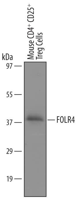
HAF016). A specific band was detected for FOLR4 at approximately 37 kDa (as indicated). This experiment was conducted under reducing conditions and using
Immunoblot Buffer Group 8." alt="Western blot shows lysates of mouse CD
+CD25
+regulatory T cells. PVDF Membrane was probed with 1 µg/mL of Sheep Anti-Mouse FOLR4 Antigen Affinity-purified Polyclonal Antibody (Catalog # AF6124) followed by HRP-conjugated Anti-Sheep IgG Secondary Antibody (Catalog #
HAF016). A specific band was detected for FOLR4 at approximately 37 kDa (as indicated). This experiment was conducted under reducing conditions and using
Immunoblot Buffer Group 8." class="big_thumb" />
402-ML), and Human TGF-beta 1 (Catalog #
100-B) for 3 days to induce T regulatory cells (Tregs) were stained with Sheep Anti-Mouse FOLR4 Antigen Affinity-purified Polyclonal Antibody (Catalog # AF6124, filled histogram) or isotype control antibody (Catalog #
5-001-A, open histogram), followed by Allophycocyanin-conjugated Anti-Sheep IgG Secondary Antibody (Catalog #
F0127). To facilitate intracellular staining, cells were fixed and permeabilized with Flow Cytometry FoxP3 Staining Buffer (Catalog #
FC011)." class="big_lightbox">
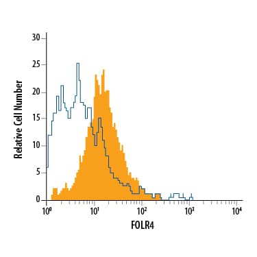
402-ML), and Human TGF-beta 1 (Catalog #
100-B) for 3 days to induce T regulatory cells (Tregs) were stained with Sheep Anti-Mouse FOLR4 Antigen Affinity-purified Polyclonal Antibody (Catalog # AF6124, filled histogram) or isotype control antibody (Catalog #
5-001-A, open histogram), followed by Allophycocyanin-conjugated Anti-Sheep IgG Secondary Antibody (Catalog #
F0127). To facilitate intracellular staining, cells were fixed and permeabilized with Flow Cytometry FoxP3 Staining Buffer (Catalog #
FC011)." class="big_thumb" />
 HAF016). A specific band was detected for FOLR4 at approximately 37 kDa (as indicated). This experiment was conducted under reducing conditions and using Immunoblot Buffer Group 8." alt="Western blot shows lysates of mouse CD+CD25+regulatory T cells. PVDF Membrane was probed with 1 µg/mL of Sheep Anti-Mouse FOLR4 Antigen Affinity-purified Polyclonal Antibody (Catalog # AF6124) followed by HRP-conjugated Anti-Sheep IgG Secondary Antibody (Catalog # HAF016). A specific band was detected for FOLR4 at approximately 37 kDa (as indicated). This experiment was conducted under reducing conditions and using Immunoblot Buffer Group 8." class="big_thumb" />
 CTS017) and counterstained with hematoxylin (blue). Specific staining was localized to the plasma membranes of cancer cells. View our protocol for Chromogenic IHC Staining of Paraffin-embedded Tissue Sections." alt="FOLR4 was detected in immersion fixed paraffin-embedded sections of human melanoma using Rat Anti-Human FOLR4 Monoclonal Antibody (Catalog # MAB6328) at 15 µg/mL overnight at 4 °C. Tissue was stained using the Anti-Rat HRP-DAB Cell & Tissue Staining Kit (brown; Catalog # CTS017) and counterstained with hematoxylin (blue). Specific staining was localized to the plasma membranes of cancer cells. View our protocol for Chromogenic IHC Staining of Paraffin-embedded Tissue Sections." class="big_thumb" />
 HAF016). A specific band was detected for FOLR4 at approximately 36 kDa (as indicated). This experiment was conducted under reducing conditions and using Immunoblot Buffer Group 8." alt="Western blot shows lysates of A431 human epithelial carcinoma cell line. PVDF Membrane was probed with 1 µg/mL of Sheep Anti-Human FOLR4 Antigen Affinity-purified Polyclonal Antibody (Catalog # AF6328) followed by HRP-conjugated Anti-Sheep IgG Secondary Antibody (Catalog # HAF016). A specific band was detected for FOLR4 at approximately 36 kDa (as indicated). This experiment was conducted under reducing conditions and using Immunoblot Buffer Group 8." class="big_thumb" />
 Immunoblot Buffer Group 1." alt="Western blot shows lysates of mouse CD4+CD25-T cells and mouse CD4+CD25+T cells. PVDF membrane was probed with 1 µg/mL of Rat Anti-Mouse FOLR4 Monoclonal Antibody (Catalog # MAB6124) followed by HRP-conjugated Anti-Rat IgG Secondary Antibody. A specific band was detected for FOLR4 at approximately 35 kDa (as indicated). This experiment was conducted under reducing conditions and using Immunoblot Buffer Group 1." class="big_thumb" />


 402-ML), and Human TGF-beta 1 (Catalog #
 ![Western Blot: FOLR4 Antibody [NBP2-99508] - Anti-FOLR4 rabbit polyclonal antibody at 1:500 dilution. Lane A: MCF7 Whole Cell Lysate. Lysates/proteins at 30 ug per lane. Secondary Goat Anti- Rabbit IgG H&L (Dylight 800) at 1/10000 dilution. Developed using the Odyssey technique. Performed under reducing conditions. Predicted band size: 29 kDa. Observed band size: 35 kDa (We are unsure as to the identity of these extra bands).](http://images.novusbio.com/fullsize/FOLR4-Antibody-Western-Blot-NBP2-99508-img0002.jpg "Western Blot: FOLR4 Antibody [NBP2-99508] - Anti-FOLR4 rabbit polyclonal antibody at 1:500 dilution. Lane A: MCF7 Whole Cell Lysate. Lysates/proteins at 30 ug per lane. Secondary Goat Anti- Rabbit IgG H&L (Dylight 800) at 1/10000 dilution. Developed using the Odyssey technique. Performed under reducing conditions. Predicted band size: 29 kDa. Observed band size: 35 kDa (We are unsure as to the identity of these extra bands).")
![Immunoprecipitation: FOLR4 Antibody [NBP2-99508] - FOLR4 was immunoprecipitated using: Lane A: 0.5 mg Hela Whole Cell Lysate. 4 ul anti-FOLR4 rabbit polyclonal antibody and 15 ul of 50 % Protein G agarose. Primary antibody: Anti-FOLR4 rabbit polyclonal antibody, at 1:100 dilution. Secondary antibody: Dylight 800-labeled antibody to rabbit IgG (H+L), at 1:5000 dilution. Developed using the Odyssey technique. Performed under reducing conditions. Predicted band size: 28 kDa. Observed band size: 34 kDa](http://images.novusbio.com/fullsize/FOLR4-Antibody-Immunoprecipitation-NBP2-99508-img0001.jpg)
![Western Blot: FOLR4 Overexpression Lysate (Adult Normal) [NBP2-08395] Left-Empty vector transfected control cell lysate (HEK293 cell lysate); Right -Over-expression Lysate for FOLR4.](http://images.novusbio.com/fullsize/FOLR4-Overexpression-Lysate-Adult-Normal-Western-Blot-NBP2-08395-img0001.jpg "Western Blot: FOLR4 Overexpression Lysate (Adult Normal) [NBP2-08395] Left-Empty vector transfected control cell lysate (HEK293 cell lysate); Right -Over-expression Lysate for FOLR4.")

![Western Blot: FOLR4 Antibody [NBP1-91496] - MCF-7 whole cell lysates, concentration 0.2-1 ug/ml.](http://images.novusbio.com/fullsize/FOLR4-Antibody-Western-Blot-NBP1-91496-img0002.jpg "Western Blot: FOLR4 Antibody [NBP1-91496] - MCF-7 whole cell lysates, concentration 0.2-1 ug/ml.")


