After August 17, 2026, Novus Biologicals products and services will no longer be available on this website; you will access all products and services on rndsystems.com. Create your R&D Systems online account today.
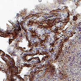
VC004). Tissue was stained using DAB (brown) and counterstained with hematoxylin (blue). Specific staining was localized to the cell surface. View our protocol for Chromogenic IHC Staining of Frozen Tissue Sections." class="big_lightbox" target="_blank">
CTS008) and counterstained with hematoxylin (blue). Specific labeling was localized to the cytoplasm of cells in the choroid plexus. View our protocol for Chromogenic IHC Staining of Frozen Tissue Sections." class="big_lightbox" target="_blank">
AF497) followed by Allophycocyanin-conjugated anti-Goat IgG Secondary Antibody (F0108). Staining was performed using our Staining Membrane-associated Proteins protocol." class="big_lightbox" target="_blank">
497-LR) was serially diluted and captured by Rat Anti-Mouse Leptin R Monoclonal Antibody (Catalog # MAB497) coated on a Clear Polystyrene Microplate (Catalog # DY990). Goat Anti-Mouse Leptin R Antigen Affinity-purified Polyclonal Antibody (Catalog # AF497) was biotinylated and incubated with the protein captured on the plate. Detection of the standard curve was achieved by incubating Streptavidin-HRP (Catalog # DY998)" class="big_lightbox" target="_blank">
389-LR) and Recombinant Mouse Leptin R Fc Chimera (Catalog # 497-LR). PVDF Membrane was probed with 1 µg/mL of Mouse Anti-Human Leptin R Monoclonal Antibody (Catalog # MAB867) followed by HRP-conjugated Anti-Mouse IgG Secondary Antibody (Catalog # HAF007). A specific band was detected for Leptin R at approximately 155-200 kDa (as indicated). This experiment was conducted under reducing conditions and using Immunoblot Buffer Group 3." class="big_lightbox" target="_blank">
VC001) or the HRP-conjugated Anti-Mouse IgG Secondary Antibody (Catalog # HAF007). Before incubation with the primary antibody, tissue was subjected to heat-induced epitope retrieval using VisUCyte Antigen Retrieval Reagent-Basic (Catalog # VCTS021). Tissue was stained using DAB (brown) and counterstained with hematoxylin (blue). Specific staining was localized to the cell surface of neurons. View our protocol for IHC Staining with VisUCyte HRP Polymer Detection Reagents." class="big_lightbox" target="_blank">
MAB004) followed by Allophycocyanin-conjugated Anti-Mouse IgG Secondary Antibody (Catalog # F0101B). View our protocol for Staining Membrane-associated Proteins. " class="big_lightbox" target="_blank">
Leptin receptor (OB-R) is a type I cytokine receptor family protein with significant amino acid sequence identity with gp130, G-CSF receptor, and LIF receptor. An OB-R transcript encodes a soluble form of the receptor. The transcript was reported to be expressed predominantly in regions of the hypothalamus previously thought to be important in body weight regulation. OB-R binds leptin with high affinity and is a potent leptin antagonist. Human OB-R encodes a 1 165 amino acid residue precursor protein with a 22 amino acid residue signal peptide, an 816 amino acid residue extracellular domain, a 21 amino acid residue transmembrane domain and a 303 amino acid residue cytoplasmic domain.
![Immunohistochemistry Leptin R Antibody [Unconjugated]](http://images.novusbio.com/images/antibody/af497_mouse-leptin-r-affinity-purified-polyclonal-ab-immunohistochemistry-251120248493..jpg)
![Western Blot Leptin R Antibody (52263) [Unconjugated]](http://images.novusbio.com/images/antibody/Leptin_R_MAB867_Western_Blot_21680.jpg)

![N/A Leptin R [HRP]](http://images.novusbio.com/fullsize/elisa/DATA_Leptin_R_DOBR00_ELISA_781.jpg)
![N/A Leptin R [Biotin]](http://images.novusbio.com/fullsize/elisa/DATA_Leptin_R_DY497_ELISA_2067.jpg)
![N/A Leptin R [Biotin]](http://images.novusbio.com/fullsize/elisa/DATA_Leptin_R_DY389_ELISA_1987.jpg)


![Immunohistochemistry Leptin R Antibody [Unconjugated]](http://images.novusbio.com/images/antibody/Leptin_R_AF497_Immunohistochemistry_6850.jpg)
![Western Blot Leptin R Antibody [Unconjugated]](http://images.novusbio.com/fullsize/af497_mouse-leptin-r-affinity-purified-polyclonal-ab-41202410481130.jpg)
![Flow Cytometry Leptin R Antibody [Unconjugated]](http://images.novusbio.com/images/antibody/af497_mouse-leptin-r-affinity-purified-polyclonal-ab-flow-cytometry-22122025104014.jpg)
![ELISA Leptin R Antibody [Unconjugated]](http://images.novusbio.com/images/af497_mouse-leptin-r-affinity-purified-polyclonal-ab-elisa-639201854768307426.jpg)
![Immunohistochemistry Leptin R Antibody (52263) [Unconjugated]](http://images.novusbio.com/images/antibody/mab867_human-leptin-r-mab-clone-52263-immunohistochemistry-278202585551.jpg)
![Flow Cytometry Leptin R Antibody (52263) [Unconjugated]](http://images.novusbio.com/images/antibody/mab867_human-leptin-r-mab-clone-52263-flow-cytometry-2842026101515.jpg)


![N/A Leptin R [HRP]](http://images.novusbio.com/fullsize/elisa/DATA_Leptin_R_DOBR00_ELISA_782.jpg)
![N/A Leptin R [HRP]](http://images.novusbio.com/fullsize/elisa/Leptin_R_DOBR00_ELISA_154.jpg)