HAF008). A specific band for p38a was detected at approximately 40 kDa (as indicated). This experiment was conducted under reducing conditions and using
Immunoblot Buffer Group 1." class="big_lightbox">
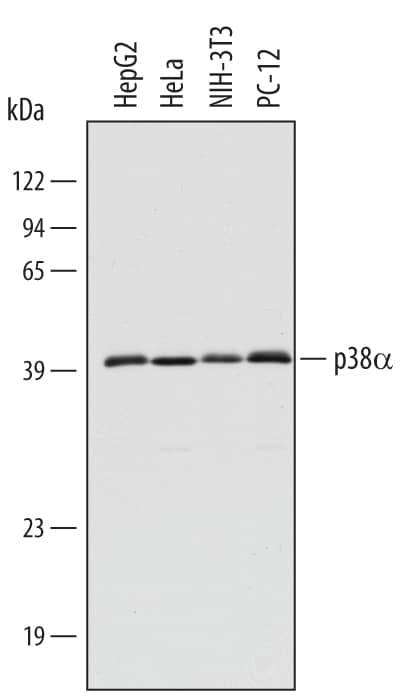
HAF008). A specific band for p38a was detected at approximately 40 kDa (as indicated). This experiment was conducted under reducing conditions and using
Immunoblot Buffer Group 1." alt="Western blot shows lysates of HepG2 human hepatocellular carcinoma cell line, HeLa human cervical epithelial carcinoma cell line, NIH-3T3 mouse embryonic fibroblast cell line, and PC-12 rat adrenal pheochromocytoma cell line. PVDF membrane was probed with 0.5 µg/mL Rabbit Anti-Human/Mouse/Rat p38a Antigen Affinity-purified Polyclonal Antibody (Catalog # AF8691) followed by HRP-conjugated Anti-Rabbit IgG Secondary Antibody (
HAF008). A specific band for p38a was detected at approximately 40 kDa (as indicated). This experiment was conducted under reducing conditions and using
Immunoblot Buffer Group 1." class="big_thumb" />
5477-KS) (2 ng/lane). PVDF membrane was probed with 0.5 µg/mL Rabbit Anti-Human/Mouse/Rat p38 alpha Antigen Affinity-purified Polyclonal Antibody (Catalog # AF8691) followed by HRP-conjugated Anti-Rabbit IgG Secondary Antibody (
HAF008). This experiment was conducted under reducing conditions and using
Immunoblot Buffer Group 1." class="big_lightbox">
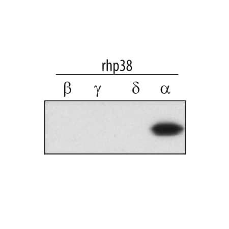
5477-KS) (2 ng/lane). PVDF membrane was probed with 0.5 µg/mL Rabbit Anti-Human/Mouse/Rat p38 alpha Antigen Affinity-purified Polyclonal Antibody (Catalog # AF8691) followed by HRP-conjugated Anti-Rabbit IgG Secondary Antibody (
HAF008). This experiment was conducted under reducing conditions and using
Immunoblot Buffer Group 1." class="big_thumb" />
MAB734), Rabbit Anti-Human/Mouse/Rat p38a Antigen Affinity-purified Polyclonal Antibody (green; Catalog # AF8691), and Mouse Integrin beta 1 Antigen Affinity-purified Polyclonal Antibody (blue;
AF2405). Tissue was incubated with primary antibodies overnight at 4 °C. Tissue was stained using NorthernLights™ 493-conjugated Anti-Rabbit IgG Secondary Antibody (green;
NL006), NorthernLights 557-conjugated Anti-Mouse IgG Secondary Antibody (red;
NL007), and NorthernLights 637-conjugated Anti-Goat IgG Secondary Antibody (blue;
NL002). The image of Integrin beta 1 is pseudo-colored for presentation. View our protocol for
Fluorescent IHC Staining of Frozen Tissue Sections." class="big_lightbox">
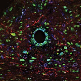
MAB734), Rabbit Anti-Human/Mouse/Rat p38a Antigen Affinity-purified Polyclonal Antibody (green; Catalog # AF8691), and Mouse Integrin beta 1 Antigen Affinity-purified Polyclonal Antibody (blue;
AF2405). Tissue was incubated with primary antibodies overnight at 4 °C. Tissue was stained using NorthernLights™ 493-conjugated Anti-Rabbit IgG Secondary Antibody (green;
NL006), NorthernLights 557-conjugated Anti-Mouse IgG Secondary Antibody (red;
NL007), and NorthernLights 637-conjugated Anti-Goat IgG Secondary Antibody (blue;
NL002). The image of Integrin beta 1 is pseudo-colored for presentation. View our protocol for
Fluorescent IHC Staining of Frozen Tissue Sections." class="big_thumb" />
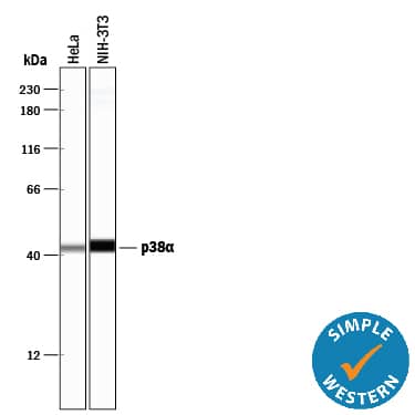
HAF008). A specific band was detected for p38a at approximately 38 kDa (as indicated) in the parental HEK293T cell line, but is not detectable in knockout HEK293T cell line. GAPDH (
AF5718) is shown as a loading control. This experiment was conducted under reducing conditions and using
Immunoblot Buffer Group 1." class="big_lightbox">
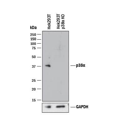
HAF008). A specific band was detected for p38a at approximately 38 kDa (as indicated) in the parental HEK293T cell line, but is not detectable in knockout HEK293T cell line. GAPDH (
AF5718) is shown as a loading control. This experiment was conducted under reducing conditions and using
Immunoblot Buffer Group 1." class="big_thumb" />
VC003). Before incubation with the primary antibody, tissue was subjected to heat-induced epitope retrieval using VisUCyte Antigen Retrieval Reagent-Basic (Catalog #
VCTS021). Tissue was stained using DAB (brown) and counterstained with hematoxylin (blue). Specific staining was localized to cytoplasm in Purkinje neurons. View our protocol for
IHC Staining with VisUCyte HRP Polymer Detection Reagents." class="big_lightbox">

VC003). Before incubation with the primary antibody, tissue was subjected to heat-induced epitope retrieval using VisUCyte Antigen Retrieval Reagent-Basic (Catalog #
VCTS021). Tissue was stained using DAB (brown) and counterstained with hematoxylin (blue). Specific staining was localized to cytoplasm in Purkinje neurons. View our protocol for
IHC Staining with VisUCyte HRP Polymer Detection Reagents." class="big_thumb" />
 HAF008). A specific band for p38a was detected at approximately 40 kDa (as indicated). This experiment was conducted under reducing conditions and using Immunoblot Buffer Group 1." alt="Western blot shows lysates of HepG2 human hepatocellular carcinoma cell line, HeLa human cervical epithelial carcinoma cell line, NIH-3T3 mouse embryonic fibroblast cell line, and PC-12 rat adrenal pheochromocytoma cell line. PVDF membrane was probed with 0.5 µg/mL Rabbit Anti-Human/Mouse/Rat p38a Antigen Affinity-purified Polyclonal Antibody (Catalog # AF8691) followed by HRP-conjugated Anti-Rabbit IgG Secondary Antibody (HAF008). A specific band for p38a was detected at approximately 40 kDa (as indicated). This experiment was conducted under reducing conditions and using Immunoblot Buffer Group 1." class="big_thumb" />
 HAF007). A specific band was detected for Phospho-p38a (T180/Y182) at approximately 45 kDa (as indicated). This experiment was conducted under reducing conditions and using Immunoblot Buffer Group 1." alt="Western blot shows lysates of HeLa human cervical epithelial carcinoma cell line untreated (-) or treated (+) with 20 mJ/cm2ultraviolet light (UV) followed by a 30 minute recovery. PVDF membrane was probed with 1 µg/mL of Mouse Anti-Human Phospho-p38a (T180/Y182) Monoclonal Antibody (Catalog # MAB8691) followed by HRP-conjugated Anti-Mouse IgG Secondary Antibody (Catalog # HAF007). A specific band was detected for Phospho-p38a (T180/Y182) at approximately 45 kDa (as indicated). This experiment was conducted under reducing conditions and using Immunoblot Buffer Group 1." class="big_thumb" />
 HAF007). A specific band was detected for p38a at approximately 38 kDa (as indicated). This experiment was conducted under reducing conditions and using Immunoblot Buffer Group 4." alt="Western blot shows lysates of HeLa human cervical epithelial carcinoma cell line, NIH-3T3 mouse embryonic fibroblast cell line, and PC-12 rat adrenal pheochromocytoma cell line. PVDF membrane was probed with 1 µg/mL of Human/Mouse/Rat p38a Monoclonal Antibody (Catalog # MAB869) followed by HRP-conjugated Anti-Mouse IgG Secondary Antibody (Catalog # HAF007). A specific band was detected for p38a at approximately 38 kDa (as indicated). This experiment was conducted under reducing conditions and using Immunoblot Buffer Group 4." class="big_thumb" />


 5477-KS) (2 ng/lane). PVDF membrane was probed with 0.5 µg/mL Rabbit Anti-Human/Mouse/Rat p38 alpha Antigen Affinity-purified Polyclonal Antibody (Catalog # AF8691) followed by HRP-conjugated Anti-Rabbit IgG Secondary Antibody (
 MAB734), Rabbit Anti-Human/Mouse/Rat p38a Antigen Affinity-purified Polyclonal Antibody (green; Catalog # AF8691), and Mouse Integrin beta 1 Antigen Affinity-purified Polyclonal Antibody (blue;
 HAF008). A specific band was detected for p38a at approximately 38 kDa (as indicated) in the parental HEK293T cell line, but is not detectable in knockout HEK293T cell line. GAPDH (
 VC003). Before incubation with the primary antibody, tissue was subjected to heat-induced epitope retrieval using VisUCyte Antigen Retrieval Reagent-Basic (Catalog #
![Western Blot: p38 [p Thr180, p Tyr182] Antibody [NB500-138] - Hela cell lysates that had been treated with UV or untreated (Control) showing specific immunolabeling of the -39 kDa p38 MAPK protein phosphorylated at Thr180/Tyr182.](http://images.novusbio.com/fullsize/p38-[p-Thr180--p-Tyr182]-Antibody-Western-Blot-NB500-138-img0005.jpg "<b>Biological Strategies Validation. </b>Western Blot: p38 [p Thr180, p Tyr182] Antibody [NB500-138] - Hela cell lysates that had been treated with UV or untreated (Control) showing specific immunolabeling of the -39 kDa p38 MAPK protein phosphorylated at Thr180/Tyr182.")
![<b>Biological Strategies Validation. </b>Immunohistochemistry-Paraffin: p38 [p Thr180, p Tyr182] Antibody [NB500-138] - Immunostaining of human breast cancer tissue showing p38 when phosphorylated at Thr180/Tyr182 in red.](http://images.novusbio.com/fullsize/p38-[p-Thr180-p-Tyr182]-Antibody-Immunohistochemistry-Paraffin-NB500-138-img0002.jpg)
![<b>Biological Strategies Validation. </b>Western Blot: p38 [p Thr180, p Tyr182] Antibody [NB500-138] - Western blot of HeLa cell lysates that had been treated with UV or untreated (Control) showing specific immunolabeling of the ~39k p38 MAPK protein phosphorylated at Thr180/Tyr182.](http://images.novusbio.com/fullsize/p38-[p-Thr180-p-Tyr182]-Antibody-Western-Blot-NB500-138-img0001.jpg)
![Western Blot: p38 alpha [p Thr180, p Tyr182] Antibody [NB500-138] - RIP2 is not essential for MAPK pathways in the early ontogenesis. (A) Immunoblot analysis of phospho-p44/42 MAPK, p44/42 MAPK, & p38 MAPK in larvae homogenate from WT & NOD1-1IS−/− zebrafish at 7 days post-fertilization (dpf). (B) Immunoblot analysis of phospho-p44/42 MAPK, p44/42 MAPK, & p38 MAPK in larvae homogenate from WT & RIP2−/− zebrafish at 7 dpf. (C) Immunoblot analysis of Atg5, p62, & LC3b in larvae homogenate from WT & NOD1-1IS−/− zebrafish at 7 dpf. (D) Immunoblot analysis of Atg5, p62, & LC3b in larvae homogenate from WT & RIP2−/− zebrafish at 7 dpf. Western blotting results were quantified using Quantity One software. Data represent the average of two independent experiments. *p < 0.05, **p < 0.01. Image collected & cropped by CiteAb from the following publication (//pubmed.ncbi.nlm.nih.gov/29692779), licensed under a CC-BY license. Not internally tested by Novus Biologicals.](http://images.novusbio.com/fullsize/nb500-138_rabbit-polyclonal-p38-alpha-p-thr180-p-tyr182-antibody-31020241534371.jpg)
![ELISA p38 alpha [p Thr180, p Tyr182] [Biotin]](http://images.novusbio.com/fullsize/dyc869b-2_phospho-p38-alpha-t180-y182-duoset-ic-elisa-2-plate-elisa-1592020145534.jpg "ELISA p38 alpha [p Thr180, p Tyr182] [Biotin]")
 HAF018). A specific band was detected for p38a at approximately 38 kDa (as indicated) in the parental HEK293T cell line, but is not detectable in knockout HEK293T cell line. GAPDH (Catalog #
![Western Blot: p38 alpha Antibody [NB110-96907] - Western blot analysis of Human cancer cell lines showing detection of p38 alpha protein using Rabbit Anti-p38 alpha Polyclonal Antibody (NB110-96907). Load: 15 ugprotein. Block: 1.5% BSA for 30 minutes at RT. Primary Antibody: Rabbit Anti-p38 alpha Polyclonal Antibody (NB110-96907) at 1:4000 for 2 hours at RT. Secondary Antibody: Donkey Anti-Rabbit IgG: HRP for 1 hour at RT.](http://images.novusbio.com/fullsize/p38-alpha-Antibody-Western-Blot-NB110-96907-img0008.jpg "Western Blot: p38 alpha Antibody [NB110-96907] - Western blot analysis of Human cancer cell lines showing detection of p38 alpha protein using Rabbit Anti-p38 alpha Polyclonal Antibody (NB110-96907). Load: 15 ugprotein. Block: 1.5% BSA for 30 minutes at RT. Primary Antibody: Rabbit Anti-p38 alpha Polyclonal Antibody (NB110-96907) at 1:4000 for 2 hours at RT. Secondary Antibody: Donkey Anti-Rabbit IgG: HRP for 1 hour at RT.")
![Immunocytochemistry/Immunofluorescence: p38 alpha Antibody [NB110-96907] - Analysis using Rabbit Anti-p38 Polyclonal Antibody . Tissue: HeLa Cells. Species: Human. Fixation: 2% Formaldehyde for 20 min at RT. Primary Antibody: Rabbit Anti-p38 Polyclonal Antibody at 1:100 for 12 hours at 4 degrees C. Secondary Antibody: FITC Goat Anti-Rabbit (green) at 1:200 for 2 hours at RT. Counterstain: DAPI (blue) nuclear stain at 1:40000 for 2 hours at RT. Localization: Mitochondrion. Cytoplasm. Nucleus. Magnification: 100x. (A) DAPI (blue) nuclear stain. (B) Anti-p38 Antibody. (C) Composite.](http://images.novusbio.com/fullsize/p38-alpha-Antibody-Immunocytochemistry-Immunofluorescence-NB110-96907-img0007.jpg)
![Western Blot: p38 alpha Antibody [NB110-96907] - CtsK & NFATc1 expression levels were decreased during osteoclastogenesis by PSTP-3,5-Me treatment. (A) mRNA expression levels of osteoclast-specific markers were determined by RT-PCR in 0 or 6 µM PSTP-3,5-Me-treated cells. *p < 0.05, & **p < 0.01 indicate the statistically significant difference between non-PSTP-3,5-Me-treated groups (0 µM) & PSTP-3,5-Me-treated groups (6 µM) on each day. Mean ± standard error. (B) Western blotting was performed to determine phosphorylation of NF-kB, p-38, ERK1/2, & JNK. Cells were pre-treated with 6 µM PSTP-3,5-Me or vehicle (Ctrl) for 2 h, & then treated with RANKL for the indicated times. beta -Actin expression level was used as a loading control. Image collected & cropped by CiteAb from the following publication (//pubmed.ncbi.nlm.nih.gov/31540026), licensed under a CC-BY license. Not internally tested by Novus Biologicals.](http://images.novusbio.com/fullsize/nb110-96907_rabbit-polyclonal-p38-alpha-antibody-31020241534327.jpg)
![Western Blot: p38 Antibody [NB100-56665] - Analysis of human pancreas lysate using p38 antibody at 1:1000.](http://images.novusbio.com/fullsize/p38-Antibody-Western-Blot-NB100-56665-img0003.jpg "Western Blot: p38 Antibody [NB100-56665] - Analysis of human pancreas lysate using p38 antibody at 1:1000.")
![Immunohistochemistry-Paraffin: p38 Antibody [NB100-56665] - Analysis of human brain, cortex using MAPK14 antibody at 1:100.](http://images.novusbio.com/fullsize/p38-Antibody-Immunohistochemistry-Paraffin-NB100-56665-img0002.jpg)
![Western Blot: p38 alpha Antibody [NBP2-19662] - Various whole cell extracts (30 ug) were separated by 10% SDS-PAGE, and the membrane was blotted with p38 alpha antibody diluted at 1:1000. HRP-conjugated anti-rabbit IgG antibody was used to detect the primary antibody.](http://images.novusbio.com/fullsize/p38-alpha-Antibody-Western-Blot-NBP2-19662-img0010.jpg "Western Blot: p38 alpha Antibody [NBP2-19662] - Various whole cell extracts (30 ug) were separated by 10% SDS-PAGE, and the membrane was blotted with p38 alpha antibody diluted at 1:1000. HRP-conjugated anti-rabbit IgG antibody was used to detect the primary antibody.")
![Immunohistochemistry-Paraffin: p38 Antibody [NBP2-19662] - Paraffin-embedded human lung adenocarcinoma. p38 antibody [N1C3-2] diluted at 1:250.](http://images.novusbio.com/fullsize/p38-Antibody-Immunohistochemistry-Paraffin-NBP2-19662-img0003.jpg)
![Immunohistochemistry-Paraffin: p38 Antibody [NBP2-19662] - Paraffin-embedded rat brain. p38 MAPK antibody [N1C3-2] diluted at 1:500.](http://images.novusbio.com/fullsize/p38-Antibody-Immunohistochemistry-Paraffin-NBP2-19662-img0002.jpg)

![p38 MAPK antibody [N1C3-2] detects p38 MAPK protein at cytoplasm and nucleus by immunofluorescent analysis.<br>Sample: HeLa cells were fixed in 4% paraformaldehyde for 10 min.<br>Green: p38 MAPK protein stained by p38 MAPK antibody [N1C3-2] (NBP2-19662) diluted at 1:100.<br>Blue: Hoechst 33342 staining.<br>Scale bar = 10 um.](http://images.novusbio.com/fullsize/nbp2-19662_rabbit-polyclonal-p38-alpha-antibody-immunocytochemistry-immunofluorescence-261120251995418.jpg)
![<b>Genetic Strategies Validation. </b>Wild-type (WT) and p38 MAPK knockout (KO) 293T cell extracts (30 ug) were separated by 10% SDS-PAGE, and the membrane was blotted with p38 MAPK antibody [N1C3-2] (NBP2-19662) diluted at 1:500. The HRP-conjugated anti-rabbit IgG antibody was used to detect the primary antibody.](http://images.novusbio.com/fullsize/nbp2-19662_rabbit-polyclonal-p38-alpha-antibody-western-blot-261120251837242.jpg)
![Western Blot: p38 alpha Antibody (8G4D11) [NBP2-37378] - Analysis using MAPK14 mouse mAb against HeLa (1), NIH/3T3 (2), Jurkat (3), Raw264.7 (4), PC-12 (5), C6 (6) and COS7 (7) cell lysate.](http://images.novusbio.com/fullsize/p38-Antibody-8G4D11-Western-Blot-NBP2-37378-img0004.jpg "Western Blot: p38 alpha Antibody (8G4D11) [NBP2-37378] - Analysis using MAPK14 mouse mAb against HeLa (1), NIH/3T3 (2), Jurkat (3), Raw264.7 (4), PC-12 (5), C6 (6) and COS7 (7) cell lysate.")
![Immunocytochemistry/Immunofluorescence: p38 alpha Antibody (8G4D11) [NBP2-37378] - Analysis of HeLa cells using MAPK14 mouse mAb (green). Blue: DRAQ5 fluorescent DNA dye. Red: Actin filaments have been labeled with Alexa Fluor-555 phalloidin.](http://images.novusbio.com/fullsize/p38-Antibody-8G4D11-Immunocytochemistry-Immunofluorescence-NBP2-37378-img0005.jpg)
![Immunohistochemistry-Paraffin: p38 alpha Antibody (8G4D11) [NBP2-37378] - Analysis of cervical cancer tissues using MAPK14 mouse mAb with DAB staining.](http://images.novusbio.com/fullsize/p38-Antibody-8G4D11-Immunohistochemistry-NBP2-37378-img0009.jpg)
![Flow Cytometry: p38 alpha Antibody (8G4D11) [NBP2-37378] - Analysis of HeLa cells using MAPK14 mouse mAb (green) and negative control (red).](http://images.novusbio.com/fullsize/p38-Antibody-8G4D11-Flow-Cytometry-NBP2-37378-img0006.jpg)
![Western Blot: p38 alpha Antibody (8G4D11) [NBP2-37378] - Analysis using MAPK14 mAb against human MAPK14 recombinant protein. (Expected MW is 33 kDa)](http://images.novusbio.com/fullsize/p38-Antibody-8G4D11-Western-Blot-NBP2-37378-img0002.jpg)
![Western Blot: p38 alpha Antibody (8G4D11) [NBP2-37378] - Analysis using MAPK14 mAb against HEK293 (1) and MAPK14 (AA: 299-360)-hIgGFc transfected HEK293 (2) cell lysate.](http://images.novusbio.com/fullsize/p38-Antibody-8G4D11-Western-Blot-NBP2-37378-img0003.jpg)
![Immunohistochemistry-Paraffin: p38 alpha Antibody (8G4D11) [NBP2-37378] - Analysis of rectum cancer tissues using MAPK14 mouse mAb with DAB staining.](http://images.novusbio.com/fullsize/p38-Antibody-8G4D11-Immunohistochemistry-NBP2-37378-img0008.jpg)
![ELISA: p38 alpha Antibody (8G4D11) [NBP2-37378] - Red: Control Antigen (100ng); Purple: Antigen (10ng); Green: Antigen (50ng); Blue: Antigen (100ng);](http://images.novusbio.com/fullsize/p38-Antibody-8G4D11-ELISA-NBP2-37378-img0001.jpg)
![Western Blot: p38 alpha [p Tyr182] Antibody [NB100-82097] - Human breast cancer cell line MDA-MB-231 was treated with different doses of p38 inhibitor and the expression of phospho-p38 and total p38 were detected by Western blot. Image from verified customer review.](http://images.novusbio.com/fullsize/p38-alpha-[p-Tyr182]-Antibody-Western-Blot-NB100-82097-img0008.jpg "<b>Biological Strategies Validation. </b>Western Blot: p38 alpha [p Tyr182] Antibody [NB100-82097] - Human breast cancer cell line MDA-MB-231 was treated with different doses of p38 inhibitor and the expression of phospho-p38 and total p38 were detected by Western blot. Image from verified customer review.")
![<b>Biological Strategies Validation. </b>Western Blot: p38 alpha [p Tyr182] Antibody [NB100-82097] - Analysis of extracts from Hela cells untreated or treated with UV for the indicated times, using P38 MAPK (Phospho-Tyr182) antibody.](http://images.novusbio.com/fullsize/p38-alpha-[p-Tyr182]-Antibody-Western-Blot-NB100-82097-img0001.jpg)
![<b>Biological Strategies Validation. </b>Western Blot: p38 alpha [p Tyr182] Antibody [NB100-82097] - Analysis of extracts from Hela cells, treated with TNFa or calf intestinal phosphatase (CIP).](http://images.novusbio.com/fullsize/p38-alpha-[p-Tyr182]-Antibody-Western-Blot-NB100-82097-img0005.jpg)
![Immunohistochemistry-Paraffin: p38 alpha [p Tyr182] Antibody [NB100-82097] - Analysis of paraffin-embedded human breast carcinoma tissue, using NB100-82097(left) or the same antibody preincubated with blocking peptide(right).](http://images.novusbio.com/fullsize/nb100-82097_rabbit-polyclonal-p38-alpha-p-tyr182-antibody-10320252332107.jpg)
![Western Blot: p38 alpha Antibody (9F12) [NB110-96875] - analysis of Human Cell lysates showing detection of p38 MAPK protein using Mouse Anti-p38 MAPK Monoclonal Antibody, Clone 9F12 . Load: 15 ug protein. Block: 1.5% BSA for 30 minutes at RT. Primary Antibody: Mouse Anti-p38 MAPK Monoclonal Antibody at 1:1000 for 2 hours at RT. Secondary Antibody: Sheep Anti-Mouse IgG: HRP for 1 hour at RT.](http://images.novusbio.com/fullsize/p38-alpha-Antibody-9F12-Western-Blot-NB110-96875-img0008.jpg "Western Blot: p38 alpha Antibody (9F12) [NB110-96875] - analysis of Human Cell lysates showing detection of p38 MAPK protein using Mouse Anti-p38 MAPK Monoclonal Antibody, Clone 9F12 . Load: 15 ug protein. Block: 1.5% BSA for 30 minutes at RT. Primary Antibody: Mouse Anti-p38 MAPK Monoclonal Antibody at 1:1000 for 2 hours at RT. Secondary Antibody: Sheep Anti-Mouse IgG: HRP for 1 hour at RT.")
![Immunohistochemistry: p38 alpha Antibody (9F12) [NB110-96875] - Immunohistochemistry analysis using Mouse Anti-p38 alpha Monoclonal Antibody, Clone 9F12 (NB110-96875). Tissue: colon carcinoma. Species: Human. Fixation: Formalin. Primary Antibody: Mouse Anti-p38 alpha Monoclonal Antibody (NB110-96875) at 1:10000 for 12 hours at 4C. Secondary Antibody: Biotin Goat Anti-Mouse at 1:2000 for 1 hour at RT. Counterstain: Mayer Hematoxylin (purple/blue) nuclear stain at 200 l for 2 minutes at RT. Magnification: 40x.](http://images.novusbio.com/fullsize/p38-alpha-Antibody-9F12-Immunohistochemistry-NB110-96875-img0009.jpg)
![Western Blot: p38 Antibody (10B11C8) [NBP2-37312] - Western blot analysis using MAPK14 mouse mAb against Hela (1), HEK293 (2), A431 (3), MCF-7 (4), RAW264.7 (5), Cos7 (6), C6 (7), Jurkat (8) and NIH/3T3 (9) cell lysate.](http://images.novusbio.com/fullsize/p38-Antibody-10B11C8-Western-Blot-NBP2-37312-img0004.jpg "Western Blot: p38 Antibody (10B11C8) [NBP2-37312] - Western blot analysis using MAPK14 mouse mAb against Hela (1), HEK293 (2), A431 (3), MCF-7 (4), RAW264.7 (5), Cos7 (6), C6 (7), Jurkat (8) and NIH/3T3 (9) cell lysate.")
![Immunohistochemistry: p38 Antibody (10B11C8) [NBP2-37312] - Immunohistochemical analysis of paraffin-embedded endometrial cancer tissues using MAPK14 mouse mAb with DAB staining.](http://images.novusbio.com/fullsize/p38-Antibody-10B11C8-Immunohistochemistry-NBP2-37312-img0005.jpg)
![Western Blot: p38 Antibody (10B11C8) [NBP2-37312] - Western blot analysis using MAPK14 mAb against human MAPK14 recombinant protein. (Expected MW is 32.6 kDa)](http://images.novusbio.com/fullsize/p38-Antibody-10B11C8-Western-Blot-NBP2-37312-img0002.jpg)
![Western Blot: p38 Antibody (10B11C8) [NBP2-37312] - Western blot analysis using MAPK14 mAb against HEK293 (1) and MAPK14 (AA: 299-360)-hIgGFc transfected HEK293 (2) cell lysate.](http://images.novusbio.com/fullsize/p38-Antibody-10B11C8-Western-Blot-NBP2-37312-img0003.jpg)
![ELISA: p38 Antibody (10B11C8) [NBP2-37312] - Red: Control Antigen (100ng); Purple: Antigen (10ng); Green: Antigen (50ng); Blue: Antigen (100ng);](http://images.novusbio.com/fullsize/p38-Antibody-10B11C8-ELISA-NBP2-37312-img0001.jpg)


