DR647RB) and counterstained with DAPI (blue; Lunaphore Catalog #
DR100). Specific staining was localized to the membrane of endothelial cells. Protocol available in
COMET™ Panel Builder." class="big_lightbox">
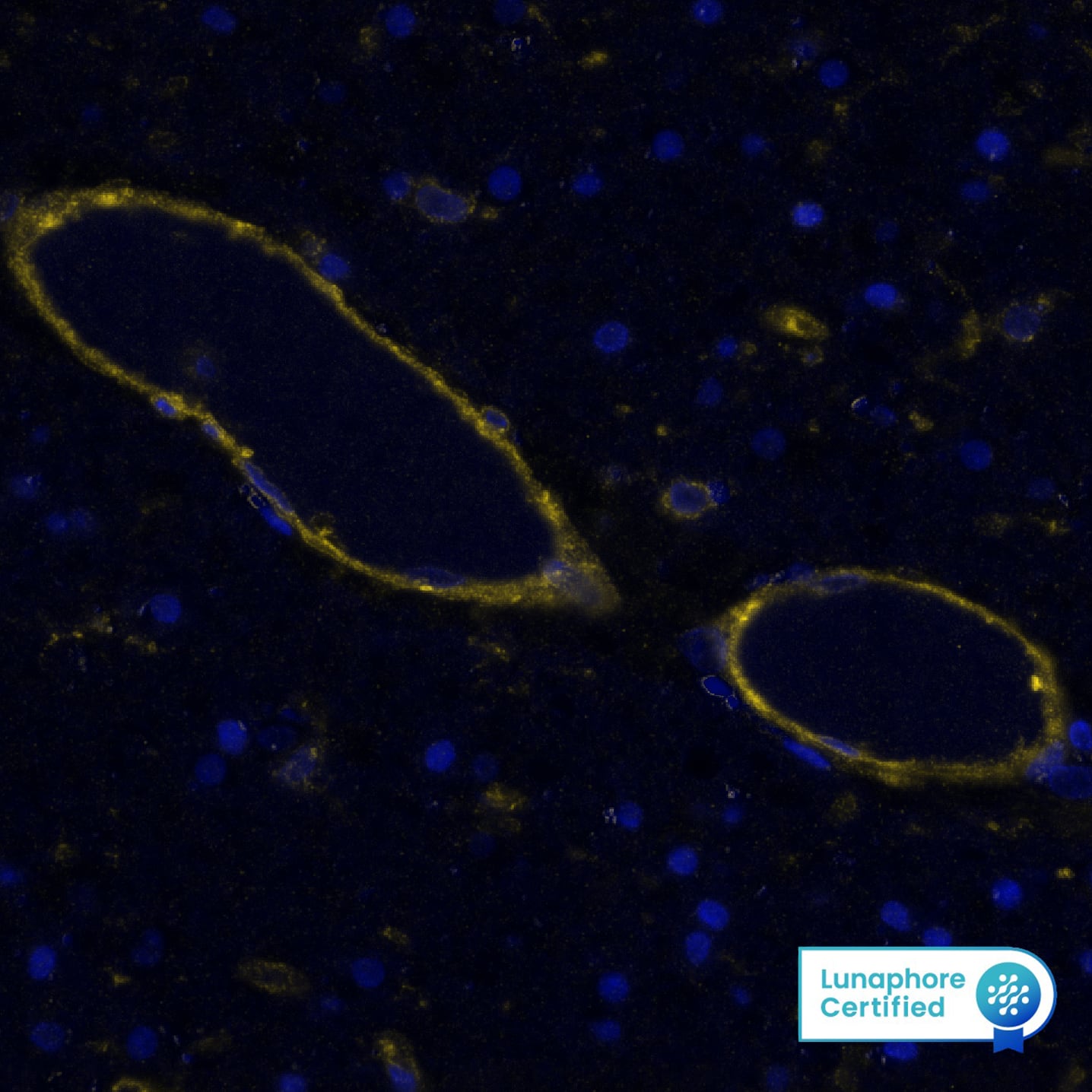
DR647RB) and counterstained with DAPI (blue; Lunaphore Catalog #
DR100). Specific staining was localized to the membrane of endothelial cells. Protocol available in
COMET™ Panel Builder." alt="CD39 was detected in immersion fixed paraffin-embedded sections of human Brain Cortex using Rabbit Anti-Human CD39, Monoclonal Antibody (Catalog# MAB11713) at 20ug/mL at 37°Celsius for 8 minutes. Before incubation with the primary antibody, tissue underwent an all-in-one dewaxing and antigen retrieval preprocessing using PreTreatment Module (PT Module) and Dewax and HIER Buffer H (pH 9; Epredia Catalog # TA-999-DHBH). Tissue was stained using the Alexa Fluor™ Plus 647 Goat anti-Rabbit IgG Secondary Antibody at 1:200 at 37 ° Celsius for 2 minutes. (Yellow; Lunaphore Catalog #
DR647RB) and counterstained with DAPI (blue; Lunaphore Catalog #
DR100). Specific staining was localized to the membrane of endothelial cells. Protocol available in
COMET™ Panel Builder." class="big_thumb" />
HAF008). A specific band was detected for CD39/ENTPD1 at approximately 75 kDa (as indicated). This experiment was conducted under reducing conditions and using
Western Blot Buffer Group 1." class="big_lightbox">
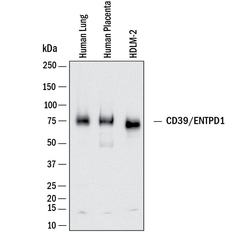
HAF008). A specific band was detected for CD39/ENTPD1 at approximately 75 kDa (as indicated). This experiment was conducted under reducing conditions and using
Western Blot Buffer Group 1." class="big_thumb" />
VC003) or the HRP-conjugated Anti-Rabbit IgG Secondary Antibody (Catalog #
HAF008). Before incubation with the primary antibody, tissue was subjected to heat-induced epitope retrieval using VisUCyte Antigen Retrieval Reagent-Basic (Catalog #
VCTS021). Tissue was stained using DAB (brown) and counterstained with hematoxylin (blue). Specific staining was localized to the cell surface of endothelial cells. View our protocol for
IHC Staining with VisUCyte HRP Polymer Detection Reagents." class="big_lightbox">
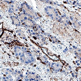
VC003) or the HRP-conjugated Anti-Rabbit IgG Secondary Antibody (Catalog #
HAF008). Before incubation with the primary antibody, tissue was subjected to heat-induced epitope retrieval using VisUCyte Antigen Retrieval Reagent-Basic (Catalog #
VCTS021). Tissue was stained using DAB (brown) and counterstained with hematoxylin (blue). Specific staining was localized to the cell surface of endothelial cells. View our protocol for
IHC Staining with VisUCyte HRP Polymer Detection Reagents." class="big_thumb" />
VC003) or the HRP-conjugated Anti-Rabbit IgG Secondary Antibody (Catalog #
HAF008). Before incubation with the primary antibody, tissue was subjected to heat-induced epitope retrieval using VisUCyte Antigen Retrieval Reagent-Basic (Catalog #
VCTS021). Tissue was stained using DAB (brown) and counterstained with hematoxylin (blue). Specific staining was localized to the cell surface of endothelial cells. View our protocol for
IHC Staining with VisUCyte HRP Polymer Detection Reagents." class="big_lightbox">
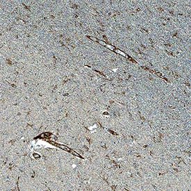
VC003) or the HRP-conjugated Anti-Rabbit IgG Secondary Antibody (Catalog #
HAF008). Before incubation with the primary antibody, tissue was subjected to heat-induced epitope retrieval using VisUCyte Antigen Retrieval Reagent-Basic (Catalog #
VCTS021). Tissue was stained using DAB (brown) and counterstained with hematoxylin (blue). Specific staining was localized to the cell surface of endothelial cells. View our protocol for
IHC Staining with VisUCyte HRP Polymer Detection Reagents." class="big_thumb" />
VC003) or the HRP-conjugated Anti-Rabbit IgG Secondary Antibody (Catalog #
HAF008). Before incubation with the primary antibody, tissue was subjected to heat-induced epitope retrieval using VisUCyte Antigen Retrieval Reagent-Basic (Catalog #
VCTS021). Tissue was stained using DAB (brown) and counterstained with hematoxylin (blue). Specific staining was localized to the cell surface of endothelial cells. View our protocol for
IHC Staining with VisUCyte HRP Polymer Detection Reagents." class="big_lightbox">
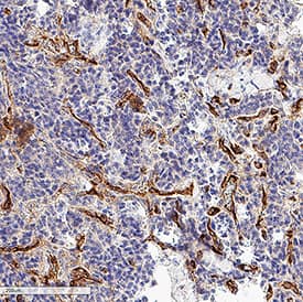
VC003) or the HRP-conjugated Anti-Rabbit IgG Secondary Antibody (Catalog #
HAF008). Before incubation with the primary antibody, tissue was subjected to heat-induced epitope retrieval using VisUCyte Antigen Retrieval Reagent-Basic (Catalog #
VCTS021). Tissue was stained using DAB (brown) and counterstained with hematoxylin (blue). Specific staining was localized to the cell surface of endothelial cells. View our protocol for
IHC Staining with VisUCyte HRP Polymer Detection Reagents." class="big_thumb" />
042-206). This experiment was conducted under reducing conditions and using the 12-230kDa separation system." class="big_lightbox">
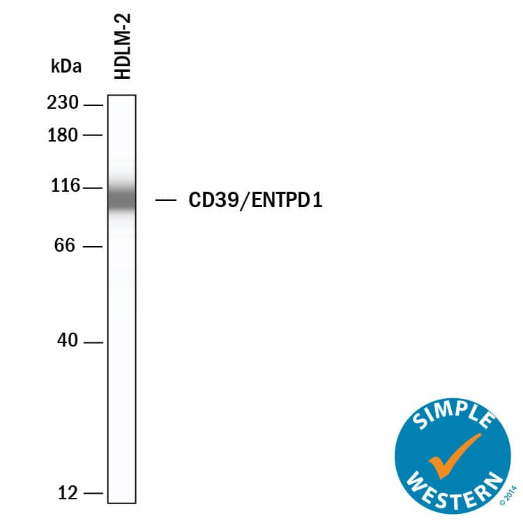
042-206). This experiment was conducted under reducing conditions and using the 12-230kDa separation system." class="big_thumb" />
 5-001-A, open histogram), followed by NorthernLights™ 637-conjugated Anti-Sheep IgG Secondary Antibody (Catalog # NL011)." alt="Mouse splenocytes were stained with Mouse CD39/ENTPD1 Antigen Affinity-purified Polyclonal Antibody (Catalog # AF4398, filled histogram) or control antibody (Catalog # 5-001-A, open histogram), followed by NorthernLights™ 637-conjugated Anti-Sheep IgG Secondary Antibody (Catalog # NL011)." class="big_thumb" />
 DR647RB) and counterstained with DAPI (blue; Lunaphore Catalog # DR100). Specific staining was localized to the membrane of endothelial cells. Protocol available in COMET™ Panel Builder." alt="CD39 was detected in immersion fixed paraffin-embedded sections of human Brain Cortex using Rabbit Anti-Human CD39, Monoclonal Antibody (Catalog# MAB11713) at 20ug/mL at 37°Celsius for 8 minutes. Before incubation with the primary antibody, tissue underwent an all-in-one dewaxing and antigen retrieval preprocessing using PreTreatment Module (PT Module) and Dewax and HIER Buffer H (pH 9; Epredia Catalog # TA-999-DHBH). Tissue was stained using the Alexa Fluor™ Plus 647 Goat anti-Rabbit IgG Secondary Antibody at 1:200 at 37 ° Celsius for 2 minutes. (Yellow; Lunaphore Catalog # DR647RB) and counterstained with DAPI (blue; Lunaphore Catalog # DR100). Specific staining was localized to the membrane of endothelial cells. Protocol available in COMET™ Panel Builder." class="big_thumb" />
 MAB005, open histogram), followed by Allophycocyanin-conjugated Anti-Rat IgG F(ab')2Secondary Antibody (Catalog # F0113)." alt="Mouse splenocytes were stained with Rat Anti-Mouse CD39/ENTPD1 Monoclonal Antibody (Catalog # MAB4398, filled histogram) or isotype control antibody (Catalog # MAB005, open histogram), followed by Allophycocyanin-conjugated Anti-Rat IgG F(ab')2Secondary Antibody (Catalog # F0113)." class="big_thumb" />
 MAB002, open histogram), followed by Phy-coerythrin-conjugated Anti-Mouse IgG F(ab')2Secondary Antibody (Catalog # F0102B)." alt="Human whole blood monocytes were stained with Mouse Anti-Human CD39/ENTPD1 Mono-clonal Antibody (Catalog # MAB4397, filled histogram) or isotype control antibody (Catalog # MAB002, open histogram), followed by Phy-coerythrin-conjugated Anti-Mouse IgG F(ab')2Secondary Antibody (Catalog # F0102B)." class="big_thumb" />


![Western Blot: CD39/ENTPD1 Antibody (17B5F11) [NBP2-25223] - Analysis of human (A), mouse (B), and rat brain (C) and a recombinant protein fragment (D) using CD39 antibody at 1 ug/mL.](http://images.novusbio.com/fullsize/CD39-ENTPD1-Antibody-17B5F11-Western-Blot-NBP2-25223-img0003.jpg "Western Blot: CD39/ENTPD1 Antibody (17B5F11) [NBP2-25223] - Analysis of human (A), mouse (B), and rat brain (C) and a recombinant protein fragment (D) using CD39 antibody at 1 ug/mL.")
![Immunohistochemistry-Paraffin: CD39/ENTPD1 Antibody (17B5F11) [NBP2-25223] - Normal human spleen tissue stained with CD39 antibody (5 ug/mL), peroxidase-conjugate and DAB chromogen.](http://images.novusbio.com/fullsize/CD39-ENTPD1-Antibody-17B5F11-Immunohistochemistry-Paraffin-NBP2-25223-img0004.jpg)

![<b>Orthogonal Strategies Validation. </b>Immunohistochemistry-Paraffin: CD39/ENTPD1 Antibody [NBP1-90071] - Staining of human placenta shows strong cytoplasmic positivity in trophoblastic cells.](http://images.novusbio.com/fullsize/CD39-ENTPD1-Antibody-Immunohistochemistry-Paraffin-NBP1-90071-img0017.jpg)
![Immunohistochemistry-Paraffin: CD39/ENTPD1 Antibody [NBP1-90071] - Staining of human skeletal muscle shows no positivity in myocytes as expected.](http://images.novusbio.com/fullsize/CD39-ENTPD1-Antibody-Immunohistochemistry-Paraffin-NBP1-90071-img0021.jpg)
![Immunohistochemistry-Paraffin: CD39/ENTPD1 Antibody [NBP1-90071] - Staining of human duodenum shows strong membranous positivity in endothelial cells.](http://images.novusbio.com/fullsize/CD39-ENTPD1-Antibody-Immunohistochemistry-Paraffin-NBP1-90071-img0019.jpg)
![Immunohistochemistry-Paraffin: CD39/ENTPD1 Antibody [NBP1-90071] - Staining of human kidney shows no positivity in cells in tubules as expected.](http://images.novusbio.com/fullsize/CD39-ENTPD1-Antibody-Immunohistochemistry-Paraffin-NBP1-90071-img0020.jpg)
![Immunohistochemistry-Paraffin: CD39/ENTPD1 Antibody [NBP1-90071] - Staining of pig aortic valve tissue. Image from verified customer review.](http://images.novusbio.com/fullsize/CD39-ENTPD1-Antibody-Immunohistochemistry-Paraffin-NBP1-90071-img0013.jpg)
 CTS019) and counterstained with hematoxylin (blue). View our protocol for
  HAF008). A specific band was detected for CD39/ENTPD1 at approximately 75 kDa (as indicated). This experiment was conducted under reducing conditions and using
  VC003) or the HRP-conjugated Anti-Rabbit IgG Secondary Antibody (Catalog #
  VC003) or the HRP-conjugated Anti-Rabbit IgG Secondary Antibody (Catalog #
  VC003) or the HRP-conjugated Anti-Rabbit IgG Secondary Antibody (Catalog #
  042-206). This experiment was conducted under reducing conditions and using the 12-230kDa separation system." class="big_thumb" />
![Western Blot: CD39/ENTPD1 Antibody (JA90-36) [NBP2-67230] - Western blot analysis of CD39/ENTPD1 on different lysates. Proteins were transferred to a PVDF membrane and blocked with 5% BSA in PBS for 1 hour at room temperature. The primary antibody (1/500) was used in 5% BSA at room temperature for 2 hours. Goat Anti-Rabbit IgG - HRP Secondary Antibody (HA1001) at 1:5,000 dilution was used for 1 hour at room temperature. Positive control: Lane 1: mouse heart tissue lysate Lane 2: mouse spleen tissue lysate](http://images.novusbio.com/fullsize/CD39-ENTPD1-Antibody-JA90-36-Western-Blot-NBP2-67230-img0006.jpg "Western Blot: CD39/ENTPD1 Antibody (JA90-36) [NBP2-67230] - Western blot analysis of CD39/ENTPD1 on different lysates. Proteins were transferred to a PVDF membrane and blocked with 5% BSA in PBS for 1 hour at room temperature. The primary antibody (1/500) was used in 5% BSA at room temperature for 2 hours. Goat Anti-Rabbit IgG - HRP Secondary Antibody (HA1001) at 1:5,000 dilution was used for 1 hour at room temperature. Positive control: Lane 1: mouse heart tissue lysate Lane 2: mouse spleen tissue lysate")
![Immunohistochemistry-Paraffin: CD39/ENTPD1 Antibody (JA90-36) [NBP2-67230] - Analysis of paraffin-embedded human endometrium tissue with Rabbit anti-CD39/ENTPDI antibody washed with ddH2O and PBS, and then probed with the primary antibody at 1/400 dilution for 1 hour at room temperature. The detection was performed using an HRP conjugated compact polymer system. DAB was used as the chromogen. Tissues were counterstained with hematoxylin and mounted with DPX.](http://images.novusbio.com/fullsize/CD39-ENTPD1-Antibody-JA90-36-Immunohistochemistry-Paraffin-NBP2-67230-img0007.jpg)
![Immunohistochemistry-Paraffin: CD39/ENTPD1 Antibody (JA90-36) [NBP2-67230] - Analysis of paraffin-embedded human placenta tissue using anti-CD39 antibody. Counter stained with hematoxylin.](http://images.novusbio.com/fullsize/CD39-ENTPD1-Antibody-JA90-36-Immunohistochemistry-Paraffin-NBP2-67230-img0001.jpg)
![Immunohistochemistry-Paraffin: CD39/ENTPD1 Antibody (JA90-36) [NBP2-67230] - Analysis of paraffin-embedded human spleen tissue using anti-CD39 antibody. Counter stained with hematoxylin.](http://images.novusbio.com/fullsize/CD39-ENTPD1-Antibody-JA90-36-Immunohistochemistry-Paraffin-NBP2-67230-img0002.jpg)
![Immunohistochemistry-Paraffin: CD39/ENTPD1 Antibody (JA90-36) [NBP2-67230] - Analysis of paraffin-embedded human tonsil tissue using anti-CD39 antibody. Counter stained with hematoxylin.](http://images.novusbio.com/fullsize/CD39-ENTPD1-Antibody-JA90-36-Immunohistochemistry-Paraffin-NBP2-67230-img0003.jpg)
![Immunohistochemistry-Paraffin: CD39/ENTPD1 Antibody (JA90-36) [NBP2-67230] - Analysis of paraffin-embedded mouse uterus tissue using anti-CD39 antibody. Counter stained with hematoxylin.](http://images.novusbio.com/fullsize/CD39-ENTPD1-Antibody-JA90-36-Immunohistochemistry-Paraffin-NBP2-67230-img0004.jpg)
![Western blot analysis on different lysates with NBP2-67230 at 1/5,000 dilution. Lane 1: Mouse lung tissue lysate (20 µg/Lane) Lane 2: Mouse placenta tissue lysate (20 µg/Lane) Predicted band size: 58 kDa Observed band size: 70 kDa Exposure time: 20 seconds; 4-20% SDS-PAGE gel. Proteins were transferred to a PVDF membrane and blocked with 5% NFDM/TBST for 1 hour at room temperature. NBP2-67230 at 1/5,000 dilution was used in 5% NFDM/TBST at 4℃ overnight. Goat Anti-Rabbit IgG [HRP] Secondary Antibody at 1/50,000 dilution was used for 1 hour at room temperature.](http://images.novusbio.com/fullsize/nbp2-67230_-western-blot-639204586670551534.jpg)


![Flow Cytometry: CD39/ENTPD1 Antibody (AC2.5) [DDX0321P-100]](http://images.novusbio.com/fullsize/CD39-ENTPD1-Antibody-AC2-5-Flow-Cytometry-DDX0321P-100-img0001.jpg "Flow Cytometry: CD39/ENTPD1 Antibody (AC2.5) [DDX0321P-100]")


