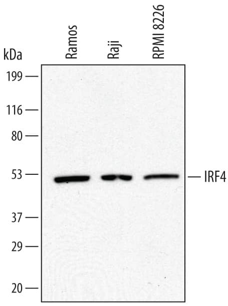
 HAF016). A specific band was detected for IRF4 at approximately 53 kDa (as indicated). This experiment was conducted under reducing conditions and using Immunoblot Buffer Group 1." alt="Western blot shows lysates of Ramos human Burkitt's lymphoma cell line, Raji human Burkitt's lymphoma cell line, and RPMI 8226 human multiple myeloma cell line. PVDF membrane was probed with 0.5 µg/mL of Sheep Anti-Human IRF4 Antigen Affinity-purified Polyclonal Antibody (Catalog # AF5525) followed by HRP-conjugated Anti-Sheep IgG Secondary Antibody (Catalog # HAF016). A specific band was detected for IRF4 at approximately 53 kDa (as indicated). This experiment was conducted under reducing conditions and using Immunoblot Buffer Group 1." class="big_thumb" />
| Formulation | Catalog # | Availability | Price | |
|---|---|---|---|---|
| Carrier Free | AF5525-SP | |||
| Formulation | Catalog # | Availability | Price | |
|---|---|---|---|---|
| BSA Free | NBP1-82814-25ul | |||
| Formulation | Catalog # | Availability | Price | |
|---|---|---|---|---|
| BSA Free | NBP1-82813-25ul | |||
| Formulation | Catalog # | Availability | Price | |
|---|---|---|---|---|
| BSA Free | NBP3-26863-25ul | |||
| Azide and BSA Free | NBP3-44146 | |||
| Formulation | Catalog # | Availability | Price | |
|---|---|---|---|---|
| BSA Free | NBP1-76574-0.025mg | |||
 HAF018). A specific band was detected for IRF4 at approximately 51 kDa (as indicated). This experiment was conducted under reducing conditions and using Western Blot Buffer Group 1." alt="Western blot shows lysates of Ramos human Burkitt's lymphoma cell line, HDLM‑2 human Hodgkin’s lymphoma cell line, U937 human histiocytic lymphoma cell line (negative control), and HUVEC human umbilical vein endothelial cells (negative control). PVDF membrane was probed with 2 µg/mL of Mouse Anti-Human IRF4 Monoclonal Antibody (Catalog # MAB55251) followed by HRP-conjugated Anti-Mouse IgG Secondary Antibody (HAF018). A specific band was detected for IRF4 at approximately 51 kDa (as indicated). This experiment was conducted under reducing conditions and using Western Blot Buffer Group 1." class="big_thumb" />
| Formulation | Catalog # | Availability | Price | |
|---|---|---|---|---|
| Carrier Free | MAB55251-SP | |||
 DR647RB) and counterstained with DAPI (blue; Lunaphore Catalog # DR100). Specific staining was localized to the nucleus. Protocol available in COMET™ Panel Builder." alt="IRF4 was detected in immersion fixed paraffin-embedded sections of human Hodgkin's Lymphoma using Rabbit Anti-Human IRF4, Monoclonal Antibody (Catalog #NBP3-20550) at 10ug/mL at 37° Celsius for 4 minutes. Before incubation with the primary antibody, tissue underwent an all-in-one dewaxing and antigen retrieval preprocessing using PreTreatment Module (PT Module) and Dewax and HIER Buffer H (pH 9; Epredia Catalog # TA-999-DHBH). Tissue was stained using the Alexa Fluor™ Plus 647 Goat anti-Rabbit IgG Secondary Antibody at 1:200 at 37 ° Celsius for 2 minutes. (Yellow; Lunaphore Catalog # DR647RB) and counterstained with DAPI (blue; Lunaphore Catalog # DR100). Specific staining was localized to the nucleus. Protocol available in COMET™ Panel Builder." class="big_thumb" />
| Formulation | Catalog # | Availability | Price | |
|---|---|---|---|---|
| BSA Free | NBP3-20718 | |||
 VC001). Before incubation with the primary antibody, tissue was subjected to heat-induced epitope retrieval using Antigen Retrieval Reagent-Basic (CTS013). Tissue was stained using DAB (brown) and counterstained with hematoxylin (blue). Specific staining was localized to cell nuclei in intestinal glands. Staining was performed using our protocol for IHC Staining with VisUCyte HRP Polymer Detection Reagents." alt="IRF4 was detected in immersion fixed paraffin-embedded sections of human intestine using Mouse Anti-Human IRF4 Monoclonal Antibody (Catalog # MAB55252) at 5 µg/mL for 1 hour at room temperature followed by incubation with the Anti-Mouse IgG VisUCyte™ HRP Polymer Antibody (VC001). Before incubation with the primary antibody, tissue was subjected to heat-induced epitope retrieval using Antigen Retrieval Reagent-Basic (CTS013). Tissue was stained using DAB (brown) and counterstained with hematoxylin (blue). Specific staining was localized to cell nuclei in intestinal glands. Staining was performed using our protocol for IHC Staining with VisUCyte HRP Polymer Detection Reagents." class="big_thumb" />
| Formulation | Catalog # | Availability | Price | |
|---|---|---|---|---|
| Carrier Free | MAB55252-SP | |||
| Formulation | Catalog # | Availability | Price | |
|---|---|---|---|---|
| BSA Free | NBP3-21820-50ul | |||


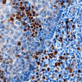 CTS013). Tissue was stained using the Anti-Sheep HRP-DAB Cell & Tissue Staining Kit (brown; Catalog #
 ![Immunohistochemistry-Paraffin: IRF4 Antibody [NBP1-82814] - Analysis in human tonsil and kidney tissues. Corresponding IRF4 RNA-seq data are presented for the same tissues.](http://images.novusbio.com/fullsize/IRF4-Antibody-Immunohistochemistry-Paraffin-NBP1-82814-img0011.jpg "Immunohistochemistry-Paraffin: IRF4 Antibody [NBP1-82814] - Analysis in human tonsil and kidney tissues. Corresponding IRF4 RNA-seq data are presented for the same tissues.")
![<b>Independent Antibodies Validation. </b>Immunohistochemistry-Paraffin: IRF4 Antibody [NBP1-82814] - Staining of human duodenum, kidney, pancreas and tonsil using Anti-IRF4 antibody NBP1-82814 (A) shows similar protein distribution across tissues to independent antibody NBP1-82813 (B).](http://images.novusbio.com/fullsize/IRF4-Antibody-Immunohistochemistry-Paraffin-NBP1-82814-img0018.jpg)
![Western Blot: IRF4 Antibody [NBP1-82814] - Analysis in human cell line HDLM-2.](http://images.novusbio.com/fullsize/IRF4-Antibody-Western-Blot-NBP1-82814-img0006.jpg)
![Immunohistochemistry-Paraffin: IRF4 Antibody [NBP1-82814] - Staining of human kidney shows no postivity in cells in tubules as expected.](http://images.novusbio.com/fullsize/IRF4-Antibody-Immunohistochemistry-Paraffin-NBP1-82814-img0014.jpg)
![Immunohistochemistry-Paraffin: IRF4 Antibody [NBP1-82814] - Staining of human tonsil shows strong nuclear positivity in non-germinal center cells.](http://images.novusbio.com/fullsize/IRF4-Antibody-Immunohistochemistry-Paraffin-NBP1-82814-img0015.jpg)
![Immunohistochemistry-Paraffin: IRF4 Antibody [NBP1-82814] - Staining of human pancreas shows no postivity in exocrine glandular cells as expected.](http://images.novusbio.com/fullsize/IRF4-Antibody-Immunohistochemistry-Paraffin-NBP1-82814-img0016.jpg)
![Immunohistochemistry-Paraffin: IRF4 Antibody [NBP1-82814] - Staining of human duodenum shows strong nuclear positivity in lymphoid cells.](http://images.novusbio.com/fullsize/IRF4-Antibody-Immunohistochemistry-Paraffin-NBP1-82814-img0017.jpg)
![Immunohistochemistry-Paraffin: IRF4 Antibody [NBP1-82813] - Analysis in human duodenum and kidney tissues. Corresponding IRF4 RNA-seq data are presented for the same tissues.](http://images.novusbio.com/fullsize/IRF4-Antibody-Immunohistochemistry-NBP1-82813-img0016.jpg "<b>Orthogonal Strategies Validation. </b>Immunohistochemistry-Paraffin: IRF4 Antibody [NBP1-82813] - Analysis in human duodenum and kidney tissues. Corresponding IRF4 RNA-seq data are presented for the same tissues.")
![<b>Independent Antibodies Validation and Orthogonal Strategies Validation.</b>Immunohistochemistry-Paraffin: IRF4 Antibody [NBP1-82813] - Staining of human duodenum, kidney, pancreas and tonsil using Anti-IRF4 antibody NBP1-82813 (A) shows similar protein distribution across tissues to independent antibody NBP1-82814 (B).](http://images.novusbio.com/fullsize/IRF4-Antibody-Immunohistochemistry-Paraffin-NBP1-82813-img0018.jpg)
![Western Blot: IRF4 Antibody [NBP1-82813] - Analysis in control (vector only transfected HEK293T lysate) and IRF4 over-expression lysate (Co-expressed with a C-terminal myc-DDK tag (3.1 kDa) in mammalian HEK293T cells).](http://images.novusbio.com/fullsize/IRF4-Antibody-Western-Blot-NBP1-82813-img0010.jpg)
![Immunohistochemistry-Paraffin: IRF4 Antibody [NBP1-82813] - Staining of human tonsil shows moderate nuclear positivity in non-germinal center cells.](http://images.novusbio.com/fullsize/IRF4-Antibody-Immunohistochemistry-Paraffin-NBP1-82813-img0022.jpg)
![Immunohistochemistry-Paraffin: IRF4 Antibody [NBP1-82813] - Staining of human duodenum shows moderate nuclear positivity in lymphoid cells.](http://images.novusbio.com/fullsize/IRF4-Antibody-Immunohistochemistry-Paraffin-NBP1-82813-img0017.jpg)
![Immunohistochemistry-Paraffin: IRF4 Antibody [NBP1-82813] - Staining of human kidney shows no positivity in cells in tubules as expected.](http://images.novusbio.com/fullsize/IRF4-Antibody-Immunohistochemistry-Paraffin-NBP1-82813-img0019.jpg)
![Immunohistochemistry-Paraffin: IRF4 Antibody [NBP1-82813] - Staining of human pancreas shows no positivity in exocrine glandular cells as expected.](http://images.novusbio.com/fullsize/IRF4-Antibody-Immunohistochemistry-Paraffin-NBP1-82813-img0021.jpg)


![Western Blot: IRF4 Antibody (2D1) [H00003662-M05] - Analysis of IRF4 expression in transfected 293T cell line by IRF4 monoclonal antibody (M05), clone 2D1. Lane 1: IRF4 transfected lysatE (51.8 KDa). Lane 2: Non-transfected lysate.](http://images.novusbio.com/fullsize/IRF4-Antibody-2D1-Western-Blot-H00003662-M05-img0006.jpg "Western Blot: IRF4 Antibody (2D1) [H00003662-M05] - Analysis of IRF4 expression in transfected 293T cell line by IRF4 monoclonal antibody (M05), clone 2D1. Lane 1: IRF4 transfected lysatE (51.8 KDa). Lane 2: Non-transfected lysate.")
![Immunocytochemistry/Immunofluorescence: IRF4 Antibody (2D1) [H00003662-M05] - Analysis of monoclonal antibody to IRF4 on HeLa cell. Antibody concentration 10 ug/ml](http://images.novusbio.com/fullsize/IRF4-Antibody-2D1-Immunocytochemistry-Immunofluorescence-H00003662-M05-img0004.jpg)
![Western Blot: IRF4 Antibody (2D1) [H00003662-M05] - Analysis of IRF4 over-expressed 293 cell line, cotransfected with IRF4 Validated Chimera RNAi or non-transfected control. Blot probed with H00003662-M05. GAPDH (36.1 kDa) used as loading control.](http://images.novusbio.com/fullsize/IRF4-Antibody-2D1-Western-Blot-H00003662-M05-img0002.jpg)
![Sandwich ELISA: IRF4 Antibody (2D1) [H00003662-M05] - Detection limit for recombinant GST tagged IRF4 is approximately 0.03ng/ml as a capture antibody.](http://images.novusbio.com/fullsize/IRF4-Antibody-2D1-Sandwich-ELISA-H00003662-M05-img0005.jpg)
![Western Blot: IRF4 Antibody [NBP1-76574] - Analysis in Ramos cell lysate with antibody at 0.5 ug/mL.](http://images.novusbio.com/fullsize/IRF4-Antibody-Western-Blot-NBP1-76574-img0005.jpg "Western Blot: IRF4 Antibody [NBP1-76574] - Analysis in Ramos cell lysate with antibody at 0.5 ug/mL.")
![Immunocytochemistry/Immunofluorescence: IRF4 Antibody [NBP1-76574] - Jurkat cells with IRF4 antibody at 10 ug/mL.](http://images.novusbio.com/fullsize/IRF4-Antibody-Immunocytochemistry-Immunofluorescence-NBP1-76574-img0002.jpg)
![Immunocytochemistry/ Immunofluorescence: IRF4 Antibody - BSA Free [NBP1-76574] - Immunofluorescence of IRF4 in Jurkat cells with IRF4 antibody at 20 ug/mL.](http://images.novusbio.com/fullsize/nbp1-76574_rabbit-polyclonal-irf4-antibody-2711202419451278.jpg)


![Western Blot: IRF4 Antibody (SR1924) [NBP3-21820] - Western blot analysis using NBP3-21820 in Daudi cell lysate.](http://images.novusbio.com/fullsize/nbp3-21820_rabbit-irf4-mab-sr1924-672023106238.jpg "Western Blot: IRF4 Antibody (SR1924) [NBP3-21820] - Western blot analysis using NBP3-21820 in Daudi cell lysate.")
![Western Blot: IRF4 Antibody (RM352) [NBP2-77422] - Western Blot of Raji cell lysate using NBP2-77422.](http://images.novusbio.com/fullsize/IRF4-Antibody-RM352-Western-Blot-NBP2-77422-img0003.jpg "Western Blot: IRF4 Antibody (RM352) [NBP2-77422] - Western Blot of Raji cell lysate using NBP2-77422.")
![Immunohistochemistry-Paraffin: IRF4 Antibody (RM352) [NBP2-77422] - Immunohistochemical staining of formalin fixed and paraffin embedded human Tonsil tissue section using NBP2-77422 at a 1:800 dilution.](http://images.novusbio.com/fullsize/IRF4-Antibody-RM352-Immunohistochemistry-Paraffin-NBP2-77422-img0005.jpg)
![Immunohistochemistry-Paraffin: IRF4 Antibody (RM352) [NBP2-77422] - Immunohistochemical staining of formalin fixed and paraffin embedded human Appendix tissue section using NBP2-77422 at a 1:800 dilution.](http://images.novusbio.com/fullsize/IRF4-Antibody-RM352-Immunohistochemistry-Paraffin-NBP2-77422-img0004.jpg)
![Immunohistochemistry: IRF4 Antibody (RM352) [NBP2-77422] - Immunohistochemical staining of formalin fixed and paraffin embedded human melanoma tissue section using NBP2-77422.](http://images.novusbio.com/fullsize/nbp2-77422_rabbit-monoclonal-irf4-antibody-rm352-immunohistochemistry-18920252343562.png)
![Western Blot: IRF4 Antibody (2F2) [H00003662-M02] - Analysis of IRF4 expression in transfected 293T cell line by IRF4 monoclonal antibody (M02), clone 2F2.Lane 1: IRF4 transfected lysate(51.8 KDa).Lane 2: Non-transfected lysate.](http://images.novusbio.com/fullsize/IRF4-Antibody-2F2-Western-Blot-H00003662-M02-img0007.jpg "Western Blot: IRF4 Antibody (2F2) [H00003662-M02] - Analysis of IRF4 expression in transfected 293T cell line by IRF4 monoclonal antibody (M02), clone 2F2.Lane 1: IRF4 transfected lysate(51.8 KDa).Lane 2: Non-transfected lysate.")
![Immunohistochemistry-Paraffin: IRF4 Antibody (2F2) [H00003662-M02] - Analysis of monoclonal antibody to IRF4 on formalin-fixed paraffin-embedded human colon. Antibody concentration 0.3 ug/ml.](http://images.novusbio.com/fullsize/IRF4-Antibody-2F2-Immunohistochemistry-Paraffin-H00003662-M02-img0005.jpg)
![Sandwich ELISA: IRF4 Antibody (2F2) [H00003662-M02] - Detection limit for recombinant GST tagged IRF4 is approximately 0.03ng/ml as a capture antibody.](http://images.novusbio.com/fullsize/IRF4-Antibody-2F2-Sandwich-ELISA-H00003662-M02-img0006.jpg)


