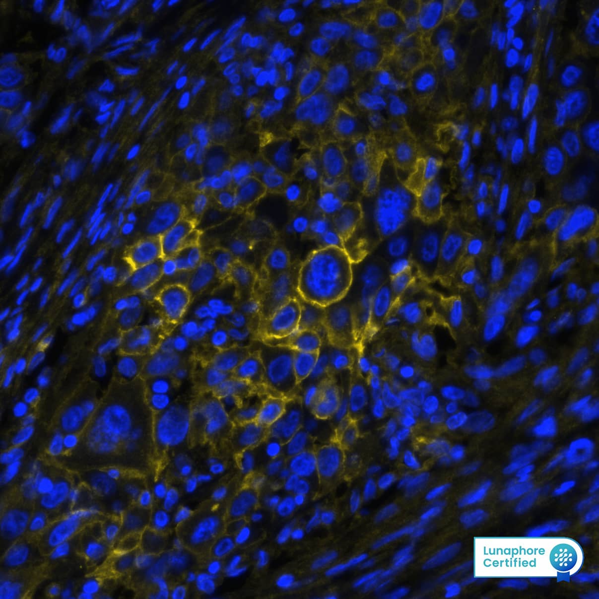
MAB11611) at 20ug/mL at 37 ° Celsius for 4 minutes. Before incubation with the primary antibody, tissue underwent an all-in-one dewaxing and antigen retrieval preprocessing using PreTreatment Module (PT Module) and Dewax and HIER Buffer H (pH 9). Tissue was stained using the Alexa Fluor™ 647 Goat anti-Mouse IgG Secondary Antibody at 1:200 at 37 ° Celsius for 2 minutes. (Yellow; Lunaphore Catalog #
DR647MS) and counterstained with DAPI (blue; Lunaphore Catalog #
DR100). Specific staining was localized to the membrane. Protocol available in
COMET™ Panel Builder." class="big_lightbox">
MAB11611) at 20ug/mL at 37 ° Celsius for 4 minutes. Before incubation with the primary antibody, tissue underwent an all-in-one dewaxing and antigen retrieval preprocessing using PreTreatment Module (PT Module) and Dewax and HIER Buffer H (pH 9). Tissue was stained using the Alexa Fluor™ 647 Goat anti-Mouse IgG Secondary Antibody at 1:200 at 37 ° Celsius for 2 minutes. (Yellow; Lunaphore Catalog #
DR647MS) and counterstained with DAPI (blue; Lunaphore Catalog #
DR100). Specific staining was localized to the membrane. Protocol available in
COMET™ Panel Builder." alt="B7-H3 was detected in immersion fixed paraffin-embedded sections of human lung cancer using Mouse Anti-Human B7-H3 Monoclonal Antibody (Catalog #
MAB11611) at 20ug/mL at 37 ° Celsius for 4 minutes. Before incubation with the primary antibody, tissue underwent an all-in-one dewaxing and antigen retrieval preprocessing using PreTreatment Module (PT Module) and Dewax and HIER Buffer H (pH 9). Tissue was stained using the Alexa Fluor™ 647 Goat anti-Mouse IgG Secondary Antibody at 1:200 at 37 ° Celsius for 2 minutes. (Yellow; Lunaphore Catalog #
DR647MS) and counterstained with DAPI (blue; Lunaphore Catalog #
DR100). Specific staining was localized to the membrane. Protocol available in
COMET™ Panel Builder." class="big_thumb" />
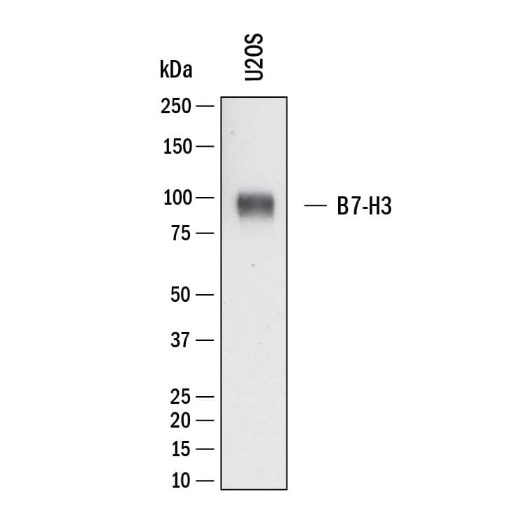
HAF018). A specific band was detected for B7‑H3 at approximately 90 kDa (as indicated). This experiment was conducted under reducing conditions and using
Western Blot Buffer Group 1." class="big_lightbox">
HAF018). A specific band was detected for B7‑H3 at approximately 90 kDa (as indicated). This experiment was conducted under reducing conditions and using
Western Blot Buffer Group 1." class="big_thumb" />
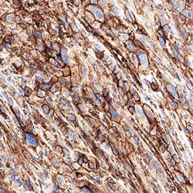
VC001). Before incubation with the primary antibody, tissue was subjected to heat-induced epitope retrieval using VisUCyte Antigen Retrieval Reagent-Basic (Catalog #
VCTS021). Tissue was stained using DAB (brown) and counterstained with hematoxylin (blue). Specific staining was localized to the cell membrane. View our protocol for
IHC Staining with VisUCyte HRP Polymer Detection Reagents." class="big_lightbox">
VC001). Before incubation with the primary antibody, tissue was subjected to heat-induced epitope retrieval using VisUCyte Antigen Retrieval Reagent-Basic (Catalog #
VCTS021). Tissue was stained using DAB (brown) and counterstained with hematoxylin (blue). Specific staining was localized to the cell membrane. View our protocol for
IHC Staining with VisUCyte HRP Polymer Detection Reagents." class="big_thumb" />
 HAF017). A specific band was detected for B7-H3 at approximately 90-110 kDa (as indicated). This experiment was conducted under reducing conditions and using Immunoblot Buffer Group 1." alt="Western blot shows lysates of LNCaP human prostate cancer cell line and U2OS human osteosarcoma cell line. PVDF membrane was probed with 1 µg/mL of Goat Anti-Human B7-H3 Antigen Affinity-purified Polyclonal Antibody (Catalog # AF1027) followed by HRP-conjugated Anti-Goat IgG Secondary Antibody (Catalog # HAF017). A specific band was detected for B7-H3 at approximately 90-110 kDa (as indicated). This experiment was conducted under reducing conditions and using Immunoblot Buffer Group 1." class="big_thumb" />
 HAF018). A specific band was detected for B7-H3 at approximately 90-110 kDa (as indicated). This experiment was conducted under reducing conditions and using Immunoblot Buffer Group 1." alt="Western blot shows lysates of LNCaP human prostate cancer cell line and U2OS human osteosarcoma cell line. PVDF membrane was probed with 2 µg/mL of Mouse Anti-Human B7-H3 Monoclonal Antibody (Catalog # MAB1027) followed by HRP-conjugated Anti-Mouse IgG Secondary Antibody (Catalog # HAF018). A specific band was detected for B7-H3 at approximately 90-110 kDa (as indicated). This experiment was conducted under reducing conditions and using Immunoblot Buffer Group 1." class="big_thumb" />
 HAF017). A specific band was detected for B7-H3 at approximately 45-55 kDa (as indicated). This experiment was conducted under reducing conditions and using Immunoblot Buffer Group 1." alt="Western blot shows lysates of MEF mouse embryonic feeder cells, P19 mouse embryonal carcinoma cell line, NIH-3T3 mouse embryonic fibroblast cell line, C2C12 mouse myoblast cell line, and 3T3-L1 mouse embryonic fibroblast adipose-like cell line. PVDF membrane was probed with 1 µg/mL of Goat Anti-Mouse B7-H3 Antigen Affinity-purified Polyclonal Antibody (Catalog # AF1397) followed by HRP-conjugated Anti-Goat IgG Secondary Antibody (Catalog # HAF017). A specific band was detected for B7-H3 at approximately 45-55 kDa (as indicated). This experiment was conducted under reducing conditions and using Immunoblot Buffer Group 1." class="big_thumb" />
 DR647RB) and counterstained with DAPI (blue; Lunaphore Catalog # DR100). Specific staining was localized to the membrane. Protocol available in COMET™ Panel Builder." alt="B7-H3 was detected in immersion fixed paraffin-embedded sections of human Lung Tumor using Rabbit Anti-Human B7-H3, Monoclonal Antibody (Catalog #MAB11721) at 1ug/mL at 37 ° Celsius for 4 minutes. Before incubation with the primary antibody, tissue underwent an all-in-one dewaxing and antigen retrieval preprocessing using PreTreatment Module (PT Module) and Dewax and HIER Buffer H (pH 9; Epredia Catalog # TA-999-DHBH). Tissue was stained using the Alexa Fluor™ Plus 647 Goat anti-Rabbit IgG Secondary Antibody at 1:200 at 37 ° Celsius for 2 minutes. (Yellow; Lunaphore Catalog # DR647RB) and counterstained with DAPI (blue; Lunaphore Catalog # DR100). Specific staining was localized to the membrane. Protocol available in COMET™ Panel Builder." class="big_thumb" />
 MAB11611) at 20ug/mL at 37 ° Celsius for 4 minutes. Before incubation with the primary antibody, tissue underwent an all-in-one dewaxing and antigen retrieval preprocessing using PreTreatment Module (PT Module) and Dewax and HIER Buffer H (pH 9). Tissue was stained using the Alexa Fluor™ 647 Goat anti-Mouse IgG Secondary Antibody at 1:200 at 37 ° Celsius for 2 minutes. (Yellow; Lunaphore Catalog # DR647MS) and counterstained with DAPI (blue; Lunaphore Catalog # DR100). Specific staining was localized to the membrane. Protocol available in COMET™ Panel Builder." alt="B7-H3 was detected in immersion fixed paraffin-embedded sections of human lung cancer using Mouse Anti-Human B7-H3 Monoclonal Antibody (Catalog # MAB11611) at 20ug/mL at 37 ° Celsius for 4 minutes. Before incubation with the primary antibody, tissue underwent an all-in-one dewaxing and antigen retrieval preprocessing using PreTreatment Module (PT Module) and Dewax and HIER Buffer H (pH 9). Tissue was stained using the Alexa Fluor™ 647 Goat anti-Mouse IgG Secondary Antibody at 1:200 at 37 ° Celsius for 2 minutes. (Yellow; Lunaphore Catalog # DR647MS) and counterstained with DAPI (blue; Lunaphore Catalog # DR100). Specific staining was localized to the membrane. Protocol available in COMET™ Panel Builder." class="big_thumb" />
 NL004) and counterstained with DAPI (blue). Specific staining was localized to plasma membrane. Staining was performed using our protocol for Fluorescent ICC Staining of Non-adherent Cells." alt="B7‑H3 was detected in immersion fixed NIH‑3T3 mouse embryonic fibroblast cell line (positive staining) and MOLT‑4 human acute lymphoblastic leukemia cell line (negative staining) using Rabbit Anti-Mouse B7‑H3 Monoclonal Antibody (Catalog # MAB10271) at 8 µg/mL for 3 hours at room temperature. Cells were stained using the NorthernLights™ 557-conjugated Anti-Rabbit IgG Secondary Antibody (red; NL004) and counterstained with DAPI (blue). Specific staining was localized to plasma membrane. Staining was performed using our protocol for Fluorescent ICC Staining of Non-adherent Cells." class="big_thumb" />


 AB-108-C, open histogram), followed by Phycoerythrin-conjugated Anti-Goat IgG Secondary Antibody (Catalog #
  CTS008) and counterstained with hematoxylin (blue). Lower panel shows a lack of labeling if primary antibodies are omitted and tissue is stained only with secondary antibody followed by incubation with detection reagents. View our protocol for
  HAF109). This experiment was conducted under reducing conditions and using the 12-230 kDa separation system." class="big_thumb" />
 HAF017). A specific band was detected for B7-H3 at approximately 95 kDa (as indicated) in the parental U2OS cell line, but is not detectable in knockout U2OS cell line. GAPDH (Catalog #


 MAB002, open histogram), followed by Phycoerythrin-conjugated Anti-Mouse IgG F(ab')2Secondary Antibody (Catalog #
  MAB002, open histogram), followed by Allophycocyanin-conjugated Anti-Mouse IgG Secondary Antibody (Catalog #
  HAF018). A specific band was detected for B7-H3 at approximately 95 kDa (as indicated) in the parental U2OS cell line, but is not detectable in knockout U2OS cell line. GAPDH (Catalog #
  MAB484) in a dose-dependent manner (orange line), as measured by the Resazurin (Catalog #


![Western Blot: B7-H3/CD276 Antibody [NBP1-88966] - Western blot shows lysates of U2OS human bone osteosarcoma parental cell line and B7-H3 knockout (KO) U2OS cell line. PVDF membrane was probed with 0.5 ug/mL of Rabbit Anti-Human B7-H3 Polyclonal Antibody (Catalog # NBP1-88966) followed by HRP-conjugated Anti-Rabbit IgG Secondary Antibody (Catalog #HAF008). Specific band was detected for B7-H3 at approximately 95 kDa (as indicated) in the parental U2OS cell line, but is not detectable in the knockout U2OS cell line. This experiment was conducted under reducing conditions.](http://images.novusbio.com/fullsize/B7-H3-CD276-Antibody-Knockout-Validated-NBP1-88966-img0016.jpg "<b>Genetic Strategies Validation. </b>Western Blot: B7-H3/CD276 Antibody [NBP1-88966] - Western blot shows lysates of U2OS human bone osteosarcoma parental cell line and B7-H3 knockout (KO) U2OS cell line. PVDF membrane was probed with 0.5 ug/mL of Rabbit Anti-Human B7-H3 Polyclonal Antibody (Catalog # NBP1-88966) followed by HRP-conjugated Anti-Rabbit IgG Secondary Antibody (Catalog #HAF008). Specific band was detected for B7-H3 at approximately 95 kDa (as indicated) in the parental U2OS cell line, but is not detectable in the knockout U2OS cell line. This experiment was conducted under reducing conditions.")
![<b>Genetic Strategies Validation. </b>Immunocytochemistry/Immunofluorescence: B7-H3/CD276 Antibody [NBP1-88966] - Staining of human cell line A-431 shows localization to vesicles. Antibody staining is shown in green.](http://images.novusbio.com/fullsize/B7-H3-CD276-Antibody-Immunocytochemistry-Immunofluorescence-NBP1-88966-img0006.jpg)
![Western Blot: B7-H3/CD276 Antibody [NBP1-88966] - Analysis in control (vector only transfected HEK293T lysate) and CD276 over-expression lysate (Co-expressed with a C-terminal myc-DDK tag (3.1 kDa) in mammalian HEK293T cells).](http://images.novusbio.com/fullsize/B7-H3-CD276-Antibody-Western-Blot-NBP1-88966-img0017.jpg)
![Immunohistochemistry-Paraffin: B7-H3/CD276 Antibody [NBP1-88966] - Staining of human placenta shows high expression.](http://images.novusbio.com/fullsize/B7-H3-CD276-Antibody-Immunohistochemistry-Paraffin-NBP1-88966-img0014.jpg)
![Immunohistochemistry-Paraffin: B7-H3/CD276 Antibody [NBP1-88966] - Staining of human endometrium shows strong membranous positivity in glandular cells.](http://images.novusbio.com/fullsize/antibody/nbp1-88966_rabbit-polyclonal-b7-h3-cd276-antibody-immunohistochemistry-paraffin-122202411124..jpg)
![Immunohistochemistry-Paraffin: B7-H3/CD276 Antibody [NBP1-88966] -Staining of human rectum shows strong cytoplasmic positivity in glandular cells.](http://images.novusbio.com/fullsize/antibody/nbp1-88966_rabbit-polyclonal-b7-h3-cd276-antibody-immunohistochemistry-paraffin-122202411220..jpg)
![Immunohistochemistry-Paraffin: B7-H3/CD276 Antibody [NBP1-88966] -Staining of human cerebellum shows negative cytoplasmic positivity in Purkinje cells as expected.](http://images.novusbio.com/fullsize/antibody/nbp1-88966_rabbit-polyclonal-b7-h3-cd276-antibody-immunohistochemistry-paraffin-122202411310..jpg)
![Western Blot: B7-H3/CD276 Antibody [NBP3-14669] - Samples: Whole cell lysate (50 ug) from TCMK-1, NIH 3T3,AlphaTC1 Clone 9, 3T3-L1, and CT26 cells prepared usingNETN lysis buffer. Antibody: Rabbit anti-B7-H3/CD276recombinant monoclonal antibody used at 1:1000. Secondary: HRP-conjugated goatanti-rabbit IgG. Chemiluminescence with anexposure time of 3 minutes.](http://images.novusbio.com/fullsize/B7-H3-CD276-Antibody-Western-Blot-NBP3-14669-img0002.jpg "Western Blot: B7-H3/CD276 Antibody [NBP3-14669] - Samples: Whole cell lysate (50 ug) from TCMK-1, NIH 3T3,AlphaTC1 Clone 9, 3T3-L1, and CT26 cells prepared usingNETN lysis buffer. Antibody: Rabbit anti-B7-H3/CD276recombinant monoclonal antibody used at 1:1000. Secondary: HRP-conjugated goatanti-rabbit IgG. Chemiluminescence with anexposure time of 3 minutes.")
![Immunocytochemistry/Immunofluorescence: B7-H3/CD276 Antibody (BLR025F) [NBP3-14669] - Section of A-172 cells. Antibody: Rabbit anti-B7-H3/CD276 recombinant monoclonal used at 1:200. Secondary: HRP-conjugated goat anti-rabbit IgG . Substrate: DAB.](http://images.novusbio.com/fullsize/B7-H3-CD276-Antibody-Immunohistochemistry-NBP3-14669-img0005.jpg)
![Immunohistochemistry: B7-H3/CD276 Antibody [NBP3-14669] - Sample: FFPE section of humanlung carcinoma. Antibody: Rabbit anti-B7-H3/CD276recombinant monoclonal antibody used at 1:250. Secondary: HRP-conjugated goat antirabbit IgG. Substrate: Opal™. Counterstain:DAPI (blue).](http://images.novusbio.com/fullsize/B7-H3-CD276-Antibody-Immunohistochemistry-NBP3-14669-img0006.jpg)
![Western Blot: B7-H3/CD276 Antibody [NBP3-14669] - Rabbit anti CD276 recombinant monoclonal antibody (NBP3-14669 lot 1) used at 1:1000. HRP-conjugated goat anti-rabbit IgG (NB7156). Chemiluminescence with an exposure time of 3 minutes. Lower Panel: Rabbit anti-GAPDH (NB300-322).](http://images.novusbio.com/fullsize/B7-H3-CD276-Antibody-Western-Blot-NBP3-14669-img0001.jpg)
![Immunohistochemistry-Paraffin: B7-H3/CD276 Antibody (BLR025F) [NBP3-14669] - Section of ovarian carcinoma. Antibody: Rabbit anti-B7-H3/CD276 recombinant monoclonal antibody used at 1:200. Secondary: HRP-conjugated goat antirabbit IgG (A120-501P). Substrate: DAB.](http://images.novusbio.com/fullsize/B7-H3-CD276-Antibody-Immunohistochemistry-NBP3-14669-img0004.jpg)
![Immunoprecipitation: B7-H3/CD276 Antibody [NBP3-14669] - Samples: Whole cell lysate (1.0 mgper IP reaction; 20% of IP loaded) from HeLa cells preparedusing NETN lysis buffer. Antibodies: Rabbit anti-B7-H3/CD276 recombinant monoclonal antibodyused for IP at 3 ul per reaction. B7-H3/CD276 was also immunoprecipitated by rabbit anti-B7-H3/CD276 recombinant monoclonal antibody. For blotting immunoprecipitated B7-H3/CD27 was used at 1:1000.Chemiluminescence with an exposure time of 30 seconds.](http://images.novusbio.com/fullsize/B7-H3-CD276-Antibody-Immunoprecipitation-NBP3-14669-img0003.jpg)
![B7-H3/CD276 Antibody (BLR025F) [NBP3-14669] - Detection of human B7-H3/CD276 (shaded) in 293T cells by flow cytometry. Antibody: Rabbit antiB7-H3/CD276 recombinant monoclonal [BLR025F]or isotype control (unshaded).Secondary: DyLight® 650-conjugated goat anti-rabbit IgG.](http://images.novusbio.com/fullsize/B7-H3-CD276-Antibody-BLR025F-NBP3-14669-img0010.jpg)
![Western Blot: B7-H3/CD276 Antibody [NBP1-88965] - Western blot shows lysates of U2OS human bone osteosarcoma parental cell line and B7-H3 knockout (KO) U2OS cell line. PVDF membrane was probed with 0.5 ug/mL of Rabbit Anti-Human B7-H3 Polyclonal Antibody (Catalog # NBP1-88965) followed by HRP-conjugated Anti-Rabbit IgG Secondary Antibody (Catalog #HAF008). Specific band was detected for B7-H3 at approximately 95 kDa (as indicated) in the parental U2OS cell line, but is not detectable in the knockout U2OS cell line. This experiment was conducted under reducing conditions.](http://images.novusbio.com/fullsize/B7-H3-CD276-Antibody-Knockout-Validated-NBP1-88965-img0014.jpg "<b>Genetic Strategies Validation. </b>Western Blot: B7-H3/CD276 Antibody [NBP1-88965] - Western blot shows lysates of U2OS human bone osteosarcoma parental cell line and B7-H3 knockout (KO) U2OS cell line. PVDF membrane was probed with 0.5 ug/mL of Rabbit Anti-Human B7-H3 Polyclonal Antibody (Catalog # NBP1-88965) followed by HRP-conjugated Anti-Rabbit IgG Secondary Antibody (Catalog #HAF008). Specific band was detected for B7-H3 at approximately 95 kDa (as indicated) in the parental U2OS cell line, but is not detectable in the knockout U2OS cell line. This experiment was conducted under reducing conditions.")
![<b>Genetic Strategies Validation and Orthogonal Strategies Validation.</b>Immunohistochemistry-Paraffin: B7-H3/CD276 Antibody [NBP1-88965] - Staining in human prostate and pancreas tissues using anti-CD276 antibody. Corresponding CD276 RNA-seq data are presented for the same tissues.](http://images.novusbio.com/fullsize/B7-H3-CD276-Antibody-Immunohistochemistry-Paraffin-NBP1-88965-img0013.jpg)
![Western Blot: B7-H3/CD276 Antibody [NBP1-88965] - Analysis in control (vector only transfected HEK293T lysate) and CD276 over-expression lysate (Co-expressed with a C-terminal myc-DDK tag (3.1 kDa) in mammalian HEK293T cells).](http://images.novusbio.com/fullsize/B7-H3-CD276-Antibody-Western-Blot-NBP1-88965-img0015.jpg)
![Western Blot: B7-H3/CD276 Antibody [NBP1-88965] - Analysis in human cell line HEK 293.](http://images.novusbio.com/fullsize/B7-H3-CD276-Antibody-Western-Blot-NBP1-88965-img0005.jpg)
![Immunohistochemistry-Paraffin: B7-H3/CD276 Antibody [NBP1-88965] - Staining of human prostate shows high expression.](http://images.novusbio.com/fullsize/B7-H3-CD276-Antibody-Immunohistochemistry-Paraffin-NBP1-88965-img0001.jpg)
![Immunohistochemistry-Paraffin: B7-H3/CD276 Antibody [NBP1-88965] - Staining of human pancreas shows low expression as expected.](http://images.novusbio.com/fullsize/B7-H3-CD276-Antibody-Immunohistochemistry-Paraffin-NBP1-88965-img0012.jpg)
 VC003) or the HRP-conjugated Anti-Rabbit IgG Secondary Antibody (Catalog #
  VC003) or the HRP-conjugated Anti-Rabbit IgG Secondary Antibody (Catalog #
  HAF008). A specific band was detected for B7‑H3 at approximately 90-110 kDa (as indicated). This experiment was conducted under reducing conditions and using
  042-206). This experiment was conducted under reducing conditions and using the 12-230kDa separation system. Right: Simple Western electropherogram showing the same Rabbit Anti-Human B7‑H3 Monoclonal Antibody (Catalog # MAB11721) tested at 2 µg/ml (blue line) and 10 µg/ml (green line) in the LNCaP human prostate cancer cell line." class="big_thumb" />

 HAF018). A specific band was detected for B7‑H3 at approximately 90 kDa (as indicated). This experiment was conducted under reducing conditions and using
  VC001). Before incubation with the primary antibody, tissue was subjected to heat-induced epitope retrieval using VisUCyte Antigen Retrieval Reagent-Basic (Catalog #
 MAB1050, open histogram), followed by Phycoerythrin-conjugated Anti-Rabbit IgG F(ab')2Secondary Antibody (
![Western Blot: B7-H3/CD276 Antibody (6A1) [NBP2-37657] - Western blot analysis using CD276 mAb against HEK293 (1) and CD276(AA: 30-130)-hIgGFc transfected HEK293 (2) cell lysate.](http://images.novusbio.com/fullsize/B7-H3-CD276-Antibody-6A1-Western-Blot-NBP2-37657-img0001.jpg "Western Blot: B7-H3/CD276 Antibody (6A1) [NBP2-37657] - Western blot analysis using CD276 mAb against HEK293 (1) and CD276(AA: 30-130)-hIgGFc transfected HEK293 (2) cell lysate.")
![Immunohistochemistry-Paraffin: B7-H3/CD276 Antibody (6A1) [NBP2-37657] - Analysis of cervical cancer tissues (left) and ovarian cancer tissues (right) using CD276 mouse mAb with DAB staining.](http://images.novusbio.com/fullsize/B7-H3-CD276-Antibody-6A1-Immunohistochemistry-NBP2-37657-img0003.jpg)
![Flow Cytometry: B7-H3/CD276 Antibody (6A1) [NBP2-37657] - Analysis of PC-3 cells using CD276 mouse mAb (green) and negative control (purple).](http://images.novusbio.com/fullsize/B7-H3-CD276-Antibody-6A1-Flow-Cytometry-NBP2-37657-img0002.jpg)
![Immunohistochemistry: B7-H3/CD276 Antibody (PD00-36) [NBP3-32138] - Immunohistochemical analysis of paraffin-embedded human lung squamous cell carcinoma tissue with Rabbit anti-B7-H3/CD276 antibody (NBP3-32138) at 1/5,000 dilution.<br/><br/> The section was pre-treated using heat mediated antigen retrieval with Tris-EDTA buffer (pH 9.0) for 20 minutes. The tissues were blocked in 1% BSA for 20 minutes at room temperature, washed with ddH2O and PBS, and then probed with the primary antibody (NBP3-32138) at 1/5,000 dilution for 1 hour at room temperature. The detection was performed using an HRP conjugated compact polymer system. DAB was used as the chromogen. Tissues were counterstained with hematoxylin and mounted with DPX.](http://images.novusbio.com/fullsize/nbp3-32138_rabbit-b7-h3-cd276-mab-pd00-36-2982024143520.jpg "Immunohistochemistry: B7-H3/CD276 Antibody (PD00-36) [NBP3-32138] - Immunohistochemical analysis of paraffin-embedded human lung squamous cell carcinoma tissue with Rabbit anti-B7-H3/CD276 antibody (NBP3-32138) at 1/5,000 dilution.<br/><br/> The section was pre-treated using heat mediated antigen retrieval with Tris-EDTA buffer (pH 9.0) for 20 minutes. The tissues were blocked in 1% BSA for 20 minutes at room temperature, washed with ddH2O and PBS, and then probed with the primary antibody (NBP3-32138) at 1/5,000 dilution for 1 hour at room temperature. The detection was performed using an HRP conjugated compact polymer system. DAB was used as the chromogen. Tissues were counterstained with hematoxylin and mounted with DPX.")
![Flow Cytometry: B7-H3/CD276 Antibody (PD00-36) [NBP3-32138] - Flow cytometric analysis of HEK-293 cells labeling B7-H3/CD276.<br/><br/> Cells were fixed and permeabilized. Then stained with the primary antibody (NBP3-32138, 1ug/ml) (red) compared with Rabbit IgG Isotype Control (green). After incubation of the primary antibody at +4℃ for an hour, the cells were stained with a iFluor™ 488 conjugate-Goat anti-Rabbit IgG Secondary antibody at 1/1,000 dilution for 30 minutes at +4℃. Unlabelled sample was used as a control (cells without incubation with primary antibody; black).](http://images.novusbio.com/fullsize/nbp3-32138_rabbit-b7-h3-cd276-mab-pd00-36-29202416453234.jpg)
![Western blot analysis on different lysates with NBP3-32138 at 1/1,000 dilution. Lane 1: HeLa cell lysate (20 µg/Lane) Lane 2: MCF7 cell lysate (20 µg/Lane) Lane 3: HEK-293 cell lysate (20 µg/Lane) Lane 4: LoVo cell lysate (20 µg/Lane) Lane 5: U-2 OS cell lysate (20 µg/Lane) Lane 6: LNCaP cell lysate (20 µg/Lane) Lane 7: SH-SY5Y cell lysate (20 µg/Lane) Lane 8: THP-1 cell lysate (20 µg/Lane) Lane 9: HCT 116 cell lysate (20 µg/Lane) Lane 10: Raji cell lysate (negative) (20 µg/Lane) Predicted band size: 57 kDa Observed band size: 100 kDa Exposure time: 6 seconds; 4-20% SDS-PAGE gel. Proteins were transferred to a PVDF membrane and blocked with 5% NFDM/TBST for 1 hour at room temperature. NBP3-32138 at 1/2,000 dilution was used in 5% NFDM/TBST at 4℃ overnight. Goat Anti-Rabbit IgG [HRP] Secondary Antibody at 1/50,000 dilution was used for 1 hour at room temperature.](http://images.novusbio.com/fullsize/nbp3-32138_-western-blot-639204594552346295.jpg)


