239-A) stimulates proliferation in the Balb/3T3 mouse embryonic fibroblast cell line in a dose-dependent manner (orange line). Proliferation elicited by Recombinant Human TGF-a (3 ng/mL) is neutralized (green line) by increasing concentrations of Goat Anti-Human TGF-a Antigen Affinity-purified Polyclonal Antibody (Catalog # AF-239-NA). The ND
50 is typically 0.15-0.75 µg/mL." class="big_lightbox">

239-A) stimulates proliferation in the Balb/3T3 mouse embryonic fibroblast cell line in a dose-dependent manner (orange line). Proliferation elicited by Recombinant Human TGF-a (3 ng/mL) is neutralized (green line) by increasing concentrations of Goat Anti-Human TGF-a Antigen Affinity-purified Polyclonal Antibody (Catalog # AF-239-NA). The ND
50 is typically 0.15-0.75 µg/mL." alt="Recombinant Human TGF-a (Catalog #
239-A) stimulates proliferation in the Balb/3T3 mouse embryonic fibroblast cell line in a dose-dependent manner (orange line). Proliferation elicited by Recombinant Human TGF-a (3 ng/mL) is neutralized (green line) by increasing concentrations of Goat Anti-Human TGF-a Antigen Affinity-purified Polyclonal Antibody (Catalog # AF-239-NA). The ND
50 is typically 0.15-0.75 µg/mL." class="big_thumb" />
CTS008) and counterstained with hematoxylin (blue). Lower panel shows a lack of labeling if primary antibodies are omitted and tissue is stained only with secondary antibody followed by incubation with detection reagents. View our protocol for
Chromogenic IHC Staining of Paraffin-embedded Tissue Sections." class="big_lightbox">
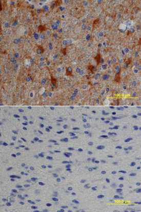
CTS008) and counterstained with hematoxylin (blue). Lower panel shows a lack of labeling if primary antibodies are omitted and tissue is stained only with secondary antibody followed by incubation with detection reagents. View our protocol for
Chromogenic IHC Staining of Paraffin-embedded Tissue Sections." class="big_thumb" />

239-A) was serially diluted and captured by Goat Anti-Human TGF‑ alpha Antigen Affinity-purified Polyclonal Antibody (Catalog # AF-239-NA) coated on a Clear Polystyrene Microplate (Catalog #
DY990). Goat Anti-Human TGF‑ alpha Antigen Affinity-purified Polyclonal Antibody (Catalog #
AF-239-NA) was biotinylated and incubated with the protein captured on the plate. Detection of the standard curve was achieved by incubating Streptavidin-HRP (Catalog #
DY998)" class="big_lightbox">

239-A) was serially diluted and captured by Goat Anti-Human TGF‑ alpha Antigen Affinity-purified Polyclonal Antibody (Catalog # AF-239-NA) coated on a Clear Polystyrene Microplate (Catalog #
DY990). Goat Anti-Human TGF‑ alpha Antigen Affinity-purified Polyclonal Antibody (Catalog #
AF-239-NA) was biotinylated and incubated with the protein captured on the plate. Detection of the standard curve was achieved by incubating Streptavidin-HRP (Catalog #
DY998)" class="big_thumb" />
239-A) was serially diluted and captured by Goat Anti-Human TGF‑ alpha Antigen Affinity-purified Polyclonal Antibody (Catalog #
AF-239-NA) coated on a Clear Polystyrene Microplate (Catalog #
DY990). Goat Anti-Human TGF‑ alpha Antigen Affinity-purified Polyclonal Antibody (Catalog # AF-239-NA) was biotinylated and incubated with the protein captured on the plate. Detection of the standard curve was achieved by incubating Streptavidin-HRP (Catalog #
DY998)" class="big_lightbox">

239-A) was serially diluted and captured by Goat Anti-Human TGF‑ alpha Antigen Affinity-purified Polyclonal Antibody (Catalog #
AF-239-NA) coated on a Clear Polystyrene Microplate (Catalog #
DY990). Goat Anti-Human TGF‑ alpha Antigen Affinity-purified Polyclonal Antibody (Catalog # AF-239-NA) was biotinylated and incubated with the protein captured on the plate. Detection of the standard curve was achieved by incubating Streptavidin-HRP (Catalog #
DY998)" class="big_thumb" />
 239-A) stimulates proliferation in the Balb/3T3 mouse embryonic fibroblast cell line in a dose-dependent manner (orange line). Proliferation elicited by Recombinant Human TGF-a (3 ng/mL) is neutralized (green line) by increasing concentrations of Goat Anti-Human TGF-a Antigen Affinity-purified Polyclonal Antibody (Catalog # AF-239-NA). The ND50 is typically 0.15-0.75 µg/mL." alt="Recombinant Human TGF-a (Catalog # 239-A) stimulates proliferation in the Balb/3T3 mouse embryonic fibroblast cell line in a dose-dependent manner (orange line). Proliferation elicited by Recombinant Human TGF-a (3 ng/mL) is neutralized (green line) by increasing concentrations of Goat Anti-Human TGF-a Antigen Affinity-purified Polyclonal Antibody (Catalog # AF-239-NA). The ND50 is typically 0.15-0.75 µg/mL." class="big_thumb" />
 239-A) stimulates proliferation in the Balb/3T3 mouse embryonic fibroblast cell line in a dose-dependent manner (orange line). Proliferation elicited by Recombinant Human TGF-a (3 ng/mL) is neutralized (green line) by increasing concentrations of Goat Anti-Human TGF-a Polyclonal Antibody (Catalog # AB-239-NA). The ND50 is typically 0.4-0.8 µg/mL." alt="Recombinant Human TGF-a (Catalog # 239-A) stimulates proliferation in the Balb/3T3 mouse embryonic fibroblast cell line in a dose-dependent manner (orange line). Proliferation elicited by Recombinant Human TGF-a (3 ng/mL) is neutralized (green line) by increasing concentrations of Goat Anti-Human TGF-a Polyclonal Antibody (Catalog # AB-239-NA). The ND50 is typically 0.4-0.8 µg/mL." class="big_thumb" />


![N/A TGF-alpha [HRP]](http://images.novusbio.com/fullsize/elisa/DATA_TGFalpha_DTGA00_ELISA_843.jpg "N/A TGF-alpha [HRP]")
![N/A TGF-alpha [HRP]](http://images.novusbio.com/fullsize/elisa/TGF-alpha_DTGA00_ELISA_192.jpg)
 CTS013). Tissue was stained with the Anti-Goat HRP-DAB Cell & Tissue Staining Kit (brown; Catalog #
  CTS008) and counterstained with hematoxylin (blue). Lower panel shows a lack of labeling if primary antibodies are omitted and tissue is stained only with secondary antibody followed by incubation with detection reagents. View our protocol for
 239-A) was serially diluted and captured by Goat Anti-Human TGF‑ alpha Antigen Affinity-purified Polyclonal Antibody (Catalog # AF-239-NA) coated on a Clear Polystyrene Microplate (Catalog #
  239-A) was serially diluted and captured by Goat Anti-Human TGF‑ alpha Antigen Affinity-purified Polyclonal Antibody (Catalog #
 ![N/A TGF-alpha [Biotin]](http://images.novusbio.com/fullsize/elisa/DATA_TGFalpha_DY239_ELISA_1820.jpg "N/A TGF-alpha [Biotin]")
![Western Blot: TGF-alpha Antibody (SPM542) [NBP2-34371] - analysis of lysate from HEK293 cells (30ug) using anti-TGF alpha antibody. Image from verified customer review.](http://images.novusbio.com/fullsize/TGF-alpha-Antibody-SPM542-Western-Blot-NBP2-34371-img0001.jpg "Western Blot: TGF-alpha Antibody (SPM542) [NBP2-34371] - analysis of lysate from HEK293 cells (30ug) using anti-TGF alpha antibody. Image from verified customer review.")
![Immunohistochemistry-Paraffin: TGF-alpha Antibody (SPM542) [NBP2-34371] - Formalin-fixed, paraffin-embedded human testicular carcinoma stained with TGF alpha Monoclonal Antibody (SPM542)](http://images.novusbio.com/fullsize/TGF-alpha-Antibody-SPM542-Immunohistochemistry-Paraffin-NBP2-34371-img0002.jpg)
![Immunohistochemistry-Paraffin: TGF-alpha Antibody (MF9) [NBP2-34296] - Formalin-fixed, paraffin-embedded human Testicular Carcinoma Stained with TGF alpha Monoclonal Antibody (MF9).](http://images.novusbio.com/fullsize/TGF-alpha-Antibody-MF9-Immunohistochemistry-Paraffin-NBP2-34296-img0001.jpg "Immunohistochemistry-Paraffin: TGF-alpha Antibody (MF9) [NBP2-34296] - Formalin-fixed, paraffin-embedded human Testicular Carcinoma Stained with TGF alpha Monoclonal Antibody (MF9).")
![Immunohistochemistry-Paraffin: TGF-alpha Antibody (TG86) [NBP2-34298] - Formalin-fixed, paraffin-embedded human Testicular Carcinoma stained with TGF-alpha Antibody (TG86)](http://images.novusbio.com/fullsize/TGF-alpha-Antibody-TG86-Immunohistochemistry-Paraffin-NBP2-34298-img0005.jpg "Immunohistochemistry-Paraffin: TGF-alpha Antibody (TG86) [NBP2-34298] - Formalin-fixed, paraffin-embedded human Testicular Carcinoma stained with TGF-alpha Antibody (TG86)")
![Immunohistochemistry-Paraffin: TGF-alpha Antibody (SPM357) [NBP3-07354] - Formalin-fixed, paraffin-embedded human Testicular Carcinoma stained with TGF-alpha Mouse Monoclonal Antibody (SPM357)](http://images.novusbio.com/fullsize/TGF-alpha-Antibody-SPM357-Immunohistochemistry-Paraffin-NBP3-07354-img0001.jpg "Immunohistochemistry-Paraffin: TGF-alpha Antibody (SPM357) [NBP3-07354] - Formalin-fixed, paraffin-embedded human Testicular Carcinoma stained with TGF-alpha Mouse Monoclonal Antibody (SPM357)")
![Immunohistochemistry-Paraffin: TGF-alpha Antibody (TG86 + P/T1) [NBP2-34300] - Formalin-fixed, paraffin-embedded human testicular carcinoma stained with TGF alpha monoclonal antibody (TG86 + P/t1)](http://images.novusbio.com/fullsize/TGF-alpha-Antibody-TG86-+-P-T1-Immunohistochemistry-Paraffin-NBP2-34300-img0004.jpg "Immunohistochemistry-Paraffin: TGF-alpha Antibody (TG86 + P/T1) [NBP2-34300] - Formalin-fixed, paraffin-embedded human testicular carcinoma stained with TGF alpha monoclonal antibody (TG86 + P/t1)")
![Immunohistochemistry-Paraffin: TGF-alpha Antibody (1E8-G6) [NBP2-34297] - Formalin-fixed, paraffin-embedded human testicular carcinoma stained with TGF alpha Monoclonal Antibody (1E8-G6)](http://images.novusbio.com/fullsize/TGF-alpha-Antibody-1E8-G6-Immunohistochemistry-Paraffin-NBP2-34297-img0004.jpg "Immunohistochemistry-Paraffin: TGF-alpha Antibody (1E8-G6) [NBP2-34297] - Formalin-fixed, paraffin-embedded human testicular carcinoma stained with TGF alpha Monoclonal Antibody (1E8-G6)")
![Immunohistochemistry-Paraffin: TGF-alpha Antibody (P/T1) [NBP2-34299] - Formalin-fixed, paraffin-embedded human testicular carcinoma stained with TGF alpha Monoclonal antibody. (P/T1)](http://images.novusbio.com/fullsize/TGF-alpha-Antibody-P-T1-Immunohistochemistry-Paraffin-NBP2-34299-img0004.jpg "Immunohistochemistry-Paraffin: TGF-alpha Antibody (P/T1) [NBP2-34299] - Formalin-fixed, paraffin-embedded human testicular carcinoma stained with TGF alpha Monoclonal antibody. (P/T1)")


