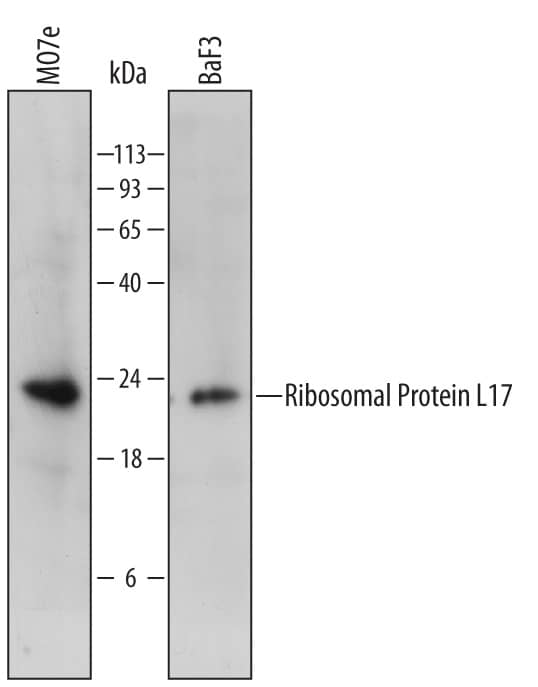
 HAF007). A specific band was detected for Ribosomal Protein L17 at approximately 22 kDa (as indicated). This experiment was conducted under reducing conditions and using Immunoblot Buffer Group 1." alt="Western blot shows lysates of MO7e human megakaryocytic leukemic cell line and BaF3 mouse pro-B cell line. PVDF membrane was probed with 1 µg/mL of Mouse Anti-Human Ribosomal Protein L17 Monoclonal Antibody (Catalog # MAB7158) followed by HRP-conjugated Anti-Mouse IgG Secondary Antibody (Catalog # HAF007). A specific band was detected for Ribosomal Protein L17 at approximately 22 kDa (as indicated). This experiment was conducted under reducing conditions and using Immunoblot Buffer Group 1." class="big_thumb" />
| Formulation | Catalog # | Availability | Price | |
|---|---|---|---|---|
| Carrier Free | MAB7158-SP | |||
| Formulation | Catalog # | Availability | Price | |
|---|---|---|---|---|
| BSA Free | NBP2-48798-25ul | |||


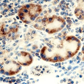 CTS013). Tissue was stained using the Anti-Mouse HRP-DAB Cell & Tissue Staining Kit (brown; Catalog #
 ![Western Blot: Ribosomal Protein L17 Antibody [NBP2-48798] - Lane 1: Marker [kDa] 250, 130, 95, 72, 55, 36, 28, 17, 10<br/>Lane 2: Human cell line RT-4<br/>Lane 3: Human cell line U-251MG sp](http://images.novusbio.com/fullsize/Ribosomal-Protein-L17-Antibody-Western-Blot-NBP2-48798-img0002.jpg "Western Blot: Ribosomal Protein L17 Antibody [NBP2-48798] - Lane 1: Marker [kDa] 250, 130, 95, 72, 55, 36, 28, 17, 10<br/>Lane 2: Human cell line RT-4<br/>Lane 3: Human cell line U-251MG sp")
![Immunocytochemistry/Immunofluorescence: Ribosomal Protein L17 Antibody [NBP2-48798] - Staining of human cell line A-431 shows localization to cytosol & endoplasmic reticulum.](http://images.novusbio.com/fullsize/Ribosomal-Protein-L17-Antibody-Immunocytochemistry-Immunofluorescence-NBP2-48798-img0003.jpg)
![Immunohistochemistry: Ribosomal Protein L17 Antibody [NBP2-48798] - Staining of human pancreas shows strong cytoplasmic positivity in exocrine glandular cells.](http://images.novusbio.com/fullsize/Ribosomal-Protein-L17-Antibody-Immunohistochemistry-NBP2-48798-img0001.jpg)
![Western Blot: Ribosomal Protein L17 Antibody [NBP2-48845] - Lane 1: Marker [kDa] 250, 130, 95, 72, 55, 36, 28, 17, 10<br/>Lane 2: Human cell line RT-4<br/>Lane 3: Human cell line U-251MG sp](http://images.novusbio.com/fullsize/Ribosomal-Protein-L17-Antibody-Western-Blot-NBP2-48845-img0002.jpg "Western Blot: Ribosomal Protein L17 Antibody [NBP2-48845] - Lane 1: Marker [kDa] 250, 130, 95, 72, 55, 36, 28, 17, 10<br/>Lane 2: Human cell line RT-4<br/>Lane 3: Human cell line U-251MG sp")
![Immunocytochemistry/Immunofluorescence: Ribosomal Protein L17 Antibody [NBP2-48845] - Staining of human cell line U-2 OS shows localization to nucleoli, cytosol & endoplasmic reticulum.](http://images.novusbio.com/fullsize/Ribosomal-Protein-L17-Antibody-Immunocytochemistry-Immunofluorescence-NBP2-48845-img0003.jpg)
![Immunohistochemistry: Ribosomal Protein L17 Antibody [NBP2-48845] - Staining of human pancreas shows strong cytoplasmic positivity in exocrine glandular cells.](http://images.novusbio.com/fullsize/Ribosomal-Protein-L17-Antibody-Immunohistochemistry-NBP2-48845-img0001.jpg)
![Western Blot: Ribosomal Protein L17 Antibody (3G11) [H00006139-M01] - Analysis of RPL17 expression in transfected 293T cell line by RPL17 monoclonal antibody (M01), clone 3G11.Lane 1: RPL17 transfected lysate(21.4 KDa).Lane 2: Non-transfected lysate.](http://images.novusbio.com/fullsize/Ribosomal-Protein-L17-Antibody-3G11-Western-Blot-H00006139-M01-img0004.jpg "Western Blot: Ribosomal Protein L17 Antibody (3G11) [H00006139-M01] - Analysis of RPL17 expression in transfected 293T cell line by RPL17 monoclonal antibody (M01), clone 3G11.Lane 1: RPL17 transfected lysate(21.4 KDa).Lane 2: Non-transfected lysate.")
![Immunocytochemistry/Immunofluorescence: Ribosomal Protein L17 Antibody (3G11) [H00006139-M01] - Analysis of monoclonal antibody to RPL17 on HeLa cell . Antibody concentration 10 ug/ml.](http://images.novusbio.com/fullsize/Ribosomal-Protein-L17-Antibody-3G11-Immunocytochemistry-Immunofluorescence-H00006139-M01-img0002.jpg)
![Sandwich ELISA: Ribosomal Protein L17 Antibody (3G11) [H00006139-M01] - Detection limit for recombinant GST tagged RPL17 is 3 ng/ml as a capture antibody.](http://images.novusbio.com/fullsize/Ribosomal-Protein-L17-Antibody-3G11-Sandwich-ELISA-H00006139-M01-img0003.jpg)
![Western Blot: Ribosomal Protein L17 Antibody [NBP1-00237] - Staining of Human Prostate lysate (35ug protein in RIPA buffer). Primary incubation was 1 hour. Detected by chemiluminescence. (0.3ug/ml)](http://images.novusbio.com/fullsize/Ribosomal-Protein-L17-Antibody-Western-Blot-NBP1-00237-img0002.jpg "Western Blot: Ribosomal Protein L17 Antibody [NBP1-00237] - Staining of Human Prostate lysate (35ug protein in RIPA buffer). Primary incubation was 1 hour. Detected by chemiluminescence. (0.3ug/ml)")
![SDS-Page: Recombinant Human Ribosomal Protein L17 Protein [H00006139-P01] - 12.5% SDS-PAGE Stained with Coomassie Blue.](http://images.novusbio.com/fullsize/qc_test-H00006139-P01-1.jpg "SDS-Page: Recombinant Human Ribosomal Protein L17 Protein [H00006139-P01] - 12.5% SDS-PAGE Stained with Coomassie Blue.")
![Western Blot: Ribosomal Protein L17 Antibody [NBP1-74075] - Mouse Liver Lysate 1ug/ml Gel Concentration 12%](http://images.novusbio.com/fullsize/Ribosomal-Protein-L17-Antibody-Western-Blot-NBP1-74075-img0002.jpg "Western Blot: Ribosomal Protein L17 Antibody [NBP1-74075] - Mouse Liver Lysate 1ug/ml Gel Concentration 12%")
![Western Blot: Ribosomal Protein L17 Overexpression Lysate (Adult Normal) [NBL1-15509] Left-Empty vector transfected control cell lysate (HEK293 cell lysate); Right -Over-expression Lysate for Ribosomal Protein L17.](http://images.novusbio.com/fullsize/Ribosomal-Protein-L17-Overexpression-Lysate-Adult-Normal-Western-Blot-NBL1-15509-img0002.jpg "Western Blot: Ribosomal Protein L17 Overexpression Lysate (Adult Normal) [NBL1-15509] Left-Empty vector transfected control cell lysate (HEK293 cell lysate); Right -Over-expression Lysate for Ribosomal Protein L17.")

