AB-108-C, open histogram) followed by Allophycocyanin-conjugated Anti-Goat IgG Secondary Antibody (Catalog #
F0108). To facilitate intracellular staining, cells were fixed with Flow Cytometry Fixation Buffer (Catalog #
FC004) and permeabilized with Flow Cytometry Permeabilization/Wash Buffer I (Catalog #
FC005). View our protocol for
Staining Intracellular Molecules." class="big_lightbox">

AB-108-C, open histogram) followed by Allophycocyanin-conjugated Anti-Goat IgG Secondary Antibody (Catalog #
F0108). To facilitate intracellular staining, cells were fixed with Flow Cytometry Fixation Buffer (Catalog #
FC004) and permeabilized with Flow Cytometry Permeabilization/Wash Buffer I (Catalog #
FC005). View our protocol for
Staining Intracellular Molecules." alt="Raji cells were stained with Goat Anti-Human IL‑16 C-terminal Peptide Antigen Affinity-purified Polyclonal Antibody (Catalog # AF-316-PB, filled histogram) or isotype control antibody (Catalog #
AB-108-C, open histogram) followed by Allophycocyanin-conjugated Anti-Goat IgG Secondary Antibody (Catalog #
F0108). To facilitate intracellular staining, cells were fixed with Flow Cytometry Fixation Buffer (Catalog #
FC004) and permeabilized with Flow Cytometry Permeabilization/Wash Buffer I (Catalog #
FC005). View our protocol for
Staining Intracellular Molecules." class="big_thumb" />
HAF017). Specific bands were detected for IL-16 at approximately 14 kDa (active) and 45-55 kDa (precursor), as indicated. This experiment was conducted under reducing conditions and using
Immunoblot Buffer Group 1." class="big_lightbox">
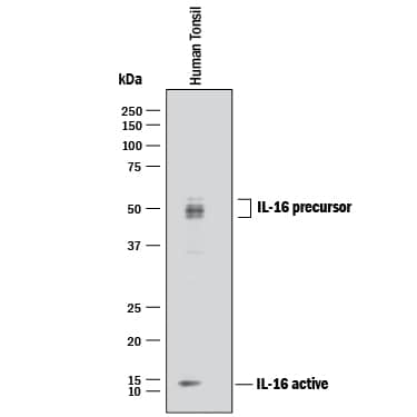
HAF017). Specific bands were detected for IL-16 at approximately 14 kDa (active) and 45-55 kDa (precursor), as indicated. This experiment was conducted under reducing conditions and using
Immunoblot Buffer Group 1." class="big_thumb" />
CTS008) and counterstained with hematoxylin (blue). Lower panel shows a lack of labeling if primary antibodies are omitted and tissue is stained only with secondary antibody followed by incubation with detection reagents. View our protocol for
Chromogenic IHC Staining of Paraffin-embedded Tissue Sections." class="big_lightbox">
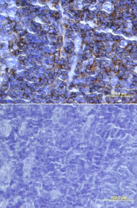
CTS008) and counterstained with hematoxylin (blue). Lower panel shows a lack of labeling if primary antibodies are omitted and tissue is stained only with secondary antibody followed by incubation with detection reagents. View our protocol for
Chromogenic IHC Staining of Paraffin-embedded Tissue Sections." class="big_thumb" />
316-IL) was serially diluted and captured by Mouse Anti-Human IL‑16 Monoclonal Antibody (Catalog #
MAB316) coated on a Clear Polystyrene Microplate (Catalog #
DY990). Goat Anti-Human IL‑16 C-terminal Peptide Antigen Affinity-purified Polyclonal Antibody (Catalog # AF-316-PB) was biotinylated and incubated with the protein captured on the plate. Detection of the standard curve was achieved by incubating Streptavidin-HRP (Catalog #
DY998)" class="big_lightbox">

316-IL) was serially diluted and captured by Mouse Anti-Human IL‑16 Monoclonal Antibody (Catalog #
MAB316) coated on a Clear Polystyrene Microplate (Catalog #
DY990). Goat Anti-Human IL‑16 C-terminal Peptide Antigen Affinity-purified Polyclonal Antibody (Catalog # AF-316-PB) was biotinylated and incubated with the protein captured on the plate. Detection of the standard curve was achieved by incubating Streptavidin-HRP (Catalog #
DY998)" class="big_thumb" />
 AB-108-C, open histogram) followed by Allophycocyanin-conjugated Anti-Goat IgG Secondary Antibody (Catalog # F0108). To facilitate intracellular staining, cells were fixed with Flow Cytometry Fixation Buffer (Catalog # FC004) and permeabilized with Flow Cytometry Permeabilization/Wash Buffer I (Catalog # FC005). View our protocol for Staining Intracellular Molecules." alt="Raji cells were stained with Goat Anti-Human IL‑16 C-terminal Peptide Antigen Affinity-purified Polyclonal Antibody (Catalog # AF-316-PB, filled histogram) or isotype control antibody (Catalog # AB-108-C, open histogram) followed by Allophycocyanin-conjugated Anti-Goat IgG Secondary Antibody (Catalog # F0108). To facilitate intracellular staining, cells were fixed with Flow Cytometry Fixation Buffer (Catalog # FC004) and permeabilized with Flow Cytometry Permeabilization/Wash Buffer I (Catalog # FC005). View our protocol for Staining Intracellular Molecules." class="big_thumb" />
 MAB004, open histogram) followed by Allophycocyanin-conjugated Anti-Mouse IgG Secondary Antibody (Catalog # F0101B). To facilitate intracellular staining, cells were fixed with Flow Cytometry Fixation Buffer (Catalog # FC004) and permeabilized with Flow Cytometry Permeabilization/Wash Buffer I (Catalog # FC005). View our protocol for Staining Intracellular Molecules." alt="Raji cells were stained with Mouse Anti-Human IL‑16 Monoclonal Antibody (Catalog # MAB3161, filled histogram) or isotype control antibody (Catalog # MAB004, open histogram) followed by Allophycocyanin-conjugated Anti-Mouse IgG Secondary Antibody (Catalog # F0101B). To facilitate intracellular staining, cells were fixed with Flow Cytometry Fixation Buffer (Catalog # FC004) and permeabilized with Flow Cytometry Permeabilization/Wash Buffer I (Catalog # FC005). View our protocol for Staining Intracellular Molecules." class="big_thumb" />


![N/A IL-16 [HRP]](http://images.novusbio.com/fullsize/elisa/DATA_IL16_D1600_ELISA_537.jpg "N/A IL-16 [HRP]")
![N/A IL-16 [HRP]](http://images.novusbio.com/fullsize/elisa/DATA_IL16_D1600_ELISA_538.jpg)
![N/A IL-16 [HRP]](http://images.novusbio.com/fullsize/elisa/IL-16_D1600_ELISA_14.jpg)
 HAF017). Specific bands were detected for IL-16 at approximately 14 kDa (active) and 45-55 kDa (precursor), as indicated. This experiment was conducted under reducing conditions and using
  NL001) and counterstained with DAPI (blue). View our protocol for
  CTS008) and counterstained with hematoxylin (blue). Lower panel shows a lack of labeling if primary antibodies are omitted and tissue is stained only with secondary antibody followed by incubation with detection reagents. View our protocol for
  316-IL) was serially diluted and captured by Mouse Anti-Human IL‑16 Monoclonal Antibody (Catalog #
 ![N/A IL-16 [Biotin]](http://images.novusbio.com/fullsize/elisa/DATA_IL16_DY1727_ELISA_1704.jpg "N/A IL-16 [Biotin]")
![N/A IL-16 [Biotin]](http://images.novusbio.com/fullsize/elisa/DATA_IL16_DY316_ELISA_1921.jpg "N/A IL-16 [Biotin]")


![Lane 1: Marker [kDa] 250, 130, 95, 72, 55, 36, 28, 17, 10 | Lane 2: RT4 | Lane 3: U-251 MG | Lane 4: Human Plasma | Lane 5: Liver | Lane 6: Tonsil](http://images.novusbio.com/fullsize/nbp3-21277_rabbit-il-16-pab-45202315481017.jpg "Lane 1: Marker [kDa] 250, 130, 95, 72, 55, 36, 28, 17, 10 | Lane 2: RT4 | Lane 3: U-251 MG | Lane 4: Human Plasma | Lane 5: Liver | Lane 6: Tonsil")

![ELISA: Mouse IL-16 ELISA Kit (Colorimetric) [NBP3-06853] - Samples were spiked with high concentrations of Mouse IL-16 and diluted with Reference Standard & Sample Diluent to produce samples with values within the range of the assay.](http://images.novusbio.com/fullsize/nbp3-06853_mouse-il-16-elisa-kit-colorimetric-132202416232382.jpg "ELISA: Mouse IL-16 ELISA Kit (Colorimetric) [NBP3-06853] - Samples were spiked with high concentrations of Mouse IL-16 and diluted with Reference Standard & Sample Diluent to produce samples with values within the range of the assay.")
![ELISA: Mouse IL-16 ELISA Kit (Colorimetric) [NBP3-06853] - Standard Curve Reference](http://images.novusbio.com/fullsize/nbp3-06853_mouse-il-16-elisa-kit-colorimetric-432024903552.jpg)
![Flow Cytometry: IL-16 Antibody (14.1) - Chimeric [NBP3-09027] - Human peripheral blood leukocytes were stimulated with PMA and Ionomycin in presence of Monensin for 4 hours. The cells were fixed using 2% PFA, permeabilized using 0.5% Triton and stained with anti-Fluorescein IgG antibody (4-4-20; isotype control, black line) or the rabbit IgG version of 14.1 (NBP3-09027, pink line) at a dilution of 1:100 for 1h at RT. The blue line shows unstimulated human peripheral blood leukocytes stained with NBP3-09027. After washing, bound antibody was detected using a goat anti-mouse IgG AlexaFluor(R) 488 antibody at a dilution of 1:1000 and cells were analyzed using a FACSCanto flow-cytometer.](http://images.novusbio.com/fullsize/IL-16-Antibody-14-1-Chimeric-Flow-Cytometry-NBP3-09027-img0001.jpg "Flow Cytometry: IL-16 Antibody (14.1) - Chimeric [NBP3-09027] - Human peripheral blood leukocytes were stimulated with PMA and Ionomycin in presence of Monensin for 4 hours. The cells were fixed using 2% PFA, permeabilized using 0.5% Triton and stained with anti-Fluorescein IgG antibody (4-4-20; isotype control, black line) or the rabbit IgG version of 14.1 (NBP3-09027, pink line) at a dilution of 1:100 for 1h at RT. The blue line shows unstimulated human peripheral blood leukocytes stained with NBP3-09027. After washing, bound antibody was detected using a goat anti-mouse IgG AlexaFluor(R) 488 antibody at a dilution of 1:1000 and cells were analyzed using a FACSCanto flow-cytometer.")


