HAF017). A specific band was detected for MMP-7 at approximately 28 kDa (as indicated). This experiment was conducted under reducing conditions and using
Immunoblot Buffer Group 1." class="big_lightbox">
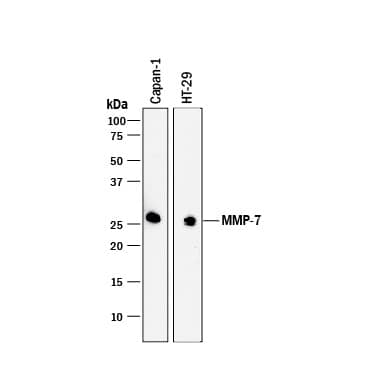
HAF017). A specific band was detected for MMP-7 at approximately 28 kDa (as indicated). This experiment was conducted under reducing conditions and using
Immunoblot Buffer Group 1." alt="Western blot shows lysates of Capan-1 human pancreatic adenocarcinoma cell line and HT-29 human colon adenocarcinoma cell line. PVDF membrane was probed with 1 µg/mL of Goat Anti-Human MMP-7 Antigen Affinity-purified Polyclonal Antibody (Catalog # AF907) followed by HRP-conjugated Anti-Goat IgG Secondary Antibody (
HAF017). A specific band was detected for MMP-7 at approximately 28 kDa (as indicated). This experiment was conducted under reducing conditions and using
Immunoblot Buffer Group 1." class="big_thumb" />
CTS008) and counterstained with hematoxylin (blue). Lower panel shows a lack of labeling if primary antibodies are omitted and tissue is stained only with secondary antibody followed by incubation with detection reagents. View our protocol for
Chromogenic IHC Staining of Paraffin-embedded Tissue Sections." class="big_lightbox">
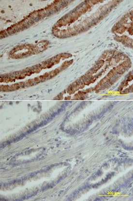
CTS008) and counterstained with hematoxylin (blue). Lower panel shows a lack of labeling if primary antibodies are omitted and tissue is stained only with secondary antibody followed by incubation with detection reagents. View our protocol for
Chromogenic IHC Staining of Paraffin-embedded Tissue Sections." class="big_thumb" />
HAF109). This experiment was conducted under reducing conditions and using the 12-230 kDa separation system." class="big_lightbox">
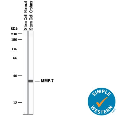
HAF109). This experiment was conducted under reducing conditions and using the 12-230 kDa separation system." class="big_thumb" />
ES001) by Recombinant Human MMP-7 (0.2 µg/mL, Catalog #
907-MP) is measured after preincubation with increasing concentrations of Goat Anti-Human MMP-7 Antigen Affinity-purified Polyclonal Antibody (Catalog # AF907). The ND
50 is typically 2 µg/mL." class="big_lightbox">
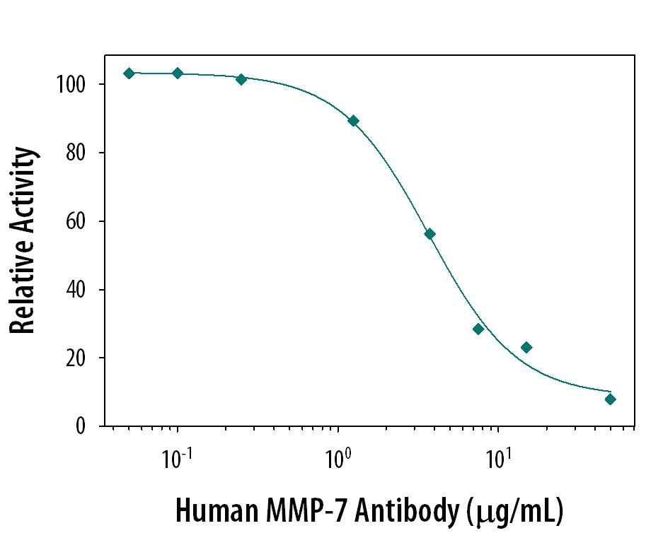
ES001) by Recombinant Human MMP-7 (0.2 µg/mL, Catalog #
907-MP) is measured after preincubation with increasing concentrations of Goat Anti-Human MMP-7 Antigen Affinity-purified Polyclonal Antibody (Catalog # AF907). The ND
50 is typically 2 µg/mL." class="big_thumb" />
AF907) at 3ug/mL with overnight incubation at 4 degrees Celsius followed by incubation with anti-goat IgG VisUCyte HRP Polymer Antibody (Catalog #
VC004) and DAB chromogen (yellow-brown). Tissue was counterstained with hematoxylin (blue). Specific staining was localized to exocrine glands. " class="big_lightbox">

AF907) at 3ug/mL with overnight incubation at 4 degrees Celsius followed by incubation with anti-goat IgG VisUCyte HRP Polymer Antibody (Catalog #
VC004) and DAB chromogen (yellow-brown). Tissue was counterstained with hematoxylin (blue). Specific staining was localized to exocrine glands. " class="big_thumb" />
907-MP) was serially diluted and captured by Mouse Anti-Human/Primate MMP‑7 Monoclonal Antibody (Catalog #
MAB9072) coated on a Clear Polystyrene Microplate (Catalog #
DY990). Goat Anti-Human MMP‑7 Antigen Affinity-purified Polyclonal Antibody (Catalog # AF907) was biotinylated and incubated with the protein captured on the plate. Detection of the standard curve was achieved by incubating Streptavidin-HRP (Catalog #
DY998)" class="big_lightbox">

907-MP) was serially diluted and captured by Mouse Anti-Human/Primate MMP‑7 Monoclonal Antibody (Catalog #
MAB9072) coated on a Clear Polystyrene Microplate (Catalog #
DY990). Goat Anti-Human MMP‑7 Antigen Affinity-purified Polyclonal Antibody (Catalog # AF907) was biotinylated and incubated with the protein captured on the plate. Detection of the standard curve was achieved by incubating Streptavidin-HRP (Catalog #
DY998)" class="big_thumb" />
 WBC016). PVDF membrane was probed with 1 µg/mL of Mouse Anti-Human MMP-7 Monoclonal Antibody (Catalog # MAB9071) followed by HRP-conjugated Anti-Mouse IgG Secondary Antibody (Catalog # HAF018). A specific band was detected for MMP-7 at approximately 28 kDa (as indicated). This experiment was conducted under reducing conditions and using Immunoblot Buffer Group 1." alt="Western blot shows lysates of Capan-1 human pancreatic adenocarcinoma cell line and SK-OV-3 human ovarian adenocarcinoma cell line and, for additional reference, Recombinant Human MMP-7 Western Blot Standard Protein (Catalog # WBC016). PVDF membrane was probed with 1 µg/mL of Mouse Anti-Human MMP-7 Monoclonal Antibody (Catalog # MAB9071) followed by HRP-conjugated Anti-Mouse IgG Secondary Antibody (Catalog # HAF018). A specific band was detected for MMP-7 at approximately 28 kDa (as indicated). This experiment was conducted under reducing conditions and using Immunoblot Buffer Group 1." class="big_thumb" />
 HAF017). A specific band was detected for MMP-7 at approximately 28 kDa (as indicated). This experiment was conducted under reducing conditions and using Immunoblot Buffer Group 1." alt="Western blot shows lysates of Capan-1 human pancreatic adenocarcinoma cell line and HT-29 human colon adenocarcinoma cell line. PVDF membrane was probed with 1 µg/mL of Goat Anti-Human MMP-7 Antigen Affinity-purified Polyclonal Antibody (Catalog # AF907) followed by HRP-conjugated Anti-Goat IgG Secondary Antibody (HAF017). A specific band was detected for MMP-7 at approximately 28 kDa (as indicated). This experiment was conducted under reducing conditions and using Immunoblot Buffer Group 1." class="big_thumb" />
 CTS008) and counterstained with hematoxylin (blue). Specific labeling was localized to the cytoplasm of cells in intestinal glands. View our protocol for Chromogenic IHC Staining of Frozen Tissue Sections." alt="MMP‑7 was detected in perfusion fixed frozen sections of mouse small intestine using Goat Anti-Mouse MMP‑7 Antigen Affinity-purified Polyclonal Antibody (Catalog # AF2967) at 15 µg/mL overnight at 4 °C. Tissue was stained using the Anti-Goat HRP-DAB Cell & Tissue Staining Kit (brown; Catalog # CTS008) and counterstained with hematoxylin (blue). Specific labeling was localized to the cytoplasm of cells in intestinal glands. View our protocol for Chromogenic IHC Staining of Frozen Tissue Sections." class="big_thumb" />
 CTS013). Tissue was stained using the Anti-Mouse HRP-DAB Cell & Tissue Staining Kit (brown; Catalog # CTS002) and counterstained with hematoxylin (blue). View our protocol for Chromogenic IHC Staining of Paraffin-embedded Tissue Sections." alt="MMP-7 was detected in immersion fixed paraffin-embedded sections of human pancreatic cancer tissue using Human Pro-MMP-7 Monoclonal Antibody (Catalog # MAB907) at 10 µg/mL overnight at 4 °C. Before incubation with the primary antibody tissue was subjected to heat-induced epitope retrieval using Antigen Retrieval Reagent-Basic (Catalog # CTS013). Tissue was stained using the Anti-Mouse HRP-DAB Cell & Tissue Staining Kit (brown; Catalog # CTS002) and counterstained with hematoxylin (blue). View our protocol for Chromogenic IHC Staining of Paraffin-embedded Tissue Sections." class="big_thumb" />
 907-MP) was serially diluted and captured by Mouse Anti-Human/Primate MMP‑7 Monoclonal Antibody (Catalog # MAB9072) coated on a Clear Polystyrene Microplate (Catalog # DY990). Goat Anti-Human MMP‑7 Antigen Affinity-purified Polyclonal Antibody (Catalog # AF907) was biotinylated and incubated with the protein captured on the plate. Detection of the standard curve was achieved by incubating Streptavidin-HRP (Catalog # DY998)" alt="Recombinant Human MMP‑7 (Catalog # 907-MP) was serially diluted and captured by Mouse Anti-Human/Primate MMP‑7 Monoclonal Antibody (Catalog # MAB9072) coated on a Clear Polystyrene Microplate (Catalog # DY990). Goat Anti-Human MMP‑7 Antigen Affinity-purified Polyclonal Antibody (Catalog # AF907) was biotinylated and incubated with the protein captured on the plate. Detection of the standard curve was achieved by incubating Streptavidin-HRP (Catalog # DY998)" class="big_thumb" />


![N/A MMP-7 [HRP]](http://images.novusbio.com/fullsize/elisa/DATA_MMP7_DMP700_ELISA_768.jpg "N/A MMP-7 [HRP]")
![N/A MMP-7 [HRP]](http://images.novusbio.com/fullsize/elisa/MMP7_DMP700_ELISA_498.jpg)


 CTS008) and counterstained with hematoxylin (blue). Lower panel shows a lack of labeling if primary antibodies are omitted and tissue is stained only with secondary antibody followed by incubation with detection reagents. View our protocol for
  HAF109). This experiment was conducted under reducing conditions and using the 12-230 kDa separation system." class="big_thumb" />
 ES001) by Recombinant Human MMP-7 (0.2 µg/mL, Catalog #
  AF907) at 3ug/mL with overnight incubation at 4 degrees Celsius followed by incubation with anti-goat IgG VisUCyte HRP Polymer Antibody (Catalog #
  907-MP) was serially diluted and captured by Mouse Anti-Human/Primate MMP‑7 Monoclonal Antibody (Catalog #
 ![N/A MMP-7 [Biotin]](http://images.novusbio.com/fullsize/elisa/DATA_MMP7_DY907_ELISA_2257.jpg "N/A MMP-7 [Biotin]")
![ELISA: Rat MMP-7 ELISA Kit (Colorimetric) [NBP3-06896] - Samples were spiked with high concentrations of Rat MMP-7 and diluted with Reference Standard & Sample Diluent to produce samples with values within the range of the assay.](http://images.novusbio.com/fullsize/nbp3-06896_rat-mmp-7-elisa-kit-colorimetric-13220241624342.jpg "ELISA: Rat MMP-7 ELISA Kit (Colorimetric) [NBP3-06896] - Samples were spiked with high concentrations of Rat MMP-7 and diluted with Reference Standard & Sample Diluent to produce samples with values within the range of the assay.")
![ELISA: Rat MMP-7 ELISA Kit (Colorimetric) [NBP3-06896] - Standard Curve Reference](http://images.novusbio.com/fullsize/nbp3-06896_rat-mmp-7-elisa-kit-colorimetric-43202497363.jpg)

![ELISA: Mouse MMP-7 ELISA Kit (Colorimetric) [NBP3-06895] - Samples were spiked with high concentrations of Mouse MMP-7 and diluted with Reference Standard & Sample Diluent to produce samples with values within the range of the assay.](http://images.novusbio.com/fullsize/nbp3-06895_mouse-mmp-7-elisa-kit-colorimetric-132202416215334.jpg "ELISA: Mouse MMP-7 ELISA Kit (Colorimetric) [NBP3-06895] - Samples were spiked with high concentrations of Mouse MMP-7 and diluted with Reference Standard & Sample Diluent to produce samples with values within the range of the assay.")
![ELISA: Mouse MMP-7 ELISA Kit (Colorimetric) [NBP3-06895] - Standard Curve Reference](http://images.novusbio.com/fullsize/nbp3-06895_mouse-mmp-7-elisa-kit-colorimetric-4320248582589.jpg)
![Immunohistochemistry: MMP-7 Antibody [NBP1-99123] - MMP7 antibody was tested in human breast cancer using DAB with hematoxylin counterstain. The image on the right had the antibody blocked by preincubation with immunizing peptide.](http://images.novusbio.com/fullsize/MMP-7-Antibody-Immunohistochemistry-NBP1-99123-img0002.jpg "Immunohistochemistry: MMP-7 Antibody [NBP1-99123] - MMP7 antibody was tested in human breast cancer using DAB with hematoxylin counterstain. The image on the right had the antibody blocked by preincubation with immunizing peptide.")


