DR647MS) and counterstained with DAPI (blue; Lunaphore Catalog #
DR100). Specific staining was localized to the cytoplasm. Protocol available in
COMET™ Panel Builder." class="big_lightbox">
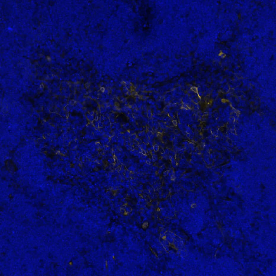
DR647MS) and counterstained with DAPI (blue; Lunaphore Catalog #
DR100). Specific staining was localized to the cytoplasm. Protocol available in
COMET™ Panel Builder." alt="Cytokeratin 17 was detected in immersion fixed paraffin-embedded sections of mouse thymus using Mouse Anti-Human/Mouse Cytokeratin 17, Monoclonal Antibody (Catalog # MAB11571) at 0.1ug/mL at 37 °Celsius for 4 minutes. Before incubation with the primary antibody, tissue underwent an all-in-one dewaxing and antigen retrieval preprocessing using PreTreatment Module (PT Module) and Dewax and HIER Buffer H (pH 9; Epredia Catalog # TA-999-DHBH). Tissue was stained using the Alexa Fluor™ 647 Goat anti-Mouse IgG Secondary Antibody at 1:200 at 37 ° Celsius for 2 minutes. (Yellow; Lunaphore Catalog #
DR647MS) and counterstained with DAPI (blue; Lunaphore Catalog #
DR100). Specific staining was localized to the cytoplasm. Protocol available in
COMET™ Panel Builder." class="big_thumb" />
VC001) or the HRP-conjugated Anti-Mouse IgG Secondary Antibody (Catalog #
HAF007). Before incubation with the primary antibody, tissue was subjected to heat-induced epitope retrieval using VisUCyte Antigen Retrieval Reagent-Basic (Catalog #
VCTS021). Tissue was stained using DAB (brown) and counterstained with hematoxylin (blue). Specific staining was localized to the cytoplasm. View our protocol for
Chromogenic IHC Staining of Paraffin-embedded Tissue Sections." class="big_lightbox">
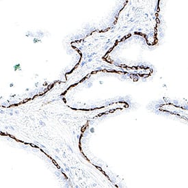
VC001) or the HRP-conjugated Anti-Mouse IgG Secondary Antibody (Catalog #
HAF007). Before incubation with the primary antibody, tissue was subjected to heat-induced epitope retrieval using VisUCyte Antigen Retrieval Reagent-Basic (Catalog #
VCTS021). Tissue was stained using DAB (brown) and counterstained with hematoxylin (blue). Specific staining was localized to the cytoplasm. View our protocol for
Chromogenic IHC Staining of Paraffin-embedded Tissue Sections." class="big_thumb" />
VC001) or the HRP-conjugated Anti-Mouse IgG Secondary Antibody (Catalog #
HAF007). Before incubation with the primary antibody, tissue was subjected to heat-induced epitope retrieval using VisUCyte Antigen Retrieval Reagent-Basic (Catalog #
VCTS021). Tissue was stained using DAB (brown) and counterstained with hematoxylin (blue). Specific staining was localized to the cytoplasm. View our protocol for
Chromogenic IHC Staining of Paraffin-embedded Tissue Sections." class="big_lightbox">
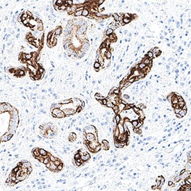
VC001) or the HRP-conjugated Anti-Mouse IgG Secondary Antibody (Catalog #
HAF007). Before incubation with the primary antibody, tissue was subjected to heat-induced epitope retrieval using VisUCyte Antigen Retrieval Reagent-Basic (Catalog #
VCTS021). Tissue was stained using DAB (brown) and counterstained with hematoxylin (blue). Specific staining was localized to the cytoplasm. View our protocol for
Chromogenic IHC Staining of Paraffin-embedded Tissue Sections." class="big_thumb" />
HAF018). A specific band was detected for Cytokeratin 17 at approximately 53 kDa (as indicated). This experiment was conducted under reducing conditions and using
Western Blot Buffer Group 1." class="big_lightbox">
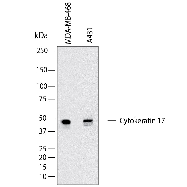
HAF018). A specific band was detected for Cytokeratin 17 at approximately 53 kDa (as indicated). This experiment was conducted under reducing conditions and using
Western Blot Buffer Group 1." class="big_thumb" />
 DR647MS) and counterstained with DAPI (blue; Lunaphore Catalog # DR100). Specific staining was localized to the cytoplasm. Protocol available in COMET™ Panel Builder." alt="Cytokeratin 17 was detected in immersion fixed paraffin-embedded sections of mouse thymus using Mouse Anti-Human/Mouse Cytokeratin 17, Monoclonal Antibody (Catalog # MAB11571) at 0.1ug/mL at 37 °Celsius for 4 minutes. Before incubation with the primary antibody, tissue underwent an all-in-one dewaxing and antigen retrieval preprocessing using PreTreatment Module (PT Module) and Dewax and HIER Buffer H (pH 9; Epredia Catalog # TA-999-DHBH). Tissue was stained using the Alexa Fluor™ 647 Goat anti-Mouse IgG Secondary Antibody at 1:200 at 37 ° Celsius for 2 minutes. (Yellow; Lunaphore Catalog # DR647MS) and counterstained with DAPI (blue; Lunaphore Catalog # DR100). Specific staining was localized to the cytoplasm. Protocol available in COMET™ Panel Builder." class="big_thumb" />
 HAF018). Specific bands were detected for Cytokeratin 17 at approximately 48 kDa and for Cytokeratin 19 at approximately 41 kDa (as indicated). This experiment was conducted under reducing conditions and using Western Blot Buffer Group 1." alt="Western Blot shows lysates of HeLa human cervical epithelial carcinoma cell line and HCC827 human non-small cell lung cancer cell line. PVDF membrane was probed with 2 µg/ml of Mouse Anti-Human Cytokeratin 17/19 Monoclonal Antibody (Catalog # MAB11568) followed by HRP-conjugated Anti-Mouse IgG Secondary Antibody (Catalog # HAF018). Specific bands were detected for Cytokeratin 17 at approximately 48 kDa and for Cytokeratin 19 at approximately 41 kDa (as indicated). This experiment was conducted under reducing conditions and using Western Blot Buffer Group 1." class="big_thumb" />


![Western Blot: Cytokeratin 17 Antibody (E3 (same as Ks17.E3)) [NBP2-29421] - Analysis of Cytokeratin 17 in human HeLa lysate using Cytokeratin 17 E3 antibody at 3 ug/ml. goat anti-mouse Ig HRP secondary antibody and PicoTect ECL substrate solution were used for this test.](http://images.novusbio.com/fullsize/Cytokeratin-17-Antibody-E3-same-as-Ks17-E3-Western-Blot-NBP2-29421-img0001.jpg "Western Blot: Cytokeratin 17 Antibody (E3 (same as Ks17.E3)) [NBP2-29421] - Analysis of Cytokeratin 17 in human HeLa lysate using Cytokeratin 17 E3 antibody at 3 ug/ml. goat anti-mouse Ig HRP secondary antibody and PicoTect ECL substrate solution were used for this test.")
![Immunohistochemistry-Paraffin: Cytokeratin 17 Antibody (E3 (same as Ks17.E3)) [NBP2-29421] - Formalin-fixed paraffin-embedded human Bladder Carcinoma stained with CK17 Monoclonal Antibody (E3)](http://images.novusbio.com/fullsize/Cytokeratin-17-Antibody-E3-same-as-Ks17-E3-Immunohistochemistry-Paraffin-NBP2-29421-img0007.jpg)
![Immunohistochemistry-Paraffin: Cytokeratin 17 Antibody (E3 (same as Ks17.E3)) [NBP2-29421] - Formalin-fixed, paraffin-embedded human cervical carcinoma stained with CK17 Monoclonal Antibody (E3).](http://images.novusbio.com/fullsize/Cytokeratin-17-Antibody-E3-same-as-Ks17-E3-Immunohistochemistry-Paraffin-NBP2-29421-img0006.jpg)
![Western Blot: Cytokeratin 17 Antibody [NBP2-38536] - Analysis in human cell lines HeLa and PC-3. Corresponding RNA-seq data are presented for the same cell lines. Loading control: Anti-HSP90B1.](http://images.novusbio.com/fullsize/Cytokeratin-17-Antibody-Western-Blot-NBP2-38536-img0011.jpg "<b>Orthogonal Strategies Validation. </b>Western Blot: Cytokeratin 17 Antibody [NBP2-38536] - Analysis in human cell lines HeLa and PC-3. Corresponding RNA-seq data are presented for the same cell lines. Loading control: Anti-HSP90B1.")
![<b>Orthogonal Strategies Validation. </b>Immunocytochemistry/Immunofluorescence: Cytokeratin 17 Antibody [NBP2-38536] - MYP3 rat urothelial cell line with CK17 (green) and DAPI (blue). Primary antibody at 1:200. Image from verified customer review.](http://images.novusbio.com/fullsize/Cytokeratin-17-Antibody-Immunocytochemistry-Immunofluorescence-NBP2-38536-img0009.jpg)

![Immunocytochemistry/Immunofluorescence: Cytokeratin 17 Antibody [NBP2-38536] - UROtsa (rat urothelial cell line). CK17 in green, DAPI in blue. Secondary antibody: Goat Polyclonal Anti-Rabbit at 1:500. Image from verified customer review.](http://images.novusbio.com/fullsize/Cytokeratin-17-Antibody-Immunocytochemistry-Immunofluorescence-NBP2-38536-img0008.jpg)
 VC001) or the HRP-conjugated Anti-Mouse IgG Secondary Antibody (Catalog #
  VC001) or the HRP-conjugated Anti-Mouse IgG Secondary Antibody (Catalog #
  HAF018). A specific band was detected for Cytokeratin 17 at approximately 53 kDa (as indicated). This experiment was conducted under reducing conditions and using
  NL007) and counterstained with DAPI (blue). Specific staining was localized to the cytoplasm. View our protocol for
![Immunocytochemistry/Immunofluorescence: Cytokeratin 17/36 Antibody (SR45-06) [NBP2-67318] - Staining Cytokeratin 17 in Hela cells (green). The nuclear counter stain is DAPI (blue). Cells were fixed in paraformaldehyde, permeabilised with 0.25% Triton X100/PBS.](http://images.novusbio.com/fullsize/Cytokeratin-17-36-Antibody-SR45-06-Immunocytochemistry-Immunofluorescence-NBP2-67318-img0003.jpg "Immunocytochemistry/Immunofluorescence: Cytokeratin 17/36 Antibody (SR45-06) [NBP2-67318] - Staining Cytokeratin 17 in Hela cells (green). The nuclear counter stain is DAPI (blue). Cells were fixed in paraformaldehyde, permeabilised with 0.25% Triton X100/PBS.")
![<b>Biological Strategies Validation. </b>Immunohistochemistry-Paraffin: Cytokeratin 17/36 Antibody (SR45-06) [NBP2-67318] - Immunohistochemical analysis of paraffin-embedded human tonsil tissue using anti-Cytokeratin 17/36 antibody. The section was pre-treated using heat mediated antigen retrieval with Tris-EDTA buffer (pH 8.0-8.4) for 20 minutes. The tissues were blocked in 5](http://images.novusbio.com/fullsize/Cytokeratin-17-36-Antibody-SR45-06-Immunohistochemistry-Paraffin-NBP2-67318-img0009.jpg)
![Flow Cytometry: Cytokeratin 17/36 Antibody (SR45-06) [NBP2-67318] - Analysis of Hela cells with Cytokeratin 17 antibody at 1/50 dilution (blue) compared with an unlabelled control (cells without incubation with primary antibody; red). Alexa Fluor 488-conjugated goat anti rabbit IgG was used as the secondary antibody.](http://images.novusbio.com/fullsize/Cytokeratin-17-36-Antibody-SR45-06-Flow-Cytometry-NBP2-67318-img0001.jpg)
![Immunocytochemistry/Immunofluorescence: Cytokeratin 17/36 Antibody (SR45-06) [NBP2-67318] - Staining Cytokeratin 17 in PC-3M cells (green). The nuclear counter stain is DAPI (blue). Cells were fixed in paraformaldehyde, permeabilised with 0.25% Triton X100/PBS.](http://images.novusbio.com/fullsize/Cytokeratin-17-36-Antibody-SR45-06-Immunocytochemistry-Immunofluorescence-NBP2-67318-img0002.jpg)
![Immunohistochemistry-Paraffin: Cytokeratin 17/36 Antibody (SR45-06) [NBP2-67318] - Analysis of paraffin-embedded human breast carcinoma tissue using anti-Cytokeratin 17 antibody. Counter stained with hematoxylin.](http://images.novusbio.com/fullsize/Cytokeratin-17-36-Antibody-SR45-06-Immunohistochemistry-Paraffin-NBP2-67318-img0004.jpg)
![Immunohistochemistry-Paraffin: Cytokeratin 17/36 Antibody (SR45-06) [NBP2-67318] - Analysis of paraffin-embedded human tonsil tissue using anti-Cytokeratin 17 antibody. Counter stained with hematoxylin.](http://images.novusbio.com/fullsize/Cytokeratin-17-36-Antibody-SR45-06-Immunohistochemistry-Paraffin-NBP2-67318-img0005.jpg)
![Immunohistochemistry-Paraffin: Cytokeratin 17/36 Antibody (SR45-06) [NBP2-67318] - Analysis of paraffin-embedded mouse prostate tissue using anti-Cytokeratin 17 antibody. Counter stained with hematoxylin.](http://images.novusbio.com/fullsize/Cytokeratin-17-36-Antibody-SR45-06-Immunohistochemistry-Paraffin-NBP2-67318-img0006.jpg)
![<b>Biological Strategies Validation. </b>Immunohistochemistry-Paraffin: Cytokeratin 17/36 Antibody (SR45-06) [NBP2-67318] - Immunohistochemical analysis of paraffin-embedded human prostate tissue using anti-Cytokeratin 17/36 antibody. The section was pre-treated using heat mediated antigen retrieval with Tris-EDTA buffer (pH 8.0-8.4) for 20 minutes. The tissues were blocked in](http://images.novusbio.com/fullsize/Cytokeratin-17-36-Antibody-SR45-06-Immunohistochemistry-Paraffin-NBP2-67318-img0007.jpg)
![<b>Biological Strategies Validation. </b>Immunohistochemistry-Paraffin: Cytokeratin 17/36 Antibody (SR45-06) [NBP2-67318] - Immunohistochemical analysis of paraffin-embedded human skin tissue using anti-Cytokeratin 17/36 antibody. The section was pre-treated using heat mediated antigen retrieval with Tris-EDTA buffer (pH 8.0-8.4) for 20 minutes. The tissues were blocked in 5%](http://images.novusbio.com/fullsize/Cytokeratin-17-36-Antibody-SR45-06-Immunohistochemistry-Paraffin-NBP2-67318-img0008.jpg)
![Western blot analysis of A431 cell lysates with NBP2-67318 at 1/2,000 dilution. Lysates/proteins at 15 µg/Lane. Predicted band size: 48 kDa Observed band size: 48 kDa Exposure time: 3 minutes; 4-20% SDS-PAGE gel. Proteins were transferred to a PVDF membrane and blocked with 5% NFDM/TBST for 1 hour at room temperature. NBP2-67318 at 1/2,000 dilution was used in 5% NFDM/TBST at 4℃ overnight. Goat Anti-Rabbit IgG [HRP] Secondary Antibody at 1/50,000 dilution was used for 1 hour at room temperature.](http://images.novusbio.com/fullsize/nbp2-67318_-western-blot-639204586675331019.jpg)
![Western blot analysis on different lysates with NBP2-67318 at 1/1,000 dilution. Lane 1: Mouse skin tissue lysate Lane 2: Rat skin tissue lysate Lysates/proteins at 40 µg/Lane. Predicted band size: 48 kDa Observed band size: 48 kDa Exposure time: 3 minutes; 4-20% SDS-PAGE gel. Proteins were transferred to a PVDF membrane and blocked with 5% NFDM/TBST for 1 hour at room temperature. NBP2-67318 at 1/1,000 dilution was used in 5% NFDM/TBST at 4℃ overnight. Goat Anti-Rabbit IgG [HRP] Secondary Antibody at 1/50,000 dilution was used for 1 hour at room temperature.](http://images.novusbio.com/fullsize/nbp2-67318_-western-blot-639204597491663928.jpg)
![Immunohistochemistry-Paraffin: Cytokeratin 17 Antibody [NBP1-32824] - Cytokeratin 17 antibody detects Cytokeratin 17 protein at cytoplasm in human cervical metaplasia (left) and carcinoma (right) by immunohistochemical analysis.Sample: Paraffin-embedded human cervix. Antibody diluted at 1:500. Antigen Retrieval: Citrate buffer, pH 6.0, 15 min](http://images.novusbio.com/fullsize/Cytokeratin-17-Antibody-Immunohistochemistry-Paraffin-NBP1-32824-img0008.jpg "Immunohistochemistry-Paraffin: Cytokeratin 17 Antibody [NBP1-32824] - Cytokeratin 17 antibody detects Cytokeratin 17 protein at cytoplasm in human cervical metaplasia (left) and carcinoma (right) by immunohistochemical analysis.Sample: Paraffin-embedded human cervix. Antibody diluted at 1:500. Antigen Retrieval: Citrate buffer, pH 6.0, 15 min")
![Western Blot: Cytokeratin 17 Antibody [NBP1-32824] - Sample (50 ug of whole cell lysate) A: Rat bladder 10% SDS PAGE; antibody diluted at 1:10000.](http://images.novusbio.com/fullsize/Cytokeratin-17-Antibody-Western-Blot-NBP1-32824-img0005.jpg)
![Immunocytochemistry/Immunofluorescence: Cytokeratin 17 Antibody [NBP1-32824] - Paraformaldehyde-fixed HeLa, using Cytokeratin 17 antibody (Green) at 1:500 dilution. Alpha-tubulin filaments were labeled with an alpha Tubulin antibody (Red) at 1:2500.](http://images.novusbio.com/fullsize/Cytokeratin-17-Antibody-Immunocytochemistry-Immunofluorescence-NBP1-32824-img0003.jpg)
![Immunohistochemistry-Paraffin: Cytokeratin 17 Antibody [NBP1-32824] - Paraffin-embedded N87 xenograft, using antibody at 1:100 dilution.](http://images.novusbio.com/fullsize/Cytokeratin-17-Antibody-Immunohistochemistry-Paraffin-NBP1-32824-img0004.jpg)
![Immunohistochemistry-Paraffin: Cytokeratin 17 Antibody [NBP1-32824] - Cytokeratin 17 antibody detects Cytokeratin 17 protein at cytoplasm on mouse esophagus by immunohistochemical analysis. Sample: Paraffin-embedded mouse esophagus.Cytokeratin 17 antibody diluted at 1:500. Antigen Retrieval: Trilogy (TM) (EDTA based, pH 8.0) buffer, 15min](http://images.novusbio.com/fullsize/Cytokeratin-17-Antibody-Immunohistochemistry-Paraffin-NBP1-32824-img0007.jpg)

![Western Blot: Cytokeratin 17 Antibody (SPM560) [NBP2-34378] - Western blot of HeLa lysate using CK17 Ab (SPM560).](http://images.novusbio.com/fullsize/Cytokeratin-17-Antibody-SPM560-Western-Blot-NBP2-34378-img0003.jpg "Western Blot: Cytokeratin 17 Antibody (SPM560) [NBP2-34378] - Western blot of HeLa lysate using CK17 Ab (SPM560).")
![Immunohistochemistry-Paraffin: Cytokeratin 17 Antibody (SPM560) [NBP2-34378] - Formalin-paraffin skin stained with CK17 MAb (SPM560).](http://images.novusbio.com/fullsize/Cytokeratin-17-Antibody-SPM560-Immunohistochemistry-Paraffin-NBP2-34378-img0002.jpg)
![Flow Cytometry: Cytokeratin 17 Antibody (SPM560) [NBP2-34378] - Flow Cytometric Analysis of PFA-fixed HeLa cells labeling CK17 with Cytokeratin 17 Antibody (SPM560) followed by Goat anti-Mouse IgG-CF488 (Blue) Isotype Control (Red)](http://images.novusbio.com/fullsize/Cytokeratin-17-Antibody-SPM560-Flow-Cytometry-NBP2-34378-img0004.jpg)
![Immunohistochemistry-Paraffin: Cytokeratin 17 Antibody (SPM560) [NBP2-34378] - Formalin-paraffin skin stained with CK17 Ab (SPM560).](http://images.novusbio.com/fullsize/Cytokeratin-17-Antibody-SPM560-Immunohistochemistry-Paraffin-NBP2-34378-img0001.jpg)
 VC001) or the HRP-conjugated Anti-Mouse IgG Secondary Antibody (Catalog #
  VC001) or the HRP-conjugated Anti-Mouse IgG Secondary Antibody (Catalog #
![Western Blot: Cytokeratin 17 Antibody (A2B10) [NBP2-89123] - Analysis of Cytokeratin 17 on SiHa cell lysates with Mouse anti-Cytokeratin 17 antibody at 1/2,000 dilution. Lysates/proteins at 10 ug/Lane. Predicted band size: 48 kDa Observed band size: 48 kDa Exposure time: 1 minute; 12% SDS-PAGE gel. Proteins were transferred to a PVDF membrane and blocked with 5% NFDM/TBST for 1 hour at room temperature. The primary antibody at 1/2,000 dilution was used in 5% NFDM/TBST at room temperature for 2 hours. Goat Anti-Mouse IgG - HRP Secondary Antibody at 1:150,000 dilution was used for 1 hour at room temperature.](http://images.novusbio.com/fullsize/Cytokeratin-17-Antibody-A2B10-Western-Blot-NBP2-89123-img0005.jpg "Western Blot: Cytokeratin 17 Antibody (A2B10) [NBP2-89123] - Analysis of Cytokeratin 17 on SiHa cell lysates with Mouse anti-Cytokeratin 17 antibody at 1/2,000 dilution. Lysates/proteins at 10 ug/Lane. Predicted band size: 48 kDa Observed band size: 48 kDa Exposure time: 1 minute; 12% SDS-PAGE gel. Proteins were transferred to a PVDF membrane and blocked with 5% NFDM/TBST for 1 hour at room temperature. The primary antibody at 1/2,000 dilution was used in 5% NFDM/TBST at room temperature for 2 hours. Goat Anti-Mouse IgG - HRP Secondary Antibody at 1:150,000 dilution was used for 1 hour at room temperature.")
![<b>Biological Strategies Validation. </b>Immunohistochemistry-Paraffin: Cytokeratin 17 Antibody (A2B10) [NBP2-89123] - Analysis of paraffin-embedded human skin tissue using anti-Cytokeratin 17 antibody. The section was pre-treated using heat mediated antigen retrieval with Tris-EDTA buffer (pH 8.0-8.4) for 20 minutes.The tissues were blocked in 5% BSA for 30 minutes at room temperature, washed with ddH2O and PBS, and then probed with the primary antibody( 1/50) for 30 minutes at room temperature. The detection was performed using an HRP conjugated compact polymer system. DAB was used as the chromogen. Tissues were counterstained with hematoxylin and mounted with DPX.](http://images.novusbio.com/fullsize/Cytokeratin-17-Antibody-A2B10-Immunohistochemistry-Paraffin-NBP2-89123-img0003.jpg)
![Flow Cytometry: Cytokeratin 17 Antibody (A2B10) [NBP2-89123] - Analysis of Cytokeratin 17 was done on SiHa cells. The cells were fixed, permeabilized and stained with the primary antibody( 1/100) (red). After incubation of the primary antibody at room temperature for an hour, the cells were stained with a Alexa Fluor 488-conjugated goat anti-Mouse IgG Secondary antibody at 1/500 dilution for 30 minutes.Unlabelled sample was used as a control (cells without incubation with primary antibody; black).](http://images.novusbio.com/fullsize/Cytokeratin-17-Antibody-A2B10-Flow-Cytometry-NBP2-89123-img0004.jpg)
![Western Blot: Cytokeratin 17 Antibody (A2B10) [NBP2-89123] - Analysis of Cytokeratin 17 on SiHa cell lysate. Proteins were transferred to a PVDF membrane and blocked with 5% BSA in PBS for 1 hour at room temperature. The primary antibody( 1/500) was used in 5% BSA at room temperature for 2 hours. Goat Anti-Mouse IgG - HRP Secondary Antibody at 1:5,000 dilution was used for 1 hour at room temperature.](http://images.novusbio.com/fullsize/Cytokeratin-17-Antibody-A2B10-Western-Blot-NBP2-89123-img0001.jpg)
![<b>Biological Strategies Validation. </b>Immunohistochemistry-Paraffin: Cytokeratin 17 Antibody (A2B10) [NBP2-89123] - Analysis of paraffin-embedded human liver carcinoma tissue using anti-Cytokeratin 17 antibody. The section was pre-treated using heat mediated antigen retrieval with Tris-EDTA buffer (pH 8.0-8.4) for 20 minutes.The tissues were blocked in 5% BSA for 30 minutes at room temperature, washed with ddH2O and PBS, and then probed with the primary antibody( 1/50) for 30 minutes at room temperature. The detection was performed using an HRP conjugated compact polymer system. DAB was used as the chromogen. Tissues were counterstained with hematoxylin and mounted with DPX.](http://images.novusbio.com/fullsize/Cytokeratin-17-Antibody-A2B10-Immunohistochemistry-Paraffin-NBP2-89123-img0002.jpg)
![Western blot analysis on different lysates with NBP2-89123 at 1/1,000 dilution. Lane 1: HeLa cell lysate Lane 2: A431 cell lysate Lane 3: MCF7 cell lysate (negative) Lane 4: HepG2 cell lysate Lysates/proteins at 20 µg/Lane. Predicted band size: 48 kDa Observed band size: 48 kDa Exposure time: 5 seconds; 4-20% SDS-PAGE gel. Proteins were transferred to a PVDF membrane and blocked with 5% NFDM/TBST for 1 hour at room temperature. NBP2-89123 at 1/1,000 dilution was used in 5% NFDM/TBST at 4℃ overnight. Goat anti-Mouse IgG [HRP] at 1/50,000 dilution was used for 1 hour at room temperature.](http://images.novusbio.com/fullsize/nbp2-89123_-western-blot-639204586671834176.jpg)
![Western Blot: Cytokeratin 17 Antibody [NBP1-87320] - Analysis in human cell lines HeLa and PC-3. Corresponding RNA-seq data are presented for the same cell lines. Loading control: Anti-HSP90B1.](http://images.novusbio.com/fullsize/Cytokeratin-17-Antibody-Western-Blot-NBP1-87320-img0006.jpg "<b>Orthogonal Strategies Validation. </b>Western Blot: Cytokeratin 17 Antibody [NBP1-87320] - Analysis in human cell lines HeLa and PC-3. Corresponding RNA-seq data are presented for the same cell lines. Loading control: Anti-HSP90B1.")
![<b>Orthogonal Strategies Validation. </b>Western Blot: Cytokeratin 17 Antibody [NBP1-87320] - Analysis in mouse cell line NIH-3T3 and rat cell line NBT-II.](http://images.novusbio.com/fullsize/Cytokeratin-17-Antibody-Western-Blot-NBP1-87320-img0009.jpg)
![Immunocytochemistry/Immunofluorescence: Cytokeratin 17 Antibody [NBP1-87320] - Staining of human cell line hTCEpi shows localization to intermediate filaments.](http://images.novusbio.com/fullsize/Cytokeratin-17-Antibody-Immunocytochemistry-Immunofluorescence-NBP1-87320-img0008.jpg)
![Immunohistochemistry-Paraffin: Cytokeratin 17 Antibody [NBP1-87320] - Staining of human skin shows distinct positivity in keratinocytes.](http://images.novusbio.com/fullsize/Cytokeratin-17-Antibody-Immunohistochemistry-Paraffin-NBP1-87320-img0007.jpg)
![Western Blot: Cytokeratin 17 Antibody (2D4-1G9) [H00003872-M01] - KRT17 monoclonal antibody (M01), clone 2D4-1G9 Analysis of KRT17 expression in A-431.](http://images.novusbio.com/fullsize/Cytokeratin-17-Antibody-2D4-1G9-Western-Blot-H00003872-M01-img0009.jpg "Western Blot: Cytokeratin 17 Antibody (2D4-1G9) [H00003872-M01] - KRT17 monoclonal antibody (M01), clone 2D4-1G9 Analysis of KRT17 expression in A-431.")
![Immunocytochemistry/Immunofluorescence: Cytokeratin 17 Antibody (2D4-1G9) [H00003872-M01] - Analysis of monoclonal antibody to KRT17 on HeLa cell. Antibody concentration 25 ug/ml.](http://images.novusbio.com/fullsize/Cytokeratin-17-Antibody-2D4-1G9-Immunocytochemistry-Immunofluorescence-H00003872-M01-img0005.jpg)
![Immunohistochemistry-Paraffin: Cytokeratin 17 Antibody (2D4-1G9) [H00003872-M01] - Analysis of monoclonal antibody to KRT17 on formalin-fixed paraffin-embedded human tonsil. Antibody concentration 3 ug/ml.](http://images.novusbio.com/fullsize/Cytokeratin-17-Antibody-2D4-1G9-Immunohistochemistry-Paraffin-H00003872-M01-img0007.jpg)
![Immunoprecipitation: Cytokeratin 17 Antibody (2D4-1G9) [H00003872-M01] - Analysis of KRT17 transfected lysate using anti-KRT17 monoclonal antibody and Protein A Magnetic Bead, and immunoblotted with KRT17 MaxPab rabbit polyclonal antibody.](http://images.novusbio.com/fullsize/Cytokeratin-17-Antibody-2D4-1G9-Immunoprecipitation-H00003872-M01-img0008.jpg)
![Western Blot: Cytokeratin 17 Antibody [NBP1-87319] - Analysis in human cell lines HeLa and PC-3. Corresponding RNA-seq data are presented for the same cell lines. Loading control: Anti-HSP90B1.](http://images.novusbio.com/fullsize/Cytokeratin-17-Antibody-Western-Blot-NBP1-87319-img0006.jpg "<b>Orthogonal Strategies Validation. </b>Western Blot: Cytokeratin 17 Antibody [NBP1-87319] - Analysis in human cell lines HeLa and PC-3. Corresponding RNA-seq data are presented for the same cell lines. Loading control: Anti-HSP90B1.")
![<b>Orthogonal Strategies Validation. </b>Immunohistochemistry-Paraffin: Cytokeratin 17 Antibody [NBP1-87319] - Staining in human urinary bladder and endometrium tissues using anti-KRT17 antibody. Corresponding KRT17 RNA-seq data are presented for the same tissues.](http://images.novusbio.com/fullsize/Cytokeratin-17-Antibody-Immunohistochemistry-Paraffin-NBP1-87319-img0009.jpg)
![Immunohistochemistry-Paraffin: Cytokeratin 17 Antibody [NBP1-87319] - Staining of human skin shows strong cytoplasmic positivity in keratinocytes.](http://images.novusbio.com/fullsize/Cytokeratin-17-Antibody-Immunohistochemistry-Paraffin-NBP1-87319-img0004.jpg)
![Immunohistochemistry-Paraffin: Cytokeratin 17 Antibody [NBP1-87319] - Staining of human endometrium shows low expression as expected.](http://images.novusbio.com/fullsize/Cytokeratin-17-Antibody-Immunohistochemistry-Paraffin-NBP1-87319-img0007.jpg)
![Immunohistochemistry-Paraffin: Cytokeratin 17 Antibody [NBP1-87319] - Staining of human urinary bladder shows high expression.](http://images.novusbio.com/fullsize/Cytokeratin-17-Antibody-Immunohistochemistry-Paraffin-NBP1-87319-img0008.jpg)
![Immunohistochemistry-Paraffin: Cytokeratin 17 Antibody (KRT17/778) [NBP2-44427] - Human Cervical Carcinoma stained with CK17 Monoclonal Antibody (KRT17/778).](http://images.novusbio.com/fullsize/Cytokeratin-17-Antibody-KRT17-778-Immunohistochemistry-Paraffin-NBP2-44427-img0002.jpg "Immunohistochemistry-Paraffin: Cytokeratin 17 Antibody (KRT17/778) [NBP2-44427] - Human Cervical Carcinoma stained with CK17 Monoclonal Antibody (KRT17/778).")
![Immunohistochemistry-Paraffin: Cytokeratin 17 Antibody (KRT17/778) [NBP2-44427] - Human Bladder Carcinoma stained with CK17 Monoclonal Antibody (KRT17/778).](http://images.novusbio.com/fullsize/Cytokeratin-17-Antibody-KRT17-778-Immunohistochemistry-Paraffin-NBP2-44427-img0001.jpg)


