HAF018). A specific band was detected for IgG
1at approximately 240 kDa (as indicated). Human IgG (Catalog #
MAB1101) is shown as a loading control.This experiment was conducted under non-reducing conditions and using
Immunoblot Buffer Group 1." class="big_lightbox">
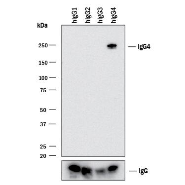
HAF018). A specific band was detected for IgG
1at approximately 240 kDa (as indicated). Human IgG (Catalog #
MAB1101) is shown as a loading control.This experiment was conducted under non-reducing conditions and using
Immunoblot Buffer Group 1." alt="Western blot shows lysates of purified human IgG
1(negative control), purified human IgG
2(negative control), purified human IgG
3(negative control), and human purified human IgG
4. PVDF membrane was probed with 2 µg/mL of Mouse Anti-Human IgG
4Monoclonal Antibody (Catalog # MAB9895) followed by HRP-conjugated Anti-Mouse IgG Secondary Antibody (Catalog #
HAF018). A specific band was detected for IgG
1at approximately 240 kDa (as indicated). Human IgG (Catalog #
MAB1101) is shown as a loading control.This experiment was conducted under non-reducing conditions and using
Immunoblot Buffer Group 1." class="big_thumb" />
DY990). Rabbit Anti-Human IgG Monoclonal Antibody (Catalog #
MAB11012) was biotinylated and incubated with the protein captured on the plate. Detection of the standard curve was achieved by incubating Streptavidin-HRP (Catalog #
DY998) followed by Substrate Solution (Catalog #
DY999) and stopping the enzymatic reaction with Stop Solution (Catalog #
DY994)." class="big_lightbox">
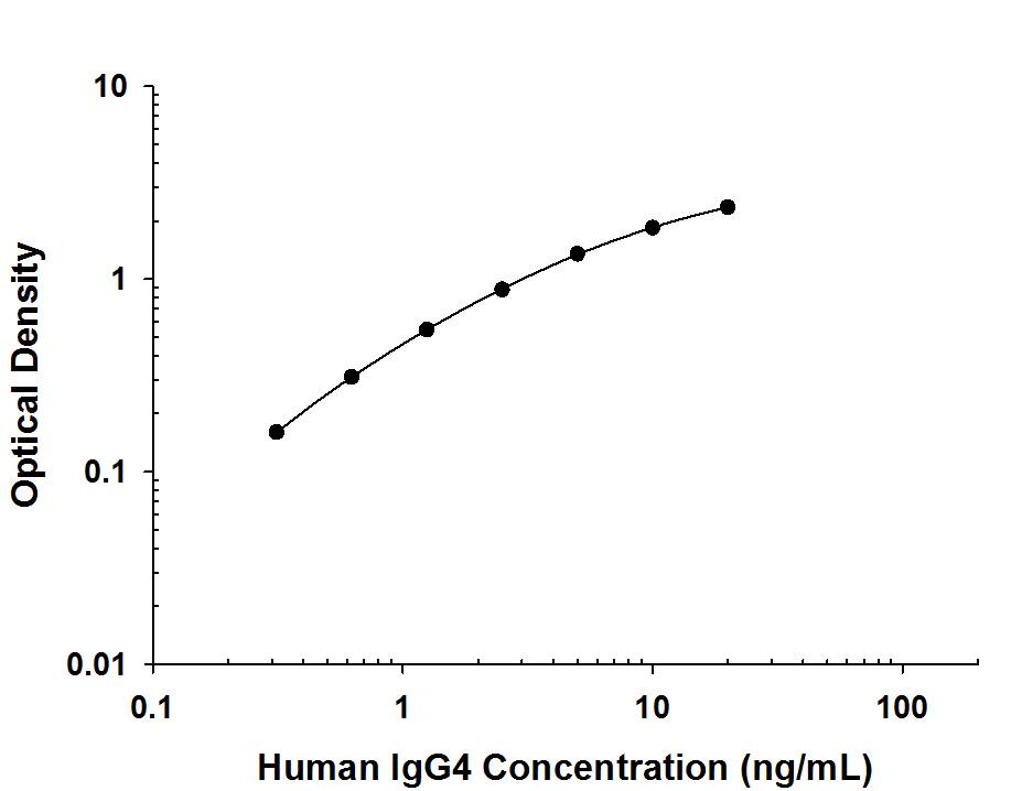
DY990). Rabbit Anti-Human IgG Monoclonal Antibody (Catalog #
MAB11012) was biotinylated and incubated with the protein captured on the plate. Detection of the standard curve was achieved by incubating Streptavidin-HRP (Catalog #
DY998) followed by Substrate Solution (Catalog #
DY999) and stopping the enzymatic reaction with Stop Solution (Catalog #
DY994)." class="big_thumb" />
 HAF018). A specific band was detected for IgG1at approximately 240 kDa (as indicated). Human IgG (Catalog # MAB1101) is shown as a loading control.This experiment was conducted under non-reducing conditions and using Immunoblot Buffer Group 1." alt="Western blot shows lysates of purified human IgG1(negative control), purified human IgG2(negative control), purified human IgG3(negative control), and human purified human IgG4. PVDF membrane was probed with 2 µg/mL of Mouse Anti-Human IgG4Monoclonal Antibody (Catalog # MAB9895) followed by HRP-conjugated Anti-Mouse IgG Secondary Antibody (Catalog # HAF018). A specific band was detected for IgG1at approximately 240 kDa (as indicated). Human IgG (Catalog # MAB1101) is shown as a loading control.This experiment was conducted under non-reducing conditions and using Immunoblot Buffer Group 1." class="big_thumb" />


 DY990). Rabbit Anti-Human IgG Monoclonal Antibody (Catalog #
 ![Immunohistochemistry-Paraffin: Ig gamma-4 Chain C Region Antibody (IGHG4/2042R) [NBP2-79716] - Formalin-fixed, paraffin-embedded human Tonsil stained with Ig gamma-4 Chain C Region Antibody (IGHG4/2042R).](http://images.novusbio.com/fullsize/Ig-gamma-4-Chain-C-Region-Antibody-IGHG4-2042R-Immunohistochemistry-Paraffin-NBP2-79716-img0001.jpg "Immunohistochemistry-Paraffin: Ig gamma-4 Chain C Region Antibody (IGHG4/2042R) [NBP2-79716] - Formalin-fixed, paraffin-embedded human Tonsil stained with Ig gamma-4 Chain C Region Antibody (IGHG4/2042R).")
![Protein Array: Ig gamma-4 Chain C Region Antibody (IGHG4/2042R) [NBP2-79716] - Analysis of Protein Array containing more than 19,000 full-length human proteins using Ig gamma-4 Chain C Region Antibody (IGHG4/2042R). Z- and S- Score: The Z-score represents the strength of a signal that a monoclonal antibody (Monoclonal Antibody) (in combination with a fluorescently-tagged anti-IgG secondary antibody) produces when binding to a particular protein on the HuProt(TM) array. Z-scores are described in units of standard deviations (SD's) above the mean value of all signals generated on that array. If targets on HuProt(TM) are arranged in descending order of the Z-score, the S-score is the difference (also in units of SD's) between the Z-score. S-score therefore represents the relative target specificity of a Monoclonal Antibody to its intended target. A Monoclonal Antibody is considered to specific to its intended target, if the Monoclonal Antibody has an S-score of at least 2.5](http://images.novusbio.com/fullsize/Ig-gamma-4-Chain-C-Region-Antibody-IGHG4-2042R-Protein-Array-NBP2-79716-img0002.jpg)

![Immunohistochemistry-Paraffin: Ig gamma-4 Chain C Region Antibody (IGHG4/1345) [NBP2-53218] - Formalin-fixed, paraffin-embedded Human Tonsil stained with IgG4 Monoclonal Antibody (IGHG4/1345).](http://images.novusbio.com/fullsize/Ig-gamma-4-Chain-C-Region-Antibody-IGHG4-1345-Immunohistochemistry-Paraffin-NBP2-53218-img0001.jpg "Immunohistochemistry-Paraffin: Ig gamma-4 Chain C Region Antibody (IGHG4/1345) [NBP2-53218] - Formalin-fixed, paraffin-embedded Human Tonsil stained with IgG4 Monoclonal Antibody (IGHG4/1345).")
![Western Blot: Mouse anti-Human Ig gamma-4 Chain C Region Secondary Antibody [H00003503-B01P] - Analysis of IGHG4 expression in human spleen.](http://images.novusbio.com/fullsize/Mouse-anti-Human-Ig-gamma-4-Chain-C-Region-Secondary-Antibody-Western-Blot-H00003503-B01P-img0001.jpg "Western Blot: Mouse anti-Human Ig gamma-4 Chain C Region Secondary Antibody [H00003503-B01P] - Analysis of IGHG4 expression in human spleen.")
![Western Blot: Mouse anti-Human Ig gamma-4 Chain C Region Secondary Antibody [H00003503-B01P] - Analysis of IGHG4 expression in transfected 293T cell line by IGHG4 polyclonal antibody. Lane 1: IGHG4 transfected lysate(52.03 KDa). Lane 2: Non-transfected lysate.](http://images.novusbio.com/fullsize/Mouse-anti-Human-Ig-gamma-4-Chain-C-Region-Secondary-Antibody-Western-Blot-H00003503-B01P-img0002.jpg)
![Immunohistochemistry-Paraffin: Ig gamma-4 Chain C Region Antibody (rIGHG4/1345) [NBP3-07432] - Formalin-fixed, paraffin-embedded human Tonsil stained with Ig gamma-4 Chain C Region Mouse Recombinant Monoclonal Antibody (rIGHG4/1345).](http://images.novusbio.com/fullsize/Ig-gamma-4-Chain-C-Region-Antibody-rIGHG4-1345-Immunohistochemistry-Paraffin-NBP3-07432-img0001.jpg "Immunohistochemistry-Paraffin: Ig gamma-4 Chain C Region Antibody (rIGHG4/1345) [NBP3-07432] - Formalin-fixed, paraffin-embedded human Tonsil stained with Ig gamma-4 Chain C Region Mouse Recombinant Monoclonal Antibody (rIGHG4/1345).")

