MAB25941) at 0.25ug/mL at 37 ° Celsius for 4 minutes. Before incubation with the primary antibody, tissue underwent an all-in-one dewaxing and antigen retrieval preprocessing using PreTreatment Module (PT Module) and Dewax and HIER Buffer H (pH 9; Epredia Catalog # TA-999-DHBH). Tissue was stained using the Alexa Fluor™ 555 Goat anti-Mouse IgG Secondary Antibody at 1:100 at 37 ° Celsius for 2 minutes. (Yellow; Lunaphore Catalog #
DR555MS) and counterstained with DAPI (blue; Lunaphore Catalog #
DR100). Specific staining was localized to the cytoplasm. Protocol available in
COMET™ Panel Builder." class="big_lightbox">
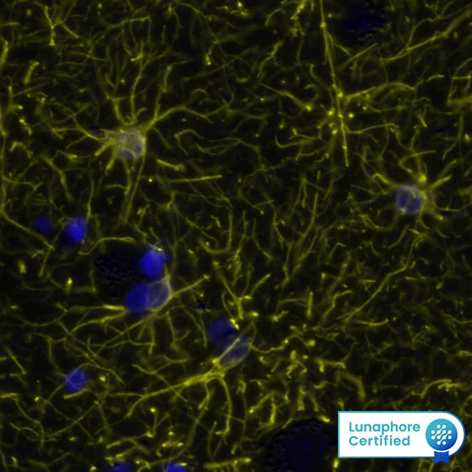
MAB25941) at 0.25ug/mL at 37 ° Celsius for 4 minutes. Before incubation with the primary antibody, tissue underwent an all-in-one dewaxing and antigen retrieval preprocessing using PreTreatment Module (PT Module) and Dewax and HIER Buffer H (pH 9; Epredia Catalog # TA-999-DHBH). Tissue was stained using the Alexa Fluor™ 555 Goat anti-Mouse IgG Secondary Antibody at 1:100 at 37 ° Celsius for 2 minutes. (Yellow; Lunaphore Catalog #
DR555MS) and counterstained with DAPI (blue; Lunaphore Catalog #
DR100). Specific staining was localized to the cytoplasm. Protocol available in
COMET™ Panel Builder." alt="GFAP was detected in immersion fixed paraffin-embedded sections of human Brain Cortex using Mouse Anti-Human GFAP Monoclonal Antibody (
MAB25941) at 0.25ug/mL at 37 ° Celsius for 4 minutes. Before incubation with the primary antibody, tissue underwent an all-in-one dewaxing and antigen retrieval preprocessing using PreTreatment Module (PT Module) and Dewax and HIER Buffer H (pH 9; Epredia Catalog # TA-999-DHBH). Tissue was stained using the Alexa Fluor™ 555 Goat anti-Mouse IgG Secondary Antibody at 1:100 at 37 ° Celsius for 2 minutes. (Yellow; Lunaphore Catalog #
DR555MS) and counterstained with DAPI (blue; Lunaphore Catalog #
DR100). Specific staining was localized to the cytoplasm. Protocol available in
COMET™ Panel Builder." class="big_thumb" />
HAF018). Specific bands were detected for GFAP at approximately 35-50 kDa (as indicated). This experiment was conducted under reducing conditions and using
Immunoblot Buffer Group 1." class="big_lightbox">
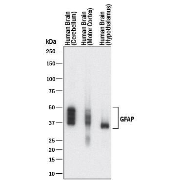
HAF018). Specific bands were detected for GFAP at approximately 35-50 kDa (as indicated). This experiment was conducted under reducing conditions and using
Immunoblot Buffer Group 1." class="big_thumb" />
NL007) and counterstained with DAPI (blue). Specific staining was localized to cytoplasm. Staining was performed using our protocol for Fluorescent ICC Staining of Non-adherent Cells." class="big_lightbox">
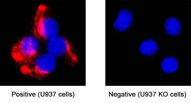
NL007) and counterstained with DAPI (blue). Specific staining was localized to cytoplasm. Staining was performed using our protocol for Fluorescent ICC Staining of Non-adherent Cells." class="big_thumb" />
 HAF016). Specific bands were detected for GFAP at approximately 35-50 kDa (as indicated). This experiment was conducted under reducing conditions and using Immunoblot Buffer Group 1." alt="Western blot shows lysates of rat cortical stem cells, rat brain tissue, human brain (cortex) tissue, and human brain (hypothalamus) tissue. PVDF membrane was probed with 0.2 µg/mL of Sheep Anti-Human GFAP Antigen Affinity-purified Polyclonal Antibody (Catalog # AF2594) followed by HRP-conjugated Anti-Sheep IgG Secondary Antibody (Catalog # HAF016). Specific bands were detected for GFAP at approximately 35-50 kDa (as indicated). This experiment was conducted under reducing conditions and using Immunoblot Buffer Group 1." class="big_thumb" />
 HAF018). Specific bands were detected for GFAP at approximately 35-50 kDa (as indicated). This experiment was conducted under reducing conditions and using Immunoblot Buffer Group 1." alt="Western blot shows lysates of human brain (motor cortex) tissue, human brain (cerebellum) tissue, and human brain (hypothalamus) tissue. PVDF membrane was probed with 1 µg/mL of Mouse Anti-Human GFAP Monoclonal Antibody (Catalog # MAB2594) followed by HRP-conjugated Anti-Mouse IgG Secondary Antibody (Catalog # HAF018). Specific bands were detected for GFAP at approximately 35-50 kDa (as indicated). This experiment was conducted under reducing conditions and using Immunoblot Buffer Group 1." class="big_thumb" />
 DR555RB) and counterstained with DAPI (blue; Lunaphore Catalog # DR100). Specific staining was localized to the astrocytes showing cytoplasmic staining. Protocol available in COMET™ Panel Builder." alt="GFAP was detected in immersion fixed paraffin-embedded sections of mouse Brain Cortex using Rabbit Anti-Mouse GFAP Monoclonal Antibody (Catalog # NBP3-07877) at 0.15ug/mL at 37 ° Celsius for 2 minutes. Before incubation with the primary antibody, tissue underwent an all-in-one dewaxing and antigen retrieval preprocessing using PreTreatment Module (PT Module) and Dewax and HIER Buffer H (pH 9; Epredia Catalog # TA-999-DHBH). Tissue was stained using the Alexa Fluor™ Plus 555 Goat anti-Rabbit IgG Secondary Antibody at 1:100 at 37 ° Celsius for 2 minutes. (Yellow; Lunaphore Catalog # DR555RB) and counterstained with DAPI (blue; Lunaphore Catalog # DR100). Specific staining was localized to the astrocytes showing cytoplasmic staining. Protocol available in COMET™ Panel Builder." class="big_thumb" />
 MAB25941) at 0.25ug/mL at 37 ° Celsius for 4 minutes. Before incubation with the primary antibody, tissue underwent an all-in-one dewaxing and antigen retrieval preprocessing using PreTreatment Module (PT Module) and Dewax and HIER Buffer H (pH 9; Epredia Catalog # TA-999-DHBH). Tissue was stained using the Alexa Fluor™ 555 Goat anti-Mouse IgG Secondary Antibody at 1:100 at 37 ° Celsius for 2 minutes. (Yellow; Lunaphore Catalog # DR555MS) and counterstained with DAPI (blue; Lunaphore Catalog # DR100). Specific staining was localized to the cytoplasm. Protocol available in COMET™ Panel Builder." alt="GFAP was detected in immersion fixed paraffin-embedded sections of human Brain Cortex using Mouse Anti-Human GFAP Monoclonal Antibody (MAB25941) at 0.25ug/mL at 37 ° Celsius for 4 minutes. Before incubation with the primary antibody, tissue underwent an all-in-one dewaxing and antigen retrieval preprocessing using PreTreatment Module (PT Module) and Dewax and HIER Buffer H (pH 9; Epredia Catalog # TA-999-DHBH). Tissue was stained using the Alexa Fluor™ 555 Goat anti-Mouse IgG Secondary Antibody at 1:100 at 37 ° Celsius for 2 minutes. (Yellow; Lunaphore Catalog # DR555MS) and counterstained with DAPI (blue; Lunaphore Catalog # DR100). Specific staining was localized to the cytoplasm. Protocol available in COMET™ Panel Builder." class="big_thumb" />


![Immunohistochemistry: GFAP Antibody [NB300-141] - Analysis of a rat cerebellum section stained with rabbit polyclonal antibody to GFAP, NB300-141, dilution 1:5000 in green and mouse monoclonal antibody to MeCP2, dilution 1:500, in red. The blue is DAPI staining of nuclear DNA. Following transcardial perfusion of rat with 4% paraformaldehyde, brain was post fixed for 1 hour, cut to 45 uM, and free-floating sections were stained with above antibodies. The GFAP antibody stains the network of astrocytic cells and the processes of Bergmann glia in the molecular layer. The MeCP2 antibody specifically labels nuclei of certain neurons.](http://images.novusbio.com/fullsize/GFAP-Antibody-Immunocytochemistry-Immunofluorescence-NB300-141-img0011.jpg "Immunohistochemistry: GFAP Antibody [NB300-141] - Analysis of a rat cerebellum section stained with rabbit polyclonal antibody to GFAP, NB300-141, dilution 1:5000 in green and mouse monoclonal antibody to MeCP2, dilution 1:500, in red. The blue is DAPI staining of nuclear DNA. Following transcardial perfusion of rat with 4% paraformaldehyde, brain was post fixed for 1 hour, cut to 45 uM, and free-floating sections were stained with above antibodies. The GFAP antibody stains the network of astrocytic cells and the processes of Bergmann glia in the molecular layer. The MeCP2 antibody specifically labels nuclei of certain neurons.")
![Immunocytochemistry/Immunofluorescence: GFAP Antibody [NB300-141] - Rat neurons stained with Neurofilament Heavy antibody NB300-217 (red) and GFAP antibody NB300-141 (green).](http://images.novusbio.com/fullsize/GFAP-Antibody-Immunocytochemistry-Immunofluorescence-NB300-141-img0002.jpg)
![Simple Western: GFAP Antibody [NB300-141] - Simple Western lane view shows a specific band for GFAP in 0.05 mg/mL of Human Brain lysate. This experiment was performed under reducing conditions using the 12-230 kDa separation system.](http://images.novusbio.com/fullsize/GFAP-Antibody-Simple-Western-NB300-141-img0004.jpg)

![Immunohistochemistry-Paraffin: GFAP Antibody [NB300-141] - Mouse brain section, 20x magnification. Antibody at 1:1000. Detection with Polymer-HRP. IHC image submitted by a verified customer review.](http://images.novusbio.com/fullsize/GFAP-Antibody-Immunohistochemistry-Paraffin-NB300-141-img0013.jpg)
![Western Blot: GFAP Antibody [NB300-141] - Analysis of Rat brain lysate. Antibody at 1:5000. Specific band running with an apparent SDS-PAGE molecular weight of ~50 kDa corresponds to rodent GFAP was observed.](http://images.novusbio.com/fullsize/GFAP-Antibody-Western-Blot-NB300-141-img0006.jpg)
![Western Blot: GFAP Antibody [NB300-141] - Analysis of GFAP expression in whole rat cerebellum homogenate.](http://images.novusbio.com/fullsize/GFAP-Antibody-Western-Blot-NB300-141-img0009.jpg)
![Western Blot: GFAP Antibody [NB300-141] - Analysis of different tissue lysates using rabbit polyclonal antibody to GFAP, NB300-141, dilution 1:5000 in green: [1] protein standard (red), [2] rat brain, [3] rat spinal cord, [4] mouse brain, [5] mouse spinal cord. Strong band at about 50 kDa corresponds to the major isotype of the GFAP protein. Smaller isotypes and proteolytic fragments of GFAP are also detected on the blot.](http://images.novusbio.com/fullsize/GFAP-Antibody-Western-Blot-NB300-141-img0012.jpg)
![Immunocytochemistry/Immunofluorescence: GFAP Antibody [NB300-141] - Analysis of mixed neuron-glial cultures using GFAP antibody NB300-141 (red) and Vimentin antibody NB300-223 (green). The fibroblastic cells contain only Vimentin and so are green. The astrocytes contain either Vimentin and GFAP (appearing golden) or predominantly GFAP (appearing red). Blue is nuclear DNA stain.](http://images.novusbio.com/fullsize/GFAP-Antibody-Immunocytochemistry-Immunofluorescence-NB300-141-img0007.jpg)
![Immunocytochemistry/Immunofluorescence: GFAP Antibody [NB300-141] - Cultured Rat hippocampal neurons. ICC/IF image submitted by a verified customer review.](http://images.novusbio.com/fullsize/GFAP-Antibody-Immunocytochemistry-Immunofluorescence-NB300-141-img0008.jpg)
![Immunohistochemistry: GFAP Antibody [NB300-141] - Xenografted mouse brain section: astocyte and human nuclei. ICC/IF image submitted by a verified customer review.](http://images.novusbio.com/fullsize/GFAP-Antibody-Immunocytochemistry-Immunofluorescence-NB300-141-img0003.jpg)
![Immunohistochemistry-Frozen: GFAP Antibody [NB300-141] - Imaging of mouse brain (cortex), 20x magnification. IHC image submitted by a verified customer review.](http://images.novusbio.com/fullsize/GFAP-Antibody-Immunohistochemistry-Frozen-NB300-141-img0010.jpg)

![Immunocytochemistry/ Immunofluorescence: GFAP Antibody [NB300-141] - EGFP+ neurons are positive for RABV antigen.Brains were collected from Cre reporter mice fifteen days post-infection, cryosectioned, & EGFP+ regions compared to cell-specific labeling, A) NeuN (blue, neuronal nuclei antibody, 20× fluorescence imaging), B) GFAP (blue, astrocyte antibody, 40× confocal imaging), or C) RABV P antigen (purple) & DAPI nuclear stain (blue, 63× confocal imaging). White arrows in (C) indicate regions positive for RABV P. Image collected & cropped by CiteAb from the following publication (//dx.plos.org/10.1371/journal.ppat.1002971), licensed under a CC-BY license. Not internally tested by Novus Biologicals.](http://images.novusbio.com/fullsize/nb300-141_rabbit-polyclonal-gfap-antibody-310202415293349.jpg)
![Western Blot: GFAP Antibody [NB300-141] - Effects of the repeated administration of NVP CXCR2 20 (NVP; 10 μg/5 μl; i.t.; 16 h & 1 h before CCI & then once a day for 7 days) on the protein levels of CXCR2, IBA1, GFAP, CXCL1, CXCL2, & CXCL3 proteins (A–I) in the spinal cord (A–F) & DRG (G–I) on the 7th day after CCI in rats. The data are presented as the mean fold changes relative to the control ± SEM (5–6 samples per group). Intergroup differences were analyzed using ANOVA with Bonferroni's multiple comparisons test. *p < 0.05, **p < 0.01, ***p < 0.001 indicate differences vs. naive rats. #p < 0.05, indicate differences between V-treated & NVP-treated rats. CCI, chronic constriction injury; N, naive; V, vehicle; NVP, NVP CXCR2 20. Image collected & cropped by CiteAb from the following publication (//pubmed.ncbi.nlm.nih.gov/31616413), licensed under a CC-BY license. Not internally tested by Novus Biologicals.](http://images.novusbio.com/fullsize/nb300-141_rabbit-polyclonal-gfap-antibody-310202415371927.jpg)
![Immunocytochemistry/ Immunofluorescence: GFAP Antibody [NB300-141] - Increased astrocyte activation in IL-1 beta -stimulated two-week old offspring is reduced by maternal LB supplementation. Representative images of fluorescence microscopy of claudin-5+ (location of the brain capillaries, red), GFAP+ astrocyte (green), & DAPI (nuclei, blue). Three to five sections per mouse were examined & at least three mice were examined in each group. Stronger than control SPF GFAP staining was observed around the blood vessel (see arrow) after IL-1 beta insult. Maternal supplemented group (LB) with or without postnatal insult had GFAP levels similar to the control group indicating that LB supplementation prevented astrocyte activation around the BBB endothelium. Image collected & cropped by CiteAb from the following publication (//pubmed.ncbi.nlm.nih.gov/32424168), licensed under a CC-BY license. Not internally tested by Novus Biologicals.](http://images.novusbio.com/fullsize/nb300-141_rabbit-polyclonal-gfap-antibody-310202415395936.jpg)
![Immunocytochemistry/ Immunofluorescence: GFAP Antibody [NB300-141] - Glial cells density is differently modulated during AD progression.a Retinal slices were immunolabeled with anti-GFAP antibody (green) & Hoechst for nuclei visualization (blue) at different ages of 3xTg-AD & non-Tg mice & density of GFAP signal was quantified as shown in b (**p < 0.01 pre vs early; n = 16 fields/four slices for each condition; two-way ANOVA, Holm-Sidak; ##p < 0.01 for comparison with age-matched non-Tg mice, two-way ANOVA, Holm-Sidak). c Representative multiarea image of retinal slice immunolabeled with anti-Iba1 antibody (green) & Hoechst for nuclei visualization (blue); density of Iba1+ cells was quantified as shown in d (**p < 0.01 vs 3xTg-AD pre; ##p < 0.01 vs age-matched non-Tg mice, n = 16 fields/four slices for each condition, two-way ANOVA, Holm-Sidak method for multiple comparison) Image collected & cropped by CiteAb from the following publication (//pubmed.ncbi.nlm.nih.gov/29880901), licensed under a CC-BY license. Not internally tested by Novus Biologicals.](http://images.novusbio.com/fullsize/nb300-141_rabbit-polyclonal-gfap-antibody-310202416205151.jpg)
![Immunohistochemistry Free-Floating: GFAP Antibody [NBP1-05198] - Staining of a section of mouse hippocampus stained with chicken pAb to GFAP, NBP1-05198, dilution 1:5000 in green and costained with rabbit pAb to FOX3/NeuN, dilution 1:5000, in red. The blue is DAPI staining of nuclear DNA. Following transcardial perfusion with 4% paraformaldehyde, mouse brain was post fixed for 24 hours, cut to 45 uM, and free-floating sections were stained with the above antibodies. The GFAP antibody stains a network of astroglial cells while the Fox3/NeuN antibody stains the nuclei and proximal perikarya of neurons.](http://images.novusbio.com/fullsize/GFAP-Antibody-NBP1-05198-img0001.jpg "Immunohistochemistry Free-Floating: GFAP Antibody [NBP1-05198] - Staining of a section of mouse hippocampus stained with chicken pAb to GFAP, NBP1-05198, dilution 1:5000 in green and costained with rabbit pAb to FOX3/NeuN, dilution 1:5000, in red. The blue is DAPI staining of nuclear DNA. Following transcardial perfusion with 4% paraformaldehyde, mouse brain was post fixed for 24 hours, cut to 45 uM, and free-floating sections were stained with the above antibodies. The GFAP antibody stains a network of astroglial cells while the Fox3/NeuN antibody stains the nuclei and proximal perikarya of neurons.")
![Western Blot: GFAP Antibody [NBP1-05198] - Analysis of whole brain lysates using chicken pAb to GFAP, NBP1-05198, dilution 1:5000 in green: [1] protein standard (red), [2] rat brain, [3] mouse brain. The strong band at about 50 kDa corresponds to the GFAP protein. Smaller proteolytic fragments and alternate transcripts of GFAP may also be detected on such blots.](http://images.novusbio.com/fullsize/GFAP-Antibody-NBP1-05198-img0002.jpg)
![Immunocytochemistry/Immunofluorescence: GFAP Antibody (GA-5) [NBP2-29415] - Cultured Rat Hippocampal Neurons. ICC/IF image submitted by a verified customer review.](http://images.novusbio.com/fullsize/GFAP-Antibody-GA-5-Immunocytochemistry-Immunofluorescence-NBP2-29415-img0007.jpg "Immunocytochemistry/Immunofluorescence: GFAP Antibody (GA-5) [NBP2-29415] - Cultured Rat Hippocampal Neurons. ICC/IF image submitted by a verified customer review.")
![Western Blot: GFAP Antibody (GA-5) [NBP2-29415] - Western Blot Analysis of human brain tissue lysate using GFAP Antibody (GA-5).](http://images.novusbio.com/fullsize/GFAP-Antibody-GA-5-Western-Blot-NBP2-29415-img0011.jpg)
![Immunohistochemistry-Paraffin: GFAP Antibody (GA-5) [NBP2-29415] - Formalin-fixed, paraffin-embedded human Cerebellum stained with GFAP Antibody (GA-5).](http://images.novusbio.com/fullsize/GFAP-Antibody-GA-5-Immunohistochemistry-Paraffin-NBP2-29415-img0010.jpg)
![Flow Cytometry: GFAP Antibody (GA-5) [NBP2-29415] - Flow Cytometric Analysis of T98G cells using GFAP Antibody (GA-5) followed by Goat anti-Mouse IgG-CF488 (Blue); Isotype Control (Red).](http://images.novusbio.com/fullsize/GFAP-Antibody-GA-5-Flow-Cytometry-NBP2-29415-img0009.jpg)
![Western Blot: GFAP Antibody (GA-5) [NBP2-29415] - Analysis of GFAP in human brain lysate using GFAP (GA5) antibody at 1 ug/mL. Goat anti-mouse Ig HRP secondary antibody and PicoTect ECL substrate solution were used for this test.](http://images.novusbio.com/fullsize/GFAP-Antibody-GA-5-Western-Blot-NBP2-29415-img0001.jpg)
![Immunohistochemistry-Paraffin: GFAP Antibody (GA-5) [NBP2-29415] - Formalin-paraffin human brain stained with GFAP Ab (GA-5). Note cytoplasmic staining.](http://images.novusbio.com/fullsize/GFAP-Antibody-GA-5-Immunohistochemistry-Paraffin-NBP2-29415-img0003.jpg)
![Flow Cytometry: GFAP Antibody (GA-5) [NBP2-29415] - Experimental autoimmune encephalomyelitis was induced in C57BL6/J mice, and mononuclear cells were isolated from the CNS at day 10 (onset of symptoms). Cells were stained for GFAP, Neun, CX3CL1, CXCL12, CCL2, CD45 and CD11b, plus for viability to exclude dead cells. GFAP staining is shown for viable cells. Flow cytometry image submitted by a verified customer review.](http://images.novusbio.com/fullsize/GFAP-Antibody-GA-5-Flow-Cytometry-NBP2-29415-img0008.jpg)
![Simple Western: GFAP Antibody (GA-5) [NBP2-29415] - Simple Western lane view shows a specific band for GFAP in 0.2 mg/mL of human brain lysate. This experiment was performed under reducing conditions using the 12-230 kDa separation system.](http://images.novusbio.com/fullsize/GFAP-Antibody-GA-5-Simple-Western-NBP2-29415-img0004.jpg)
![Simple Western: GFAP Antibody (GA-5) [NBP2-29415] - Electropherogram image of the corresponding Simple Western lane. GFAP antibody was used at 10 ug/mL dilution of human brain lysate.](http://images.novusbio.com/fullsize/GFAP-Antibody-GA-5-Simple-Western-NBP2-29415-img0006.jpg)
![Immunohistochemistry-Paraffin: Mouse Monoclonal GFAP Antibody (GA-5) [NBP2-29415] - Analysis of GFAP on adult human brain tissue. Antigen retrieval in a basic buffer x200 (1ug/mL). Image from a verified customer review.](http://images.novusbio.com/fullsize/antibody/nbp2-29415_mouse-monoclonal-gfap-antibody-ga-5-immunohistochemistry-paraffin-3102023141238..jpg)
![Immunocytochemistry/ Immunofluorescence: GFAP Antibody (GA-5) [NBP2-29415] - Advancing super‐resolution microscopy for astroglial research. (A) STED images showing CA1 stratum radiatum astrocytic processes (green, whole‐cell loading with Alexa Fluor 488) adjacent to synaptic structures in organotypic slices (red; Thy1‐YFP; dendritic spines, arrows) at lower (A1) & higher (A2, area indicted by square in A1) magnification; asterisk, O‐ring structures indicating tentative cell process envelopes; adapted from (Panatier et al., 2014). (B) STED image of P2Y1 receptors (red) along a multi‐branched astrocytic process (glutamine synthase, grey) in the adult mouse hippocampus. Adapted from (Volterra et al., 2014). (C) STORM imaging of pre‐ (Bassoon; red) & postsynaptic (Homer1; green) scaffolding proteins in the mouse main olfactory bulb glomeruli imaged using conventional fluorescence imaging (C1) & STORM (C2); adapted from (Dani et al., 2010). (D, E) dSTORM images of cultured (14 DIV) mixed glial cells from rat hippocampus (ProLong Diamond in Zeiss Elyra PS.1 microscope; Fiji Plugin ThunderSTORM, 3,000 frames); unpublished data by J. Heller. (D) GFAP stained with monoclonal antibody (Novus, GA5, secondary Alexa Fluor 488 donkey anti‐mouse antibody, Life Technologies) shown at lower (D1) & higher (D2, fragment indicated in D1) magnification. (E) GLT‐1 (polyclonal, Millipore) visualized with Alexa Fluor 568 goat anti‐guinea pig antibody (Life Technologies) shown at lower (E1) & higher (E2, fragment indicated in E1) magnification. Note that the cells were permeabilized, & therefore GLT‐1 was stained throughout cellular compartments. Image collected & cropped by CiteAb from the following publication (//pubmed.ncbi.nlm.nih.gov/25782611), licensed under a CC-BY license. Not internally tested by Novus Biologicals.](http://images.novusbio.com/fullsize/nbp2-29415_mouse-monoclonal-gfap-antibody-ga-5-imgenex-nbo-107a-20ug-310202416114755.jpg)


![Immunohistochemistry Free-Floating: GFAP Antibody (5C10) [NBP1-05197] - Analysis of rat cerebellum section stained with mouse GFAP mAb, dilution 1:1,000 (Green), costained with rabbit neurofilament NF-L pAb, dilution 1:2,000 (Red). Following transcardial perfusion with 4% paraformaldehyde, brain was post fixed for 24hrs, cut to 45uM, and free-floating sections were stained with antibodies. The GFAP antibody stains a network of astroglial cells, while the NF-L antibody labels neuronal cells and their processes.](http://images.novusbio.com/fullsize/GFAP-Antibody-5C10-Immunohistochemistry-Free-Floating-NBP1-05197-img0007.jpg "Immunohistochemistry Free-Floating: GFAP Antibody (5C10) [NBP1-05197] - Analysis of rat cerebellum section stained with mouse GFAP mAb, dilution 1:1,000 (Green), costained with rabbit neurofilament NF-L pAb, dilution 1:2,000 (Red). Following transcardial perfusion with 4% paraformaldehyde, brain was post fixed for 24hrs, cut to 45uM, and free-floating sections were stained with antibodies. The GFAP antibody stains a network of astroglial cells, while the NF-L antibody labels neuronal cells and their processes.")
![Simple Western: GFAP Antibody (5C10) [NBP1-05197] - Simple Western lane view shows a specific band for GFAP in 0.05 mg/ml of Human Brain lysate. This experiment was performed under reducing conditions using the 12-230 kDa separation system.](http://images.novusbio.com/fullsize/GFAP-Antibody-5C10-Simple-Western-NBP1-05197-img0004.jpg)

![Immunohistochemistry: GFAP Antibody (5C10) [NBP1-05197] - Analysis of rat cerebellum section stained with mouse mAb to GFAP, NBP1-05197, dilution 1:1,000, in green, costained with rabbit pAb to neurofilament NF-L, dilution 1:2,000, in red. Following transcardial perfusion of rat with 4% paraformaldehyde, brain was post fixed for 24 hours, cut to 45uM, and free-floating sections were stained with above antibodies. The NBP1-05197 antibody stains a network of astroglial cells, while the NF-L antibody labels neuronal cells and their processes.](http://images.novusbio.com/fullsize/GFAP-Antibody-5C10-Immunocytochemistry-Immunofluorescence-NBP1-05197-img0005.jpg)
![Western Blot: GFAP Antibody (5C10) [NBP1-05197] - Analysis of whole tissue lysates using mouse mAb to GFAP, NBP1-05197, dilution 1:2,000, in green: [1] protein standard (red), [2] rat brain, [3] rat spinal cord, [4] mouse brain, [5] mouse spinal cord. The strong band at about 50kDa corresponds to the GFAP protein.](http://images.novusbio.com/fullsize/GFAP-Antibody-5C10-Western-Blot-NBP1-05197-img0006.jpg)
![Western Blot: GFAP Antibody [NB100-53809] - (0.005ug/ml) staining of Human Cerebellum (A) and (0.01ug/ml) Cerebral Cortex (B) (35ug protein in RIPA buffer). Detected by chemiluminescence.](http://images.novusbio.com/fullsize/GFAP-Antibody-Western-Blot-NB100-53809-img0002.jpg "Western Blot: GFAP Antibody [NB100-53809] - (0.005ug/ml) staining of Human Cerebellum (A) and (0.01ug/ml) Cerebral Cortex (B) (35ug protein in RIPA buffer). Detected by chemiluminescence.")
![Western Blot: GFAP Antibody [NB100-53809] - (0.1ug/ml) staining of Rat Brain lysate (35ug protein in RIPA buffer). Detected by chemiluminescence.](http://images.novusbio.com/fullsize/GFAP-Antibody-Western-Blot-NB100-53809-img0001.jpg)
 MAB1195). GFAP was detected in rat astrocytes using 10 µg/mL Sheep Anti-Human GFAP Antigen Affinity-purified Poly-clonal Antibody (Catalog # AF2594). Cells were incubated with primary antibodies for 3 hours at room temperature. Cells were stained for beta-III Tubulin using the Northern-Lights™ 557-conjugated Anti-Mouse IgG Secondary Antibody (red; Catalog #
  NL010) and counterstained with DAPI (blue). View our protocol for
  NL010) and counterstained with DAPI (blue). View our protocol for
  HAF016). This experiment was conducted under reducing conditions and using the 12-230 kDa separation system." class="big_thumb" />
 HAF016). This experiment was conducted under reducing conditions and using the 12-230 kDa separation system." class="big_thumb" />


 NL007) and counterstained with DAPI (blue). Specific staining was localized to cytoplasm. View our protocol for
 NL007) and counterstained with DAPI (blue). Specific staining was localized to cytoplasm. Staining was performed using our protocol for Fluorescent ICC Staining of Non-adherent Cells." class="big_thumb" />
![Western Blot: GFAP Antibody (012) [NBP2-89203] - Lane A: Mouse brain tissue lysate Lysates/proteins at 30 ug per lane. Secondary Goat Anti-Rabbit IgG H&L (DyLight 800) at 1:10000 dilution. Developed using the Odyssey technique. Performed under reducing conditions. Predicted band size: 50 kDa. Observed band size: 45 kDa.](http://images.novusbio.com/fullsize/GFAP-Antibody-012-Western-Blot-NBP2-89203-img0001.jpg "Western Blot: GFAP Antibody (012) [NBP2-89203] - Lane A: Mouse brain tissue lysate Lysates/proteins at 30 ug per lane. Secondary Goat Anti-Rabbit IgG H&L (DyLight 800) at 1:10000 dilution. Developed using the Odyssey technique. Performed under reducing conditions. Predicted band size: 50 kDa. Observed band size: 45 kDa.")
![Immunocytochemistry/Immunofluorescence: GFAP Antibody (ASTRO/1974R) [NBP3-07877] - Immunofluorescence Analysis of methanol-fixed human cerebral cortex cryosection stained with CF405S GFAP antibody (ASTRO/1974R) (blue) and CF647 Monoclonal Mouse Anti-Histone H1 (HH1/957) (magenta).](http://images.novusbio.com/fullsize/GFAP-Antibody-ASTRO-1974R-Immunocytochemistry-Immunofluorescence-NBP3-07877-img0003.jpg)
![Immunohistochemistry-Paraffin: GFAP Antibody (ASTRO/1974R) [NBP3-07877] - Formalin-fixed, paraffin-embedded human Cerebellum stained with GFAP Rabbit Recombinant Monoclonal Antibody (ASTRO/1974R).](http://images.novusbio.com/fullsize/GFAP-Antibody-ASTRO-1974R-Immunohistochemistry-Paraffin-NBP3-07877-img0002.jpg)
![Flow Cytometry: GFAP Antibody (ASTRO/1974R) [NBP3-07877] - Flow Cytometric Analysis of T98G cells using GFAP Rabbit Recombinant Monoclonal Antibody (ASTRO/1974R) followed by Goat anti-Rabbit IgG-CF488 (Blue); Isotype Control (Red).](http://images.novusbio.com/fullsize/GFAP-Antibody-ASTRO-1974R-Flow-Cytometry-NBP3-07877-img0001.jpg)

![Western Blot: GFAP Antibody (SPM248) [NBP2-34353] - Western Blot Analysis of human brain tissue lysate using GFAP Antibody (SPM248).](http://images.novusbio.com/fullsize/GFAP-Antibody-SPM248-Western-Blot-NBP2-34353-img0003.jpg "Western Blot: GFAP Antibody (SPM248) [NBP2-34353] - Western Blot Analysis of human brain tissue lysate using GFAP Antibody (SPM248).")
![Immunohistochemistry-Paraffin: GFAP Antibody (SPM248) [NBP2-34353] - Formalin-fixed, paraffin-embedded human schwannoma stained with GFAP monoclonal antibody (SPM248).](http://images.novusbio.com/fullsize/GFAP-Antibody-SPM248-Immunohistochemistry-Paraffin-NBP2-34353-img0001.jpg)
![Flow Cytometry: GFAP Antibody (SPM248) [NBP2-34353] - Flow Cytometric Analysis of T98G cells using GFAP Antibody (SPM248) followed by Goat anti-Mouse IgG-CF488 (Blue); Isotype Control (Red).](http://images.novusbio.com/fullsize/GFAP-Antibody-SPM248-Flow-Cytometry-NBP2-34353-img0002.jpg)
 HAF018). Specific bands were detected for GFAP at approximately 35-50 kDa (as indicated). This experiment was conducted under reducing conditions and using
  NL007) and counterstained with DAPI (blue). Specific staining was localized to cytoplasm. View our protocol for
  VC001). Tissue was stained using DAB (brown) and counterstained with hematoxylin (blue). Specific staining was localized to glial cells. View our protocol for
  NL007) and counterstained with DAPI (blue). Specific staining was localized to cytoplasm. Staining was performed using our protocol for Fluorescent ICC Staining of Non-adherent Cells." class="big_thumb" />

![Western Blot: GFAP Antibody [NB110-93480] - Rat cortex lysate showing specific immunolabeling of the ~ 50k GFAP protein.](http://images.novusbio.com/fullsize/GFAP-Antibody-Western-Blot-NB110-93480-img0001.jpg "Western Blot: GFAP Antibody [NB110-93480] - Rat cortex lysate showing specific immunolabeling of the ~ 50k GFAP protein.")
![Immunocytochemistry/Immunofluorescence: GFAP Antibody [NB110-93480] - Immunolabeling of mouse hippocampal cultures labeled with Anti-GFAP (1:1000, green) and anti-MAP2 (1:1000, red).](http://images.novusbio.com/fullsize/GFAP-Antibody-Immunocytochemistry-Immunofluorescence-NB110-93480-img0006.jpg)
![Immunocytochemistry/Immunofluorescence: GFAP Antibody [NB110-93480] - Showing specific labeling of GFAP (red) and vimentin (green) cultured neurons and glia. Cells containing GFAP and vimentin appear yellow.](http://images.novusbio.com/fullsize/GFAP-Antibody-Immunocytochemistry-Immunofluorescence-NB110-93480-img0002.jpg)
![Immunocytochemistry/Immunofluorescence: GFAP Antibody [NB110-93480] - Immunofluorescence of a section of rat cerebellum labeled with anti-GFAP (1:5000, green), colabeled with anti-MeCP2 (1:500, red), and DAPI staining of nuclear DNA. The anti-GFAP labels the astrocytic cells and the processes of Bergmann glia in the molecular layer.](http://images.novusbio.com/fullsize/GFAP-Antibody-Immunocytochemistry-Immunofluorescence-NB110-93480-img0003.jpg)
![Immunocytochemistry/Immunofluorescence: GFAP Antibody [NB110-93480] - Immunofluorescence of a section of rat cerebellum labeled with anti-GFAP (1:5000,green), colabeled with anti-calbindin (1:2,000, red), and DAPI staining of nuclear DNA. The anti-calbindin prominently labels the dendrites and perikarya of Purkinje cells in the molecular layer of the cerebellum. The anti-GFAP labels the processes of Bergmann glia in the molecular layer and the astroglia in the granular and white layers of the cerebellum.](http://images.novusbio.com/fullsize/GFAP-Antibody-Immunocytochemistry-Immunofluorescence-NB110-93480-img0004.jpg)
![Immunocytochemistry/Immunofluorescence: GFAP Antibody [NB110-93480] - Immunolabeling of mouse cortical cultures labeled with Anti-GFAP (1:1000, green) and anti-MAP2 (1:1000, red).](http://images.novusbio.com/fullsize/GFAP-Antibody-Immunocytochemistry-Immunofluorescence-NB110-93480-img0005.jpg)


