MAB6330) at 20 µg/mL at 37 ° Celsius for 4 minutes. Before incubation with the primary antibody, tissue underwent an all-in-one dewaxing and antigen retrieval preprocessing using PreTreatment Module (PT Module) and Dewax and HIER Buffer H (pH 9). Tissue was stained using the Alexa Fluor™ 647 Goat anti-Mouse IgG Secondary Antibody at 1:200 at 37 ° Celsius for 2 minutes. (Yellow; Lunaphore Catalog #
DR647MS) and counterstained with DAPI (blue; Lunaphore Catalog #
DR100). Specific staining was localized to the nucleus with dim cytoplasmic background signal. Protocol available in
COMET™ Panel Builder." class="big_lightbox">

MAB6330) at 20 µg/mL at 37 ° Celsius for 4 minutes. Before incubation with the primary antibody, tissue underwent an all-in-one dewaxing and antigen retrieval preprocessing using PreTreatment Module (PT Module) and Dewax and HIER Buffer H (pH 9). Tissue was stained using the Alexa Fluor™ 647 Goat anti-Mouse IgG Secondary Antibody at 1:200 at 37 ° Celsius for 2 minutes. (Yellow; Lunaphore Catalog #
DR647MS) and counterstained with DAPI (blue; Lunaphore Catalog #
DR100). Specific staining was localized to the nucleus with dim cytoplasmic background signal. Protocol available in
COMET™ Panel Builder." alt="GATA-3 was detected in immersion fixed paraffin-embedded sections of human breast cancer using Mouse Anti-Human GATA‑3 Monoclonal Antibody (
MAB6330) at 20 µg/mL at 37 ° Celsius for 4 minutes. Before incubation with the primary antibody, tissue underwent an all-in-one dewaxing and antigen retrieval preprocessing using PreTreatment Module (PT Module) and Dewax and HIER Buffer H (pH 9). Tissue was stained using the Alexa Fluor™ 647 Goat anti-Mouse IgG Secondary Antibody at 1:200 at 37 ° Celsius for 2 minutes. (Yellow; Lunaphore Catalog #
DR647MS) and counterstained with DAPI (blue; Lunaphore Catalog #
DR100). Specific staining was localized to the nucleus with dim cytoplasmic background signal. Protocol available in
COMET™ Panel Builder." class="big_thumb" />
HAF007). Specific bands were detected for full length (FL) GATA-3 at approximately 52 kDa and the splice form (SF) found in MCF-7 cells at approximately 40 kDa (as indicated). This experiment was conducted under reducing conditions and using
Immunoblot Buffer Group 1." class="big_lightbox">
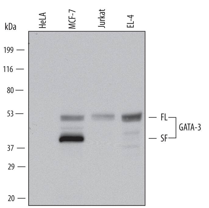
HAF007). Specific bands were detected for full length (FL) GATA-3 at approximately 52 kDa and the splice form (SF) found in MCF-7 cells at approximately 40 kDa (as indicated). This experiment was conducted under reducing conditions and using
Immunoblot Buffer Group 1." class="big_thumb" />
VC001). Tissue was stained using ACD RNAscope® 2.5 HD Duplex Detection Reagents (Catalog # 322500)." class="big_lightbox">
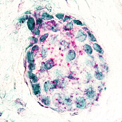
VC001). Tissue was stained using ACD RNAscope® 2.5 HD Duplex Detection Reagents (Catalog # 322500)." class="big_thumb" />
 HAF109). Specific bands were detected for GATA-3 full length (FL) at approximately 55 kDa and the splice form (SF) at approximately 40 kDa (as indicated). This experiment was conducted under reducing conditions and using Immunoblot Buffer Group 1." alt="Western blot shows lysates of MCF-7 human breast cancer cell line, Jurkat human acute T cell leukemia cell line, and EL-4 mouse lymphoblast cell line. PVDF membrane was probed with 0.25 µg/mL of Goat Anti-Human GATA-3 Antigen Affinity-purified Polyclonal Antibody (Catalog # AF2605) followed by HRP-conjugated Anti-Goat IgG Secondary Antibody (Catalog # HAF109). Specific bands were detected for GATA-3 full length (FL) at approximately 55 kDa and the splice form (SF) at approximately 40 kDa (as indicated). This experiment was conducted under reducing conditions and using Immunoblot Buffer Group 1." class="big_thumb" />
 MAB6330) at 20 µg/mL at 37 ° Celsius for 4 minutes. Before incubation with the primary antibody, tissue underwent an all-in-one dewaxing and antigen retrieval preprocessing using PreTreatment Module (PT Module) and Dewax and HIER Buffer H (pH 9). Tissue was stained using the Alexa Fluor™ 647 Goat anti-Mouse IgG Secondary Antibody at 1:200 at 37 ° Celsius for 2 minutes. (Yellow; Lunaphore Catalog # DR647MS) and counterstained with DAPI (blue; Lunaphore Catalog # DR100). Specific staining was localized to the nucleus with dim cytoplasmic background signal. Protocol available in COMET™ Panel Builder." alt="GATA-3 was detected in immersion fixed paraffin-embedded sections of human breast cancer using Mouse Anti-Human GATA‑3 Monoclonal Antibody (MAB6330) at 20 µg/mL at 37 ° Celsius for 4 minutes. Before incubation with the primary antibody, tissue underwent an all-in-one dewaxing and antigen retrieval preprocessing using PreTreatment Module (PT Module) and Dewax and HIER Buffer H (pH 9). Tissue was stained using the Alexa Fluor™ 647 Goat anti-Mouse IgG Secondary Antibody at 1:200 at 37 ° Celsius for 2 minutes. (Yellow; Lunaphore Catalog # DR647MS) and counterstained with DAPI (blue; Lunaphore Catalog # DR100). Specific staining was localized to the nucleus with dim cytoplasmic background signal. Protocol available in COMET™ Panel Builder." class="big_thumb" />
 HAF018). Specific bands were detected for GATA-3 full length (FL) at approximately 50 kDa and the splice form (SF) at approximately 37 kDa (as indicated). This experiment was conducted under reducing conditions and using Immunoblot Buffer Group 3." alt="Western blot shows lysates of MCF-7 human breast cancer cell line. PVDF membrane was probed with 2 µg/mL of Mouse Anti-Human GATA-3 Monoclonal Antibody (Catalog # MAB26051) followed by HRP-conjugated Anti-Mouse IgG Secondary Antibody (Catalog # HAF018). Specific bands were detected for GATA-3 full length (FL) at approximately 50 kDa and the splice form (SF) at approximately 37 kDa (as indicated). This experiment was conducted under reducing conditions and using Immunoblot Buffer Group 3." class="big_thumb" />
 HAF018). Specific bands were detected for GATA-3 full length (FL) at approximately 50 kDa and the splice form (SF) at approximately 37 kDa (as indicated). This experiment was conducted under reducing conditions and using Immunoblot Buffer Group 3." alt="Western blot shows lysates of MCF-7 human breast cancer cell line. PVDF membrane was probed with 2 µg/mL of Mouse Anti-Human GATA-3 Monoclonal Antibody (Catalog # MAB26052) followed by HRP-conjugated Anti-Mouse IgG Secondary Antibody (HAF018). Specific bands were detected for GATA-3 full length (FL) at approximately 50 kDa and the splice form (SF) at approximately 37 kDa (as indicated). This experiment was conducted under reducing conditions and using Immunoblot Buffer Group 3." class="big_thumb" />


 CTS013). Tissue was stained using the Anti-Goat HRP-DAB Cell & Tissue Staining Kit (brown; Catalog #
  NL001) and counterstained with DAPI (blue, lower panel). View our protocol for
  CTS013). Tissue was stained using the Anti-Goat HRP-DAB Cell & Tissue Staining Kit (brown; Catalog #
  HAF109). This experiment was conducted under reducing conditions and using the 12-230 kDa separation system." class="big_thumb" />

 HAF007). Specific bands were detected for full length (FL) GATA-3 at approximately 52 kDa and the splice form (SF) found in MCF-7 cells at approximately 40 kDa (as indicated). This experiment was conducted under reducing conditions and using
  NL007) and counterstained with DAPI (blue). Specific staining was localized to nuclei. View our protocol for
  CTS013). Tissue was stained using the Anti-Mouse HRP-DAB Cell & Tissue Staining Kit (brown
 VC001). Tissue was stained using ACD RNAscope® 2.5 HD Duplex Detection Reagents (Catalog # 322500)." class="big_thumb" />
![Western Blot: GATA-3 Antibody (OTI8H4) [NBP2-45810] - Analysis (35 ug) from 3 different cell lines by using anti-GATA3 monoclonal antibody at 1:200.](http://images.novusbio.com/fullsize/GATA-3-Antibody-8H4-Western-Blot-NBP2-45810-img0013.jpg "Western Blot: GATA-3 Antibody (OTI8H4) [NBP2-45810] - Analysis (35 ug) from 3 different cell lines by using anti-GATA3 monoclonal antibody at 1:200.")
![Immunohistochemistry-Paraffin: GATA-3 Antibody (OTI8H4) [NBP2-45810] - Mouse lung tissue section stained with GATA-3 antibody. IHC-P image submitted by a verified customer review.](http://images.novusbio.com/fullsize/GATA-3-Antibody-OTI8H4-Immunohistochemistry-Paraffin-NBP2-45810-img0014.jpg)
![Flow Cytometry: GATA-3 Antibody (OTI8H4) [NBP2-45810] - Flow cytometric Analysis of penetrated MCF-7 cells, compared to a nonspecific negative control antibody, (Blue). The right is HUVEC cells as negtive control (1:100).](http://images.novusbio.com/fullsize/GATA-3-Antibody-8H4-Flow-Cytometry-NBP2-45810-img0011.jpg)
![Immunohistochemistry: GATA-3 Antibody (OTI8H4) [NBP2-45810] - Analysis of Human breast tissue. Heat-induced epitope retrieval by 1 mM EDTA in 10 mM Tris buffer (pH 8.5) at 120C for 3 minutes.](http://images.novusbio.com/fullsize/GATA-3-Antibody-8H4-Immunohistochemistry-NBP2-45810-img0001.jpg)
![Immunohistochemistry: GATA-3 Antibody (OTI8H4) [NBP2-45810] - Analysis of Adenocarcinoma of Human breast tissue. Heat-induced epitope retrieval by 1 mM EDTA in 10 mM Tris buffer (pH 8.5) at 120C for 3 minutes.](http://images.novusbio.com/fullsize/GATA-3-Antibody-8H4-Immunohistochemistry-NBP2-45810-img0002.jpg)
![Immunohistochemistry: GATA-3 Antibody (OTI8H4) [NBP2-45810] - Analysis of Human colon tissue. Heat-induced epitope retrieval by 1 mM EDTA in 10 mM Tris buffer (pH 8.5) at 120C for 3 minutes.](http://images.novusbio.com/fullsize/GATA-3-Antibody-8H4-Immunohistochemistry-NBP2-45810-img0003.jpg)
![Immunohistochemistry: GATA-3 Antibody (OTI8H4) [NBP2-45810] - Analysis of Human Kidney tissue. Heat-induced epitope retrieval by 1 mM EDTA in 10 mM Tris buffer (pH 8.5) at 120C for 3 minutes.](http://images.novusbio.com/fullsize/GATA-3-Antibody-8H4-Immunohistochemistry-NBP2-45810-img0004.jpg)
![Immunohistochemistry: GATA-3 Antibody (OTI8H4) [NBP2-45810] - Analysis of Carcinoma of Human kidney tissue. Heat-induced epitope retrieval by 1 mM EDTA in 10 mM Tris buffer (pH 8.5) at 120C for 3 minutes.](http://images.novusbio.com/fullsize/GATA-3-Antibody-8H4-Immunohistochemistry-NBP2-45810-img0005.jpg)
![Immunohistochemistry: GATA-3 Antibody (OTI8H4) [NBP2-45810] - Analysis of Adenocarcinoma of Human endometrium tissue. Heat-induced epitope retrieval by 1 mM EDTA in 10 mM Tris buffer (pH 8.5) at 120C for 3 minutes.](http://images.novusbio.com/fullsize/GATA-3-Antibody-8H4-Immunohistochemistry-NBP2-45810-img0006.jpg)
![Immunohistochemistry: GATA-3 Antibody (OTI8H4) [NBP2-45810] - Analysis of Human prostate tissue. Heat-induced epitope retrieval by 1 mM EDTA in 10 mM Tris buffer (pH 8.5) at 120C for 3 minutes.](http://images.novusbio.com/fullsize/GATA-3-Antibody-8H4-Immunohistochemistry-NBP2-45810-img0007.jpg)
![Immunohistochemistry: GATA-3 Antibody (OTI8H4) [NBP2-45810] - Analysis of Carcinoma of Human prostate tissue. Heat-induced epitope retrieval by 1 mM EDTA in 10 mM Tris buffer (pH 8.5) at 120C for 3 minutes.](http://images.novusbio.com/fullsize/GATA-3-Antibody-8H4-Immunohistochemistry-NBP2-45810-img0008.jpg)
![Immunohistochemistry: GATA-3 Antibody (OTI8H4) [NBP2-45810] - Analysis of Human bladder tissue. Heat-induced epitope retrieval by 1 mM EDTA in 10 mM Tris buffer (pH 8.5) at 120C for 3 minutes.](http://images.novusbio.com/fullsize/GATA-3-Antibody-8H4-Immunohistochemistry-NBP2-45810-img0009.jpg)
![Immunohistochemistry: GATA-3 Antibody (OTI8H4) [NBP2-45810] - Analysis of Human lymphoma tissue. Heat-induced epitope retrieval by 1 mM EDTA in 10 mM Tris buffer (pH 8.5) at 120C for 3 minutes.](http://images.novusbio.com/fullsize/GATA-3-Antibody-8H4-Immunohistochemistry-NBP2-45810-img0010.jpg)
 NL007) and counterstained with DAPI (blue, lower panel). View our protocol for
 ![Immunohistochemistry-Paraffin: GATA-3 Antibody (CL8660) [NBP2-88917] - Analysis in human skin and pancreas tissues using NBP2-88917 antibody. Corresponding GATA3 RNA-seq data are presented for the same tissues.](http://images.novusbio.com/fullsize/GATA-3-Antibody-CL8660-Immunohistochemistry-Paraffin-NBP2-88917-img0007.jpg "<b>Orthogonal Strategies Validation. </b>Immunohistochemistry-Paraffin: GATA-3 Antibody (CL8660) [NBP2-88917] - Analysis in human skin and pancreas tissues using NBP2-88917 antibody. Corresponding GATA3 RNA-seq data are presented for the same tissues.")
![<b>Orthogonal Strategies Validation. </b>Western Blot: GATA-3 Antibody (CL8660) [NBP2-88917] - Analysis in human cell line TD47D.](http://images.novusbio.com/fullsize/GATA-3-Antibody-CL8660-Western-Blot-NBP2-88917-img0008.jpg)
![Immunocytochemistry/Immunofluorescence: GATA-3 Antibody (CL8660) [NBP2-88917] - Staining of MCF7 cells using the Anti-GATA3 monoclonal antibody, showing specific staining in the nucleoplasm in green. Microtubule- and nuclear probes are visualized in red and blue, respectively (where available).](http://images.novusbio.com/fullsize/GATA-3-Antibody-CL8660-Immunocytochemistry-Immunofluorescence-NBP2-88917-img0001.jpg)
![Immunohistochemistry-Paraffin: GATA-3 Antibody (CL8660) [NBP2-88917] - Staining of human breast cancer shows moderate to strong nuclear positivity in tumor cells.](http://images.novusbio.com/fullsize/GATA-3-Antibody-CL8660-Immunohistochemistry-Paraffin-NBP2-88917-img0002.jpg)
![Immunohistochemistry-Paraffin: GATA-3 Antibody (CL8660) [NBP2-88917] - Staining of human kidney shows strong nuclear positivity in a small subset of cells in glomeruli.](http://images.novusbio.com/fullsize/GATA-3-Antibody-CL8660-Immunohistochemistry-Paraffin-NBP2-88917-img0003.jpg)
![Immunohistochemistry-Paraffin: GATA-3 Antibody (CL8660) [NBP2-88917] - Staining of human pancreas shows no positivity in exocrine glandular cells as expected.](http://images.novusbio.com/fullsize/GATA-3-Antibody-CL8660-Immunohistochemistry-Paraffin-NBP2-88917-img0004.jpg)
![Immunohistochemistry-Paraffin: GATA-3 Antibody (CL8660) [NBP2-88917] - Staining of human placenta shows moderate to strong nuclear positivity in trophoblastic cells.](http://images.novusbio.com/fullsize/GATA-3-Antibody-CL8660-Immunohistochemistry-Paraffin-NBP2-88917-img0005.jpg)
![Immunohistochemistry-Paraffin: GATA-3 Antibody (CL8660) [NBP2-88917] - Staining of human skin shows moderate to strong nuclear positivity in squamous epithelial cells.](http://images.novusbio.com/fullsize/GATA-3-Antibody-CL8660-Immunohistochemistry-Paraffin-NBP2-88917-img0006.jpg)

![Western Blot: GATA-3 Antibody [NBP1-77115] - Analysis in K562 cell lysate with antibody at 1 ug/mL.](http://images.novusbio.com/fullsize/GATA-3-Antibody-Western-Blot-NBP1-77115-img0010.jpg "Western Blot: GATA-3 Antibody [NBP1-77115] - Analysis in K562 cell lysate with antibody at 1 ug/mL.")
![Immunocytochemistry/ Immunofluorescence: GATA-3 Antibody - BSA Free [NBP1-77115] - Immunofluorescence of GATA-3 in Rat Brain tissue with GATA-3 antibody at 20 u/mL.](http://images.novusbio.com/fullsize/nbp1-77115_rabbit-polyclonal-gata-3-antibody-2711202420212392.jpg)
![Immunohistochemistry: GATA-3 Antibody - BSA Free [NBP1-77115] - Immunohistochemistry of GATA-3 in mouse brain tissue with GATA-3 antibody at 2.5 ug/ml.](http://images.novusbio.com/fullsize/nbp1-77115_rabbit-polyclonal-gata-3-antibody-2711202419463470.gif)
![Immunohistochemistry: GATA-3 Antibody - BSA Free [NBP1-77115] - Immunohistochemistry of GATA-3 in rat brain tissue with GATA-3 antibody at 2.5 ug/mL.](http://images.novusbio.com/fullsize/nbp1-77115_rabbit-polyclonal-gata-3-antibody-2711202419172230.jpg)
![Immunocytochemistry/ Immunofluorescence: GATA-3 Antibody - BSA Free [NBP1-77115] - Immunofluorescence of GATA-3 in mouse brain tissue with GATA-3 Antibody at 20 ug/mL.](http://images.novusbio.com/fullsize/nbp1-77115_rabbit-polyclonal-gata-3-antibody-271120242042572.gif)
![Western Blot: GATA-3 Antibody (BLR121H) [NBP3-14705] - Whole cell lysate (50 ug) from BW5147.3, mIMCD-3, A20, NIH 3T3, CT26, EL4, RenCa, CH27, C2C12, and TCMK-1 cells prepared using NETN lysis buffer. Antibody: Rabbit anti-GATA3 recombinant monoclonal antibody [BLR121H] used at 1:1000. Secondary: HRPconjugated goat anti-rabbit IgG. Chemiluminescence with an exposure time of 3 minutes.](http://images.novusbio.com/fullsize/GATA-3-Antibody-BLR121H-Western-Blot-NBP3-14705-img0003.jpg "Western Blot: GATA-3 Antibody (BLR121H) [NBP3-14705] - Whole cell lysate (50 ug) from BW5147.3, mIMCD-3, A20, NIH 3T3, CT26, EL4, RenCa, CH27, C2C12, and TCMK-1 cells prepared using NETN lysis buffer. Antibody: Rabbit anti-GATA3 recombinant monoclonal antibody [BLR121H] used at 1:1000. Secondary: HRPconjugated goat anti-rabbit IgG. Chemiluminescence with an exposure time of 3 minutes.")
![Immunocytochemistry/Immunofluorescence: GATA-3 Antibody (BLR121H) [NBP3-14705] - Rabbit anti-GATA3 recombinant monoclonal antibody [BLR121H]. Secondary: HRP-conjugated goat anti-rabbit IgG. Substrate: DAB.](http://images.novusbio.com/fullsize/GATA-3-Antibody-BLR121H-Immunocytochemistry-Immunofluorescence-NBP3-14705-img0005.jpg)
![Immunohistochemistry-Paraffin: GATA-3 Antibody (BLR121H) [NBP3-14705] - Rabbit anti-GATA3 recombinant monoclonal antibody [BLR121H]. Secondary: HRP-conjugated goat anti-rabbit IgG. Substrate: DAB.](http://images.novusbio.com/fullsize/GATA-3-Antibody-BLR121H-Immunohistochemistry-Paraffin-NBP3-14705-img0004.jpg)
![Western Blot: GATA-3 Antibody (BLR121H) [NBP3-14705] - Whole cell lysate (10 ug) from HEK293T, Hep-G2, Jurkat, RKO, K-562, MCF-7, and HeLa cells prepared using NETN lysis buffer. Antibody: Rabbit anti-GATA3 recombinant monoclonal antibody [BLR121H] used at 1:1000. Secondary: HRP-conjugated goat anti-rabbit IgG. Chemiluminescence with an exposure timeof 30 seconds.](http://images.novusbio.com/fullsize/GATA-3-Antibody-BLR121H-Western-Blot-NBP3-14705-img0002.jpg)
![Immunoprecipitation: GATA-3 Antibody [NBP3-14705] - Whole cell lysate (1.0 mgper IP reaction; 20% of IP loaded) from MCF-7 cellsprepared using NETN lysis buffer. Antibodies: Rabbit antiGATA3 recombinant monoclonal antibody used for IP at 6 ul per reaction. GATA3was also immunoprecipitated by rabbit anti-GATA3antibody For blotting immunoprecipitatedGATA3, A700-121 was used at 1:1000.Chemiluminescence with an exposure time of 3 seconds.](http://images.novusbio.com/fullsize/GATA-3-Antibody-Immunoprecipitation-NBP3-14705-img0001.jpg)
 FAB3791P) and either (A) Mouse Anti-Human GATA-3 Monoclonal Antibody (Catalog # MAB26052) or (B) Mouse IgG2BFlow Cytometry Isotype Control (
 MAB004, open histogram), followed by Allophycocyanin-conjugated Anti-Mouse IgG Secondary Antibody (Catalog #
 ![Immunohistochemistry: GATA-3 Antibody (A3G3-R) [NBP3-32375] - Immunohistochemical analysis of paraffin-embedded human breast carcinoma tissue with Mouse anti-GATA-3 antibody (NBP3-32375) at 1/200 dilution.<br/><br/> The section was pre-treated using heat mediated antigen retrieval with sodium citrate buffer (pH 6.0) for 2 minutes. The tissues were blocked in 1% BSA for 20 minutes at room temperature, washed with ddH2O and PBS, and then probed with the primary antibody (NBP3-32375) at 1/200 dilution for 1 hour at room temperature. The detection was performed using an HRP conjugated compact polymer system. DAB was used as the chromogen. Tissues were counterstained with hematoxylin and mounted with DPX.](http://images.novusbio.com/fullsize/nbp3-32375_mouse-gata-3-mab-a3g3-r-298202414161838.jpg "Immunohistochemistry: GATA-3 Antibody (A3G3-R) [NBP3-32375] - Immunohistochemical analysis of paraffin-embedded human breast carcinoma tissue with Mouse anti-GATA-3 antibody (NBP3-32375) at 1/200 dilution.<br/><br/> The section was pre-treated using heat mediated antigen retrieval with sodium citrate buffer (pH 6.0) for 2 minutes. The tissues were blocked in 1% BSA for 20 minutes at room temperature, washed with ddH2O and PBS, and then probed with the primary antibody (NBP3-32375) at 1/200 dilution for 1 hour at room temperature. The detection was performed using an HRP conjugated compact polymer system. DAB was used as the chromogen. Tissues were counterstained with hematoxylin and mounted with DPX.")
![Immunocytochemistry/ Immunofluorescence: GATA-3 Antibody (A3G3-R) [NBP3-32375] - Immunocytochemistry analysis of SH-SY5Y cells labeling GATA-3 with Mouse anti-GATA-3 antibody (NBP3-32375) at 1/100 dilution.<br/><br/> Cells were fixed in 4% paraformaldehyde for 20 minutes at room temperature, permeabilized with 0.1% Triton X-100 in PBS for 5 minutes at room temperature, then blocked with 1% BSA in 10% negative goat serum for 1 hour at room temperature. Cells were then incubated with Mouse anti-GATA-3 antibody (NBP3-32375) at 1/100 dilution in 1% BSA in PBST overnight at 4 ℃. Goat Anti-Mouse IgG H&L (iFluor™ 488) was used as the secondary antibody at 1/1,000 dilution. PBS instead of the primary antibody was used as the secondary antibody only control. Nuclear DNA was labelled in blue with DAPI.<br/><br/> beta Tubulin (red) was stained at 1/100 dilution overnight at +4℃. Goat Anti-Rabbit IgG H&L (iFluor™ 594) were used as the secondary antibody at 1/1,000 dilution.](http://images.novusbio.com/fullsize/nbp3-32375_mouse-gata-3-mab-a3g3-r-29202410234242.jpg)
![Flow Cytometry: GATA-3 Antibody (A3G3-R) [NBP3-32375] - Flow cytometric analysis of SH-SY5Y cells labeling GATA-3.<br/><br/> Cells were fixed and permeabilized. Then stained with the primary antibody (NBP3-32375, 1μg/mL) (red) compared with Mouse IgG1 Isotype Control (green). After incubation of the primary antibody at +4℃ for an hour, the cells were stained with a iFluor™ 488 conjugate-Goat anti-Mouse IgG Secondary antibody at 1/1,000 dilution for 30 minutes at +4℃. Unlabelled sample was used as a control (cells without incubation with primary antibody; black).](http://images.novusbio.com/fullsize/nbp3-32375_mouse-gata-3-mab-a3g3-r-29202416372173.jpg)
![Western blot analysis on different lysates with NBP3-32375 at 1/1,000 dilution. Lane 1: SH-SY5Y cell lysate Lane 2: MCF7 cell lysate Lane 3: Jurkat cell lysate Lysates/proteins at 20 µg/Lane. Predicted band size: 48 kDa Observed band size: 48/37/25 kDa Exposure time: 1 minute; 4-20% SDS-PAGE gel. Proteins were transferred to a PVDF membrane and blocked with 5% NFDM/TBST for 1 hour at room temperature. NBP3-32375 at 1/1,000 dilution was used in 5% NFDM/TBST at 4℃ overnight. Goat anti-Mouse IgG [HRP] at 1/50,000 dilution was used for 1 hour at room temperature.](http://images.novusbio.com/fullsize/nbp3-32375_-western-blot-639204590075300249.jpg)
![Western Blot: GATA-3 Antibody (7B5) [NBP1-47397] - Analysis using GATA3 mouse mAb against GATA3(AA: full length)-hIgGFc transfected HEK293 cell lysate.](http://images.novusbio.com/fullsize/GATA-3-Antibody-7B5-Western-Blot-NBP1-47397-img0005.jpg "Western Blot: GATA-3 Antibody (7B5) [NBP1-47397] - Analysis using GATA3 mouse mAb against GATA3(AA: full length)-hIgGFc transfected HEK293 cell lysate.")
![Immunohistochemistry-Paraffin: GATA-3 Antibody (7B5) [NBP1-47397] - Analysis of human Small Intestine, muscularis propria tissues using anti-GATA3 mouse mAb](http://images.novusbio.com/fullsize/GATA-3-Antibody-7B5-Immunohistochemistry-Paraffin-NBP1-47397-img0004.jpg)
![SDS-Page: Recombinant Human GATA-3 Protein [H00002625-P02] - 12.5% SDS-PAGE Stained with Coomassie Blue.](http://images.novusbio.com/fullsize/qc_test-H00002625-P02-1.jpg "SDS-Page: Recombinant Human GATA-3 Protein [H00002625-P02] - 12.5% SDS-PAGE Stained with Coomassie Blue.")
![Immunohistochemistry: GATA-3 Antibody (PD00-41) [NBP3-32374] - Immunohistochemical analysis of paraffin-embedded human breast carcinoma tissue with Mouse anti-GATA-3 antibody (NBP3-32374) at 1/1,000 dilution.<br/><br/> The section was pre-treated using heat mediated antigen retrieval with sodium citrate buffer (pH 6.0) for 2 minutes. The tissues were blocked in 1% BSA for 20 minutes at room temperature, washed with ddH2O and PBS, and then probed with the primary antibody (NBP3-32374) at 1/1,000 dilution for 1 hour at room temperature. The detection was performed using an HRP conjugated compact polymer system. DAB was used as the chromogen. Tissues were counterstained with hematoxylin and mounted with DPX.](http://images.novusbio.com/fullsize/nbp3-32374_mouse-gata-3-mab-pd00-41-298202414262623.jpg "Immunohistochemistry: GATA-3 Antibody (PD00-41) [NBP3-32374] - Immunohistochemical analysis of paraffin-embedded human breast carcinoma tissue with Mouse anti-GATA-3 antibody (NBP3-32374) at 1/1,000 dilution.<br/><br/> The section was pre-treated using heat mediated antigen retrieval with sodium citrate buffer (pH 6.0) for 2 minutes. The tissues were blocked in 1% BSA for 20 minutes at room temperature, washed with ddH2O and PBS, and then probed with the primary antibody (NBP3-32374) at 1/1,000 dilution for 1 hour at room temperature. The detection was performed using an HRP conjugated compact polymer system. DAB was used as the chromogen. Tissues were counterstained with hematoxylin and mounted with DPX.")
![Immunocytochemistry/ Immunofluorescence: GATA-3 Antibody (PD00-41) [NBP3-32374] - Immunocytochemistry analysis of SH-SY5Y cells labeling GATA-3 with Mouse anti-GATA-3 antibody (NBP3-32374) at 1/50 dilution.<br/><br/> Cells were fixed in 4% paraformaldehyde for 30 minutes, permeabilized with 0.05% Triton X-100 in PBS for 20 minutes, and then blocked with 2% negative goat serum for 30 minutes at room temperature. Cells were then incubated with Mouse anti-GATA-3 antibody (NBP3-32374) at 1/50 dilution in 2% negative goat serum overnight at 4 ℃. Goat Anti-Mouse IgG H&L (iFluor™ 488) was used as the secondary antibody at 1/1,000 dilution. Nuclear DNA was labelled in blue with DAPI.<br/><br/> beta Tubulin (red) was stained at 1/100 dilution overnight at +4℃. Goat Anti-Rabbit IgG H&L (iFluor™ 594) were used as the secondary antibody at 1/1,000 dilution.](http://images.novusbio.com/fullsize/nbp3-32374_mouse-gata-3-mab-pd00-41-2920241023423.jpg)
![Western blot analysis on different lysates with NBP3-32374 at 1/1,000 dilution. Lane 1: MCF7 cell lysate (10 µg/Lane) Lane 2: Jurkat cell lysate (20 µg/Lane) Predicted band size: 48 kDa Observed band size: 48/35 kDa Exposure time: 1 minute; 4-20% SDS-PAGE gel. Proteins were transferred to a PVDF membrane and blocked with 5% NFDM/TBST for 1 hour at room temperature. NBP3-32374 at 1/1,000 dilution was used in 5% NFDM/TBST at room temperature for 2 hours. Goat anti-Mouse IgG [HRP] at 1:150,000 dilution was used for 1 hour at room temperature.](http://images.novusbio.com/fullsize/nbp3-32374_-western-blot-639204590074588015.jpg)


