BT-002-AFL) as the standard IL-2 protein or engineered Recombinant Human IL-2 (CD122-Directed) Protein (
BT-002DBR), refreshed every 2-3 days. The average fold expansion of the cells was determined every 2-3 days. The IL-2 (CD122-Directed) Protein supports greater than 6x more CD4+/CD8+ T cell growth than the standard IL-2 protein." class="big_lightbox">
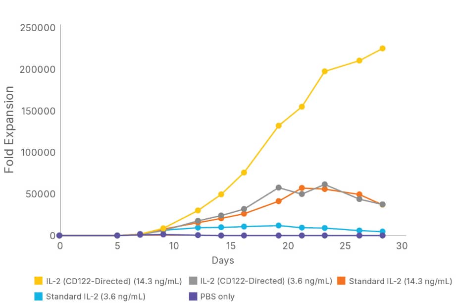
BT-002-AFL) as the standard IL-2 protein or engineered Recombinant Human IL-2 (CD122-Directed) Protein (
BT-002DBR), refreshed every 2-3 days. The average fold expansion of the cells was determined every 2-3 days. The IL-2 (CD122-Directed) Protein supports greater than 6x more CD4+/CD8+ T cell growth than the standard IL-2 protein." alt="CD4
+ and CD8
+ T cells from two different donors were activated with anti-CD3-, anti-CD28-conjugated beads, and grown for 28 days in media containing either 3.6 or 14.3 ng/mL of Animal-free Recombinant Human IL-2 Protein (
BT-002-AFL) as the standard IL-2 protein or engineered Recombinant Human IL-2 (CD122-Directed) Protein (
BT-002DBR), refreshed every 2-3 days. The average fold expansion of the cells was determined every 2-3 days. The IL-2 (CD122-Directed) Protein supports greater than 6x more CD4+/CD8+ T cell growth than the standard IL-2 protein." class="big_thumb" />
BT-002-AFL) as the standard IL-2 protein or Recombinant Human IL-2 (CD122-Directed) Protein (
BT-002DBR), refreshed every 2-3 days. A) After 28 days, the phenotypes of the cells were compared by flow cytometry using a Brilliant Violet 711™ anti-human CD197 (CCR7) antibody and PE/Dazzle™ 594 anti-human CD45RA antibody to determine the number of CD4
+ and CD8
+ naive (CD45RA
+/CCR7
+) Tscm cells. The percentage of Tscm cells was found to be higher when the cells were expanded with the IL-2 (CD122-Directed) Protein at 14.3 ng/mL. B) The number of cells expressing the T cell exhaustion markers, PD-1, LAG-3, and TIM-3 was determined by flow cytometry. The results showed that the percentages of CD4
+/CD8
+ T cells that were also PD-1, LAG-3, or TIM-3 positive were equivalent or reduced with the IL-2 (CD122-Directed) Protein. These results demonstrate that the IL-2 (CD122-Directed) Protein promotes increased T cell expansion with enhanced expression of naive phenotype markers and equivalent or reduced expression of cell exhaustion markers. " class="big_lightbox">
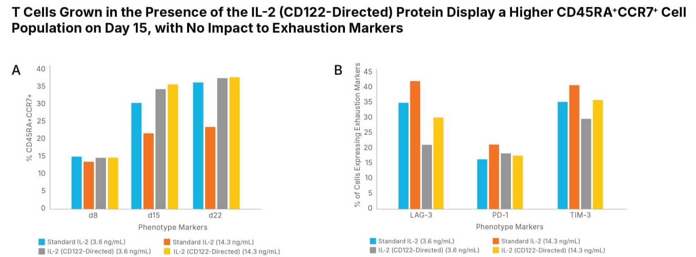
BT-002-AFL) as the standard IL-2 protein or Recombinant Human IL-2 (CD122-Directed) Protein (
BT-002DBR), refreshed every 2-3 days. A) After 28 days, the phenotypes of the cells were compared by flow cytometry using a Brilliant Violet 711™ anti-human CD197 (CCR7) antibody and PE/Dazzle™ 594 anti-human CD45RA antibody to determine the number of CD4
+ and CD8
+ naive (CD45RA
+/CCR7
+) Tscm cells. The percentage of Tscm cells was found to be higher when the cells were expanded with the IL-2 (CD122-Directed) Protein at 14.3 ng/mL. B) The number of cells expressing the T cell exhaustion markers, PD-1, LAG-3, and TIM-3 was determined by flow cytometry. The results showed that the percentages of CD4
+/CD8
+ T cells that were also PD-1, LAG-3, or TIM-3 positive were equivalent or reduced with the IL-2 (CD122-Directed) Protein. These results demonstrate that the IL-2 (CD122-Directed) Protein promotes increased T cell expansion with enhanced expression of naive phenotype markers and equivalent or reduced expression of cell exhaustion markers. " class="big_thumb" />
10919-2B) was captured on a Sensor Chip Protein AGL, and binding to Recombinant Human IL-2 (CD122-Directed) Protein (
BT-002DBR) was measured at a concentration range between 0.097nM and 200 nM. The double-referenced sensorgram was fit to a 1:1 binding model to determine the binding kinetics and affinity, with an affinity constant of K
d=49.3 nM. Note: The IL-2 (CD122-Directed) Protein was modeled to engage only the IL-2 dimeric beta gamma receptor complex. In comparison, wild type recombinant human IL-2 (
BT-002) affinity to Recombinant Human IL 2 R beta Fc Chimera (
10919-2B) has a binding constant K
d=544 nm." class="big_lightbox">
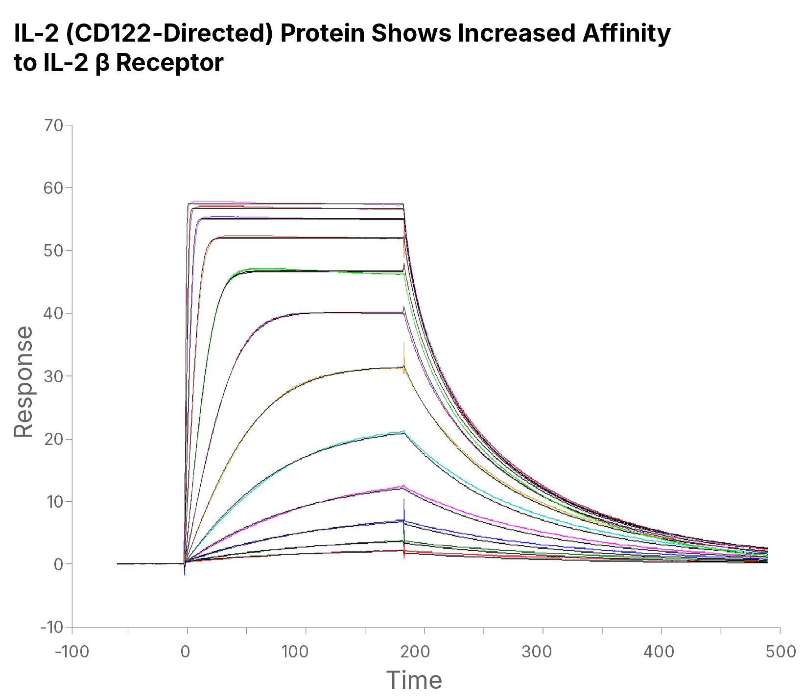
10919-2B) was captured on a Sensor Chip Protein AGL, and binding to Recombinant Human IL-2 (CD122-Directed) Protein (
BT-002DBR) was measured at a concentration range between 0.097nM and 200 nM. The double-referenced sensorgram was fit to a 1:1 binding model to determine the binding kinetics and affinity, with an affinity constant of K
d=49.3 nM. Note: The IL-2 (CD122-Directed) Protein was modeled to engage only the IL-2 dimeric beta gamma receptor complex. In comparison, wild type recombinant human IL-2 (
BT-002) affinity to Recombinant Human IL 2 R beta Fc Chimera (
10919-2B) has a binding constant K
d=544 nm." class="big_thumb" />
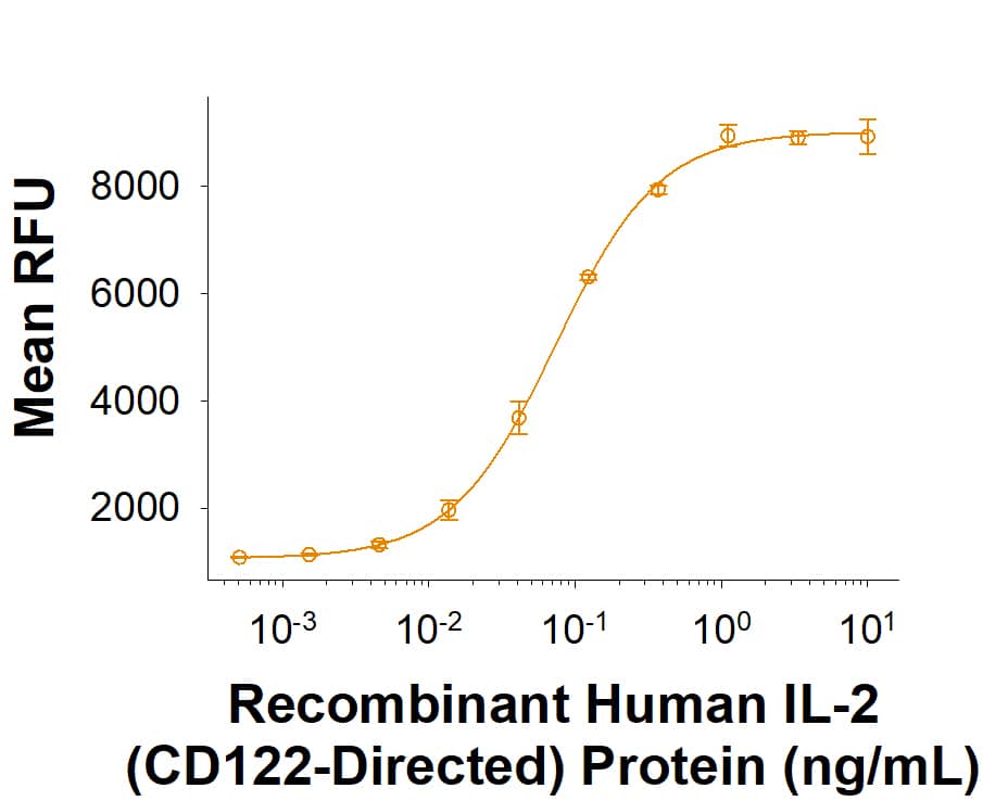
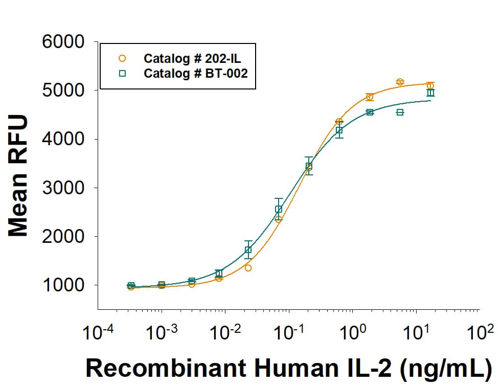 BT-002). It has equivalent bioactivity to Recombinant Human IL-2 (Catalog # 202-IL). It combines R&D Systems quality with scalability that allows for a solid supply chain. Both Recombinant Human IL-2 proteins are measured in a cell proliferation assay using CTLL 2 mouse cytotoxic T cells." alt="As an alternative, please consider our next generation Recombinant Human IL-2 (BT-002). It has equivalent bioactivity to Recombinant Human IL-2 (Catalog # 202-IL). It combines R&D Systems quality with scalability that allows for a solid supply chain. Both Recombinant Human IL-2 proteins are measured in a cell proliferation assay using CTLL 2 mouse cytotoxic T cells." class="big_thumb" />
 BT-002-AFL) as the standard IL-2 protein or engineered Recombinant Human IL-2 (CD122-Directed) Protein (BT-002DBR), refreshed every 2-3 days. The average fold expansion of the cells was determined every 2-3 days. The IL-2 (CD122-Directed) Protein supports greater than 6x more CD4+/CD8+ T cell growth than the standard IL-2 protein." alt="CD4+ and CD8+ T cells from two different donors were activated with anti-CD3-, anti-CD28-conjugated beads, and grown for 28 days in media containing either 3.6 or 14.3 ng/mL of Animal-free Recombinant Human IL-2 Protein (BT-002-AFL) as the standard IL-2 protein or engineered Recombinant Human IL-2 (CD122-Directed) Protein (BT-002DBR), refreshed every 2-3 days. The average fold expansion of the cells was determined every 2-3 days. The IL-2 (CD122-Directed) Protein supports greater than 6x more CD4+/CD8+ T cell growth than the standard IL-2 protein." class="big_thumb" />
 PPK-002-GMP) stimulates proliferation of CTLL‑2 mouse cytotoxic T cells. The ED50 for this effect is 0.0300-0.250 ng/mL. Three independent lots were tested for activity and plotted on the same graph to show lot-to-lot consistency of ProPak™ GMP IL-2." alt="ProPak™ GMP-grade Recombinant Human IL-2 (PPK-002-GMP) stimulates proliferation of CTLL‑2 mouse cytotoxic T cells. The ED50 for this effect is 0.0300-0.250 ng/mL. Three independent lots were tested for activity and plotted on the same graph to show lot-to-lot consistency of ProPak™ GMP IL-2." class="big_thumb" />
 1020-RL) is coated at 1 µg/mL, Recombinant Biotinylated Human IL-2 (Catalog # BT202) binds with an ED50 of 4-24 ng/mL." alt="When Recombinant Human IL-2 Ra Fc Chimera (Catalog # 1020-RL) is coated at 1 µg/mL, Recombinant Biotinylated Human IL-2 (Catalog # BT202) binds with an ED50 of 4-24 ng/mL." class="big_thumb" />


![SDS-Page: Human IL-2 Protein [NBP2-35054]](http://images.novusbio.com/fullsize/Human-IL-2-Protein-SDS-Page-NBP2-35054-img0001.jpg "SDS-Page: Human IL-2 Protein [NBP2-35054]")


 BT-002HS) Provides consistent CD4+/CD8+ T Cell Proliferation in Side-by-Side Testing with the Standard IL-2 Protein. CD4+ and CD8+ T cells were activated with anti-CD3- , anti-CD28- conjugated beads, and grown for 14 days in media containing 10 ng/mL Animal-free Recombinant Human IL-2 (R&D Systems, Catalog #
  BT-002-AFL) as the standard IL-2 protein or Heat Stable IL-2 (Cat #
  BT-002-AFL) as the standard IL-2 protein or Recombinant Human IL-2 (CD122-Directed) Protein (
 10919-2B) was captured on a Sensor Chip Protein AGL, and binding to Recombinant Human IL-2 (CD122-Directed) Protein (


 10453-IL) stimulates cell proliferation of the CTLL-2 mouse cytotoxic T cell line. The ED50 for this effect is 0.05-0.3 ng/mL. Mammalian-expressed Recombinant Human IL-2 has similar activity to E.coli-derived Recombinant Human IL-2 (
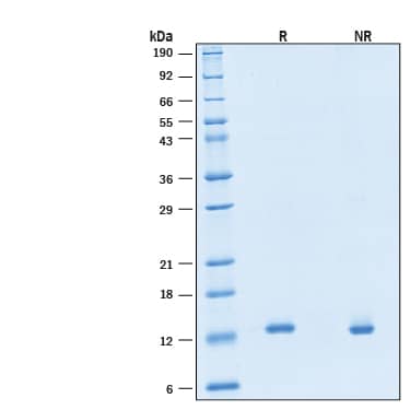
 10453-IL) was resolved with SDS-PAGE under reducing (R) and non-reducing (NR) conditions and visualized by Coomassie® Blue staining, showing bands at 14-18 kDa." class="big_thumb" />
 BT-002-GMP) was added to Xeno-free Human T cell media in a bottle (
 BT-002-GMP) was added to Xeno-free Human T cell media in a bottle (
 BT-002-GMP) was added to Xeno-free Human T cell media in a bottle (

 BT-002-GMP) Animal-Free (Catalog # BT-002-AFL) and RUO (
 BT202, 500 ng/mL) binds to HEK293 human embryonic kidney cell line transfected with recombinant human CD25/IL-2R alpha (black dotted line). Binding is completely blocked (orange histogram) by 0.5 µg/mL of Mouse Anti-Human CD25/IL-2R alpha Monoclonal Antibody (Catalog # MAB1020). Mouse IgG2A Isotype Control (Catalog #


