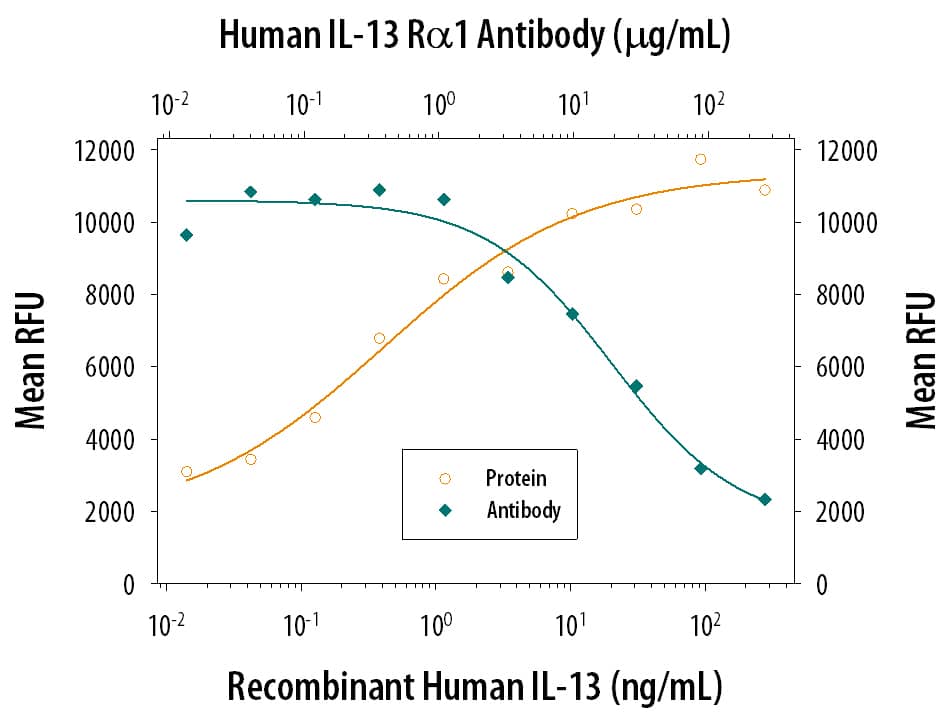
213-ILB) stimulates prolif-eration in the TF-1 human erythroleukemic cell line in a dose-dependent manner (orange line). Proliferation elicited by Recombinant Human IL-13 (10 ng/mL) is neutralized (green line) by increasing concentrations of Goat Anti-Human IL-13 Ra1 Antigen Affinity-purified Polyclonal Antibody (Catalog # AF152). The ND
50 is typically 1.00-12.0 µg/mL." class="big_lightbox">
213-ILB) stimulates prolif-eration in the TF-1 human erythroleukemic cell line in a dose-dependent manner (orange line). Proliferation elicited by Recombinant Human IL-13 (10 ng/mL) is neutralized (green line) by increasing concentrations of Goat Anti-Human IL-13 Ra1 Antigen Affinity-purified Polyclonal Antibody (Catalog # AF152). The ND
50 is typically 1.00-12.0 µg/mL." alt="Recombinant Human IL-13 (
213-ILB) stimulates prolif-eration in the TF-1 human erythroleukemic cell line in a dose-dependent manner (orange line). Proliferation elicited by Recombinant Human IL-13 (10 ng/mL) is neutralized (green line) by increasing concentrations of Goat Anti-Human IL-13 Ra1 Antigen Affinity-purified Polyclonal Antibody (Catalog # AF152). The ND
50 is typically 1.00-12.0 µg/mL." class="big_thumb" />
AB-108-C, open histogram), followed by Phycoerythrin-conjugated Anti-Goat IgG Secondary Antibody (
F0107)." class="big_lightbox">

AB-108-C, open histogram), followed by Phycoerythrin-conjugated Anti-Goat IgG Secondary Antibody (
F0107)." class="big_thumb" />
VC004). Before incubation with the primary antibody, tissue was subjected to heat-induced epitope retrieval using VisUCyte Antigen Retrieval Reagent-Basic (Catalog #
VCTS021). Tissue was stained using DAB (brown) and counterstained with hematoxylin (blue). Specific staining was localized to cytoplasm in myocardial cells. View our protocol for
IHC Staining with VisUCyte HRP Polymer Detection Reagents." class="big_lightbox">

VC004). Before incubation with the primary antibody, tissue was subjected to heat-induced epitope retrieval using VisUCyte Antigen Retrieval Reagent-Basic (Catalog #
VCTS021). Tissue was stained using DAB (brown) and counterstained with hematoxylin (blue). Specific staining was localized to cytoplasm in myocardial cells. View our protocol for
IHC Staining with VisUCyte HRP Polymer Detection Reagents." class="big_thumb" />
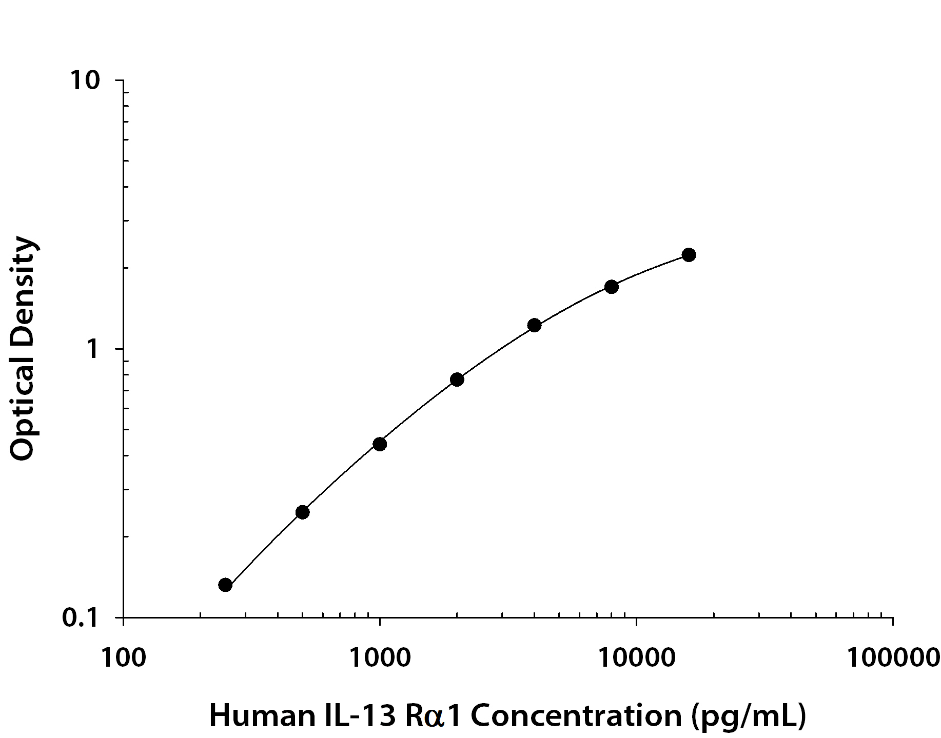
146-IR) was serially diluted and captured by Mouse Anti-Human IL‑13 R alpha 1 Monoclonal Antibody (Catalog #
MAB146) coated on a Clear Polystyrene Microplate (Catalog #
DY990). Goat Anti-Human IL‑13 R alpha 1 Antigen Affinity-purified Polyclonal Antibody (Catalog # AF152) was biotinylated and incubated with the protein captured on the plate. Detection of the standard curve was achieved by incubating Streptavidin-HRP (Catalog #
DY998)" class="big_lightbox">
146-IR) was serially diluted and captured by Mouse Anti-Human IL‑13 R alpha 1 Monoclonal Antibody (Catalog #
MAB146) coated on a Clear Polystyrene Microplate (Catalog #
DY990). Goat Anti-Human IL‑13 R alpha 1 Antigen Affinity-purified Polyclonal Antibody (Catalog # AF152) was biotinylated and incubated with the protein captured on the plate. Detection of the standard curve was achieved by incubating Streptavidin-HRP (Catalog #
DY998)" class="big_thumb" />
 213-ILB) stimulates prolif-eration in the TF-1 human erythroleukemic cell line in a dose-dependent manner (orange line). Proliferation elicited by Recombinant Human IL-13 (10 ng/mL) is neutralized (green line) by increasing concentrations of Goat Anti-Human IL-13 Ra1 Antigen Affinity-purified Polyclonal Antibody (Catalog # AF152). The ND50 is typically 1.00-12.0 µg/mL." alt="Recombinant Human IL-13 (213-ILB) stimulates prolif-eration in the TF-1 human erythroleukemic cell line in a dose-dependent manner (orange line). Proliferation elicited by Recombinant Human IL-13 (10 ng/mL) is neutralized (green line) by increasing concentrations of Goat Anti-Human IL-13 Ra1 Antigen Affinity-purified Polyclonal Antibody (Catalog # AF152). The ND50 is typically 1.00-12.0 µg/mL." class="big_thumb" />
 HAF007). A specific band was detected for IL-13 Ra1 at approximately 65 kDa (as indicated). This experiment was conducted under reducing conditions and using Immunoblot Buffer Group 1." alt="Western blot shows lysates of human heart tissue and human skeletal muscle tissue. PVDF membrane was probed with 2 µg/mL of Mouse Anti-Human IL-13 Ra1 Monoclonal Antibody (Catalog # MAB1462) followed by HRP-conjugated Anti-Mouse IgG Secondary Antibody (Catalog # HAF007). A specific band was detected for IL-13 Ra1 at approximately 65 kDa (as indicated). This experiment was conducted under reducing conditions and using Immunoblot Buffer Group 1." class="big_thumb" />
 146-IR) was serially diluted and captured by Mouse Anti-Human IL‑13 R alpha 1 Monoclonal Antibody (Catalog # MAB146) coated on a Clear Polystyrene Microplate (Catalog # DY990). Goat Anti-Human IL‑13 R alpha 1 Antigen Affinity-purified Polyclonal Antibody (Catalog # AF152) was biotinylated and incubated with the protein captured on the plate. Detection of the standard curve was achieved by incubating Streptavidin-HRP (Catalog # DY998)" alt="Recombinant Human IL‑13 R alpha 1 Fc Chimera (Catalog # 146-IR) was serially diluted and captured by Mouse Anti-Human IL‑13 R alpha 1 Monoclonal Antibody (Catalog # MAB146) coated on a Clear Polystyrene Microplate (Catalog # DY990). Goat Anti-Human IL‑13 R alpha 1 Antigen Affinity-purified Polyclonal Antibody (Catalog # AF152) was biotinylated and incubated with the protein captured on the plate. Detection of the standard curve was achieved by incubating Streptavidin-HRP (Catalog # DY998)" class="big_thumb" />


 AB-108-C, open histogram), followed by Phycoerythrin-conjugated Anti-Goat IgG Secondary Antibody (
 CTS008) and counterstained with hematoxylin (blue). View our protocol for
  VC004). Before incubation with the primary antibody, tissue was subjected to heat-induced epitope retrieval using VisUCyte Antigen Retrieval Reagent-Basic (Catalog #

 146-IR) was serially diluted and captured by Mouse Anti-Human IL‑13 R alpha 1 Monoclonal Antibody (Catalog #
  FAB1137A) and (B) Mouse Anti-Human IL-13 Ra1 Monoclonal Antibody (Catalog # MAB1462, filled histogram) or isotype control antibody (Catalog #
 ![Immunohistochemistry-Paraffin: IL-13 R alpha 1 Antibody [NBP2-16942] - Analysis of paraffin-embedded Hepatoma, using antibody at 1:500 dilution.](http://images.novusbio.com/fullsize/IL-13-R-alpha-1-Antibody-Immunohistochemistry-Paraffin-NBP2-16942-img0003.jpg "Immunohistochemistry-Paraffin: IL-13 R alpha 1 Antibody [NBP2-16942] - Analysis of paraffin-embedded Hepatoma, using antibody at 1:500 dilution.")
![Immunohistochemistry-Paraffin: IL-13R alpha 1 Antibody [NBP2-16942] - IL-13R alpha 1 antibody detects IL-13R alpha 1 protein at cell membrane and cytoplasm by immunohistochemical analysis.</br>Sample: Paraffin-embedded mouse lung.</br>IL-13R alpha 1 stained by IL-13R alpha 1 antibody (NBP2-16942) diluted at 1:500.</br>Antigen Retrieval: Citrate buffer, pH 6.0, 15 min](http://images.novusbio.com/fullsize/nbp2-16942_rabbit-polyclonal-il-13r-alpha-1-antibody-91020242075969.jpg)
![Immunohistochemistry-Paraffin: IL-13R alpha 1 Antibody [NBP2-16942] - IL-13R alpha 1 antibody detects IL-13R alpha 1 protein at cytoplasm by immunohistochemical analysis.</br>Sample: Paraffin-embedded rat liver.</br>IL-13R alpha 1 stained by IL-13R alpha 1 antibody (NBP2-16942) diluted at 1:500.</br>Antigen Retrieval: Citrate buffer, pH 6.0, 15 min](http://images.novusbio.com/fullsize/nbp2-16942_rabbit-polyclonal-il-13r-alpha-1-antibody-91020242091459.jpg)


![Western Blot: IL-13 R alpha 1 Antibody [NBP2-55120] - Analysis in human cell line HDLM-2.](http://images.novusbio.com/fullsize/IL-13-R-alpha-1-Antibody-Western-Blot-NBP2-55120-img0003.jpg "Western Blot: IL-13 R alpha 1 Antibody [NBP2-55120] - Analysis in human cell line HDLM-2.")
![Immunocytochemistry/Immunofluorescence: IL-13 R alpha 1 Antibody [NBP2-55120] - Staining of human cell line A-431 shows localization to nucleoplasm & the Golgi apparatus. Antibody staining is shown in green.](http://images.novusbio.com/fullsize/IL-13-R-alpha-1-Antibody-Immunocytochemistry-Immunofluorescence-NBP2-55120-img0001.jpg)
![Western Blot: IL-13R alpha 1 Antibody (5A3W5) [NBP3-16207] - Western blot analysis of extracts of various cell lines, using IL-13R alpha 1 antibody (NBP3-16207) at 1:1000 dilution. Secondary antibody: HRP Goat Anti-Rabbit IgG (H+L) at 1:10000 dilution. Lysates/proteins: 25ug per lane. Blocking buffer: 3% nonfat dry milk in TBST. Detection: ECL Enhanced Kit. Exposure time: 180s.](http://images.novusbio.com/fullsize/IL-13R-alpha-1-Antibody-5A3W5-Western-Blot-NBP3-16207-img0002.jpg "Western Blot: IL-13R alpha 1 Antibody (5A3W5) [NBP3-16207] - Western blot analysis of extracts of various cell lines, using IL-13R alpha 1 antibody (NBP3-16207) at 1:1000 dilution. Secondary antibody: HRP Goat Anti-Rabbit IgG (H+L) at 1:10000 dilution. Lysates/proteins: 25ug per lane. Blocking buffer: 3% nonfat dry milk in TBST. Detection: ECL Enhanced Kit. Exposure time: 180s.")
![Immunohistochemistry-Paraffin: IL-13R alpha 1 Antibody (5A3W5) [NBP3-16207] - Immunohistochemistry of paraffin-embedded human lung cancer using IL-13R alpha 1 Rabbit mAb (NBP3-16207) at dilution of 1:100 (40x lens).Perform microwave antigen retrieval with 10 mM Tris/EDTA buffer pH 9.0 before commencing with IHC staining protocol.](http://images.novusbio.com/fullsize/IL-13R-alpha-1-Antibody-5A3W5-Immunohistochemistry-Paraffin-NBP3-16207-img0001.jpg)
![Western Blot: IL-13R alpha 1 Antibody [NBP2-16943] - Non-transfected (-) and transfected (+) 293T whole cell extracts (30 ug) were separated by 10% SDS-PAGE, and the membrane was blotted with IL-13R alpha 1 antibody [N1C1] (NBP2-16943) diluted at 1:3000. The HRP-conjugated anti-rabbit IgG antibody was used to detect the primary antibody.](http://images.novusbio.com/fullsize/nbp2-16943_rabbit-polyclonal-il-13r-alpha-1-antibody-910202420569.jpg "Western Blot: IL-13R alpha 1 Antibody [NBP2-16943] - Non-transfected (-) and transfected (+) 293T whole cell extracts (30 ug) were separated by 10% SDS-PAGE, and the membrane was blotted with IL-13R alpha 1 antibody [N1C1] (NBP2-16943) diluted at 1:3000. The HRP-conjugated anti-rabbit IgG antibody was used to detect the primary antibody.")
![IL13 Receptor alpha 1 antibody [N1C1] detects IL13 Receptor alpha 1 protein at cell membrane and cytoplasm by immunohistochemical analysis.</br>Sample: Paraffin-embedded human breast carcinoma.</br>IL13 Receptor alpha 1 stained by IL13 Receptor alpha 1 antibody [N1C1] (NBP2-16943) diluted at 1:500.</br>Antigen Retrieval: Citrate buffer, pH 6.0, 15 min](http://images.novusbio.com/fullsize/nbp2-16943_rabbit-polyclonal-il-13r-alpha-1-antibody-immunohistochemistry-paraffin-261120251848482.jpg)


