DR647MS) and counterstained with DAPI (blue; Lunaphore Catalog #
DR100). Specific staining was localized to the membrane. Protocol available in
COMET™ Panel Builder." class="big_lightbox">
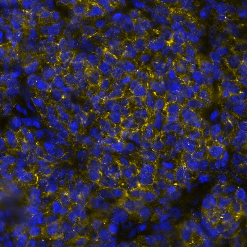
DR647MS) and counterstained with DAPI (blue; Lunaphore Catalog #
DR100). Specific staining was localized to the membrane. Protocol available in
COMET™ Panel Builder." alt="CD19 was detected in immersion fixed paraffin-embedded sections of B-Cell Lymphoma using Mouse Anti-Human CD19 Monoclonal Antibody (Catalog # MAB11717) at 1 µg/mL at 37 ° Celsius for 4 minutes. Before incubation with the primary antibody, tissue underwent an all-in-one dewaxing and antigen retrieval preprocessing using PreTreatment Module (PT Module) and Dewax and HIER Buffer H (pH 9; Epredia Catalog # TA-999-DHBH). Tissue was stained using the Alexa Fluor™ 647 Goat anti-Mouse IgG Secondary Antibody at 1:200 at 37 ° Celsius for 2 minutes. (Yellow; Lunaphore Catalog #
DR647MS) and counterstained with DAPI (blue; Lunaphore Catalog #
DR100). Specific staining was localized to the membrane. Protocol available in
COMET™ Panel Builder." class="big_thumb" />
DR647MS) and counterstained with DAPI (blue; Lunaphore Catalog #
DR100). Specific staining was localized to the membrane. Protocol available in
COMET™ Panel Builder." class="big_lightbox">
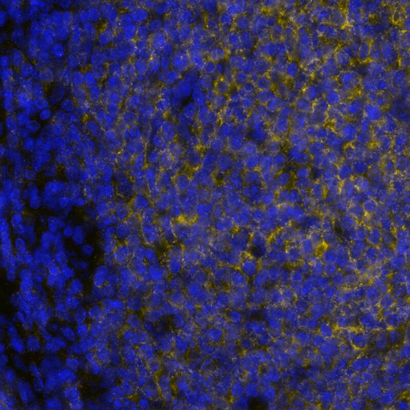
DR647MS) and counterstained with DAPI (blue; Lunaphore Catalog #
DR100). Specific staining was localized to the membrane. Protocol available in
COMET™ Panel Builder." class="big_thumb" />
DR647MS) and counterstained with DAPI (blue; Lunaphore Catalog #
DR100). Specific staining was localized to the membrane. Protocol available in
COMET™ Panel Builder." class="big_lightbox">
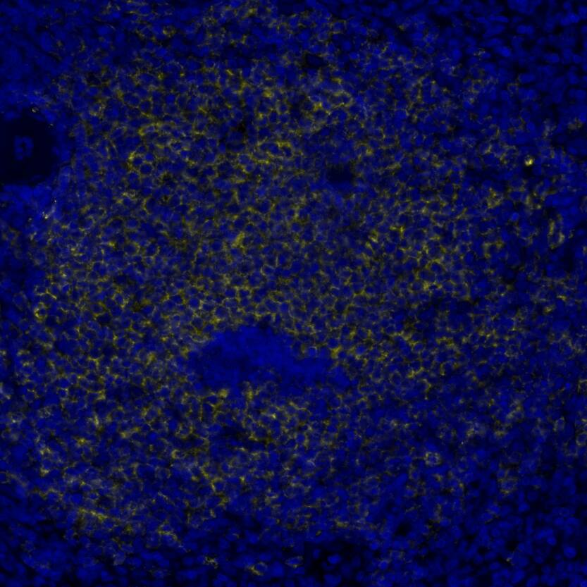
DR647MS) and counterstained with DAPI (blue; Lunaphore Catalog #
DR100). Specific staining was localized to the membrane. Protocol available in
COMET™ Panel Builder." class="big_thumb" />
HAF018). A specific band was detected for CD19 at approximately 95 kDa (as indicated). This experiment was conducted under reducing conditions and using
Western Blot Buffer Group 1." class="big_lightbox">

HAF018). A specific band was detected for CD19 at approximately 95 kDa (as indicated). This experiment was conducted under reducing conditions and using
Western Blot Buffer Group 1." class="big_thumb" />
VC001) or the HRP-conjugated Anti-Mouse IgG Secondary Antibody (Catalog #
HAF007). Before incubation with the primary antibody, tissue was subjected to heat-induced epitope retrieval using VisUCyte Antigen Retrieval Reagent-Basic (Catalog #
VCTS021). Tissue was stained using DAB (brown) and counterstained with hematoxylin (blue). Specific staining was localized to the membrane. View our protocol for
IHC Staining with VisUCyte HRP Polymer Detection Reagents." class="big_lightbox">

VC001) or the HRP-conjugated Anti-Mouse IgG Secondary Antibody (Catalog #
HAF007). Before incubation with the primary antibody, tissue was subjected to heat-induced epitope retrieval using VisUCyte Antigen Retrieval Reagent-Basic (Catalog #
VCTS021). Tissue was stained using DAB (brown) and counterstained with hematoxylin (blue). Specific staining was localized to the membrane. View our protocol for
IHC Staining with VisUCyte HRP Polymer Detection Reagents." class="big_thumb" />
VC001) or the HRP-conjugated Anti-Mouse IgG Secondary Antibody (Catalog #
HAF007). Before incubation with the primary antibody, tissue was subjected to heat-induced epitope retrieval using VisUCyte Antigen Retrieval Reagent-Basic (Catalog #
VCTS021). Tissue was stained using DAB (brown) and counterstained with hematoxylin (blue). Specific staining was localized to the membrane in the white pulp of spleen. View our protocol for
IHC Staining with VisUCyte HRP Polymer Detection Reagents." class="big_lightbox">

VC001) or the HRP-conjugated Anti-Mouse IgG Secondary Antibody (Catalog #
HAF007). Before incubation with the primary antibody, tissue was subjected to heat-induced epitope retrieval using VisUCyte Antigen Retrieval Reagent-Basic (Catalog #
VCTS021). Tissue was stained using DAB (brown) and counterstained with hematoxylin (blue). Specific staining was localized to the membrane in the white pulp of spleen. View our protocol for
IHC Staining with VisUCyte HRP Polymer Detection Reagents." class="big_thumb" />
VC001) or the HRP-conjugated Anti-Mouse IgG Secondary Antibody (Catalog #
HAF007). Before incubation with the primary antibody, tissue was subjected to heat-induced epitope retrieval using VisUCyte Antigen Retrieval Reagent-Basic (Catalog #
VCTS021). Tissue was stained using DAB (brown) and counterstained with hematoxylin (blue). Specific staining was localized to the membrane . View our protocol for
IHC Staining with VisUCyte HRP Polymer Detection Reagents." class="big_lightbox">

VC001) or the HRP-conjugated Anti-Mouse IgG Secondary Antibody (Catalog #
HAF007). Before incubation with the primary antibody, tissue was subjected to heat-induced epitope retrieval using VisUCyte Antigen Retrieval Reagent-Basic (Catalog #
VCTS021). Tissue was stained using DAB (brown) and counterstained with hematoxylin (blue). Specific staining was localized to the membrane . View our protocol for
IHC Staining with VisUCyte HRP Polymer Detection Reagents." class="big_thumb" />
 FAB100P) and either (A) Mouse Anti-Human CD19 Monoclonal Antibody (Catalog # MAB4867) or (B) Mouse IgG1Isotype Control (Catalog # MAB002) followed by Allophycocyanin-conjugated Anti-Mouse IgG Secondary Antibody (Catalog # F0101B)." alt="Human peripheral blood mononuclear cells (PBMCs) were stained with Mouse Anti-Human CD3e PE‑conjugated Monoclonal Antibody (Catalog # FAB100P) and either (A) Mouse Anti-Human CD19 Monoclonal Antibody (Catalog # MAB4867) or (B) Mouse IgG1Isotype Control (Catalog # MAB002) followed by Allophycocyanin-conjugated Anti-Mouse IgG Secondary Antibody (Catalog # F0101B)." class="big_thumb" />
 DR647MS) and counterstained with DAPI (blue; Lunaphore Catalog # DR100). Specific staining was localized to the membrane. Protocol available in COMET™ Panel Builder." alt="CD19 was detected in immersion fixed paraffin-embedded sections of B-Cell Lymphoma using Mouse Anti-Human CD19 Monoclonal Antibody (Catalog # MAB11717) at 1 µg/mL at 37 ° Celsius for 4 minutes. Before incubation with the primary antibody, tissue underwent an all-in-one dewaxing and antigen retrieval preprocessing using PreTreatment Module (PT Module) and Dewax and HIER Buffer H (pH 9; Epredia Catalog # TA-999-DHBH). Tissue was stained using the Alexa Fluor™ 647 Goat anti-Mouse IgG Secondary Antibody at 1:200 at 37 ° Celsius for 2 minutes. (Yellow; Lunaphore Catalog # DR647MS) and counterstained with DAPI (blue; Lunaphore Catalog # DR100). Specific staining was localized to the membrane. Protocol available in COMET™ Panel Builder." class="big_thumb" />


![Immunocytochemistry/Immunofluorescence: CD19 Antibody (CB19) [NBP2-25196] - CD19 antibody (CB19) was tested in Non-Hodgkin's lymphoma cells at 1:200 dilution. Green: Alexa Flour 488. Image from verified customer review.](http://images.novusbio.com/fullsize/CD19-Antibody-CB19-Immunocytochemistry-Immunofluorescence-NBP2-25196-img0014.jpg "Immunocytochemistry/Immunofluorescence: CD19 Antibody (CB19) [NBP2-25196] - CD19 antibody (CB19) was tested in Non-Hodgkin's lymphoma cells at 1:200 dilution. Green: Alexa Flour 488. Image from verified customer review.")
![Flow Cytometry: CD19 Antibody (CB19) [NBP2-25196] - A surface stain was performed on Ramos cells with CD19 Antibody (CB19) NBP2-26646 (blue) and a matched isotype control (orange). Cells were incubated in an antibody dilution of 2.5 ug/mL for 20 minutes at room temperature. Both antibodies were conjugated to phycoerythrin.](http://images.novusbio.com/fullsize/CD19-Antibody-CB19-Flow-Cytometry-NBP2-25196-img0013.jpg)
![Immunocytochemistry/Immunofluorescence: CD19 Antibody (CB19) [NBP2-25196] - The CD19 (CB19) antibody was tested in Daudi cells at a 1:40 dilution against Dylight 488 (Green). Actin and nuclei were counterstained against Phalloidin 568 (Red) and DAPI (Blue), respectively.](http://images.novusbio.com/fullsize/CD19-Antibody-CB19-Immunocytochemistry-Immunofluorescence-NBP2-25196-img0002.jpg)
![Flow Cytometry: CD19 Antibody (CB19) [NBP2-25196] - Surface staining of 10^6 human lymphocytes using 0.2 ug of CD19 antibody (red) and isotype control (green). Cell surface staining kit was used for this test.](http://images.novusbio.com/fullsize/CD19-Antibody-CB19-Flow-Cytometry-NBP2-25196-img0001.jpg)
![Flow Cytometry: CD19 Antibody (CB19) [NBP2-25196] - Analysis using the FITC conjugate of NBP2-25196. Staining of CD19 in 10^6 human lymphocytes using 10 ul (0.25 ug) of this antibody. Shaded histogram represents cells without antibody; green represents isotype control ; red represents anti-CD19 antibody.](http://images.novusbio.com/fullsize/CD19-Antibody-CB19-Flow-Cytometry-NBP2-25196-img0007.jpg)
![Flow Cytometry: CD19 Antibody (CB19) [NBP2-25196] - Analysis using the PE conjugate of NBP2-25196. Staining of CD19 in 1x10^6 human PBMC using 10 ul (0.25 ug) of was used to test this product. Propidium iodide negative lymphocyte population gated for analysis.](http://images.novusbio.com/fullsize/CD19-Antibody-CB19-Flow-Cytometry-NBP2-25196-img0008.jpg)
![Flow Cytometry: CD19 Antibody (CB19) [NBP2-25196] - Analysis using the Alexa Fluor (R) 700 conjugate of NBP2-25196. Staining of human PBMC. Image from verified customer review.](http://images.novusbio.com/fullsize/CD19-Antibody-CB19-Flow-Cytometry-NBP2-25196-img0009.jpg)
![Flow Cytometry: CD19 Antibody (CB19) [NBP2-25196] - Analysis using the DyLight 405 conjugate of NBP2-25196. Staining of CD19 in human PBMCs using anti-CD19 antibody. Image from verified customer review.](http://images.novusbio.com/fullsize/CD19-Antibody-CB19-Flow-Cytometry-NBP2-25196-img0010.jpg)
![Flow Cytometry: CD19 Antibody (CB19) [NBP2-25196] - A surface stain was performed on human peripheral blood lymphocytes with TACI/TNFRSF13B/CVID (1A1) antibody NB600-1189PE and a matched isotype control. Cells were incubated in an antibody dilution of 2 ug/mL for 20 minutes at room temperature. A co-stain was performed with NBP2-25196AF647.](http://images.novusbio.com/fullsize/CD19-Antibody-CB19-Flow-Cytometry-NBP2-25196-img0011.jpg)
![Flow Cytometry: CD19 Antibody (CB19) [NBP2-25196] - A surface stain was performed on human peripheral blood lymphocytes with CD9 (HI9a) antibody NB100-77915AF647 and a matched isotype control NBP2-27287AF488. Cells were incubated in an antibody dilution of 1 ug/mL for 20 minutes at room temperature. A co-stain was performed with NBP2-26646.](http://images.novusbio.com/fullsize/CD19-Antibody-CB19-Flow-Cytometry-NBP2-25196-img0012.jpg)


![Flow Cytometry: CD19 Antibody (1D3) [NBP2-24965] - Cell surface analysis of CD19 in BALB/c mouse splenocytes using 1 ug of this antibody. Green: isotype control; red: anti-CD19 PE. Image using the PE form of this antibody.](http://images.novusbio.com/fullsize/CD19-Antibody-1D3-Flow-Cytometry-NBP2-24965-img0011.jpg "Flow Cytometry: CD19 Antibody (1D3) [NBP2-24965] - Cell surface analysis of CD19 in BALB/c mouse splenocytes using 1 ug of this antibody. Green: isotype control; red: anti-CD19 PE. Image using the PE form of this antibody.")
![Flow Cytometry: CD19 Antibody (1D3) [NBP2-24965] - Cell surface analysis of CD19 in mouse splenocytes, 25 ug/10^6 cells. Anti-rat PE secondary and cell surface staining kit were used in this test.](http://images.novusbio.com/fullsize/CD19-Antibody-1D3-Flow-Cytometry-NBP2-24965-img0001.jpg)
![Flow (Cell Surface): CD19 Antibody (1D3) [NBP2-24965] - Analysis using the Allophycocyanin conjugate of NBP2-24965. Staining of CD19 in BALB/c mouse splenocytes using 1 ug of this antibody. Green represents isotype control; red represents anti-CD19 APC.](http://images.novusbio.com/fullsize/CD19-Antibody-1D3-Flow-Cell-Surface-NBP2-24965-img0006.jpg)
![Flow Cytometry: CD19 Antibody (1D3) [NBP2-24965] - Analysis using the Allophycocyanin conjugate of NBP2-24965. Staining of CD19 in BALB/c mouse splenocytes. Cells were stained with anti-CD4 PE this antibody and either 1 ug of isotype control antibody (left) or 1 ug of anti-CD19 APC (right).](http://images.novusbio.com/fullsize/CD19-Antibody-1D3-Flow-Cytometry-NBP2-24965-img0007.jpg)
![Flow Cytometry: CD19 Antibody (1D3) [NBP2-24965] - Analysis using the PE conjugate of NBP2-24965. Staining of CD19 in BALB/c mouse splenocytes. Cells were stained with anti-CD4 FITC this antibody and either 1 ug of isotype control antibody (left, ) or 1 ug of anti-CD19 PE (right).](http://images.novusbio.com/fullsize/CD19-Antibody-1D3-Flow-Cytometry-NBP2-24965-img0010.jpg)
![Western Blot: CD19 Antibody [NBP2-15782] - Various whole cell extracts (30 ug) were separated by 7.5% SDS-PAGE, and the membrane was blotted with CD19 antibody [C1C3] diluted at 1:1000. The HRP-conjugated anti-rabbit IgG antibody (NBP2-19301) was used to detect the primary antibody.](http://images.novusbio.com/fullsize/CD19-Antibody-Western-Blot-NBP2-15782-img0016.jpg "Western Blot: CD19 Antibody [NBP2-15782] - Various whole cell extracts (30 ug) were separated by 7.5% SDS-PAGE, and the membrane was blotted with CD19 antibody [C1C3] diluted at 1:1000. The HRP-conjugated anti-rabbit IgG antibody (NBP2-19301) was used to detect the primary antibody.")
![Immunohistochemistry-Paraffin: CD19 Antibody [NBP2-15782] - Human tonsil. CD19 stained by CD19 antibody [C1C3] diluted at 1:500.Antigen Retrieval: Citrate buffer, pH 6.0, 15 min.](http://images.novusbio.com/fullsize/CD19-Antibody-Immunohistochemistry-Paraffin-NBP2-15782-img0011.jpg)
![Flow Cytometry: CD19 Antibody [NBP2-15782] - Mouse splenocytes cell fixed in 4% paraformaldehyde at 4-C for 15 min. Black: Unlabelled sample was used as a control. Red: CD19 antibody [C1C3] dilution: 1:50. Acquisition of 20,000 events were collected using a Dylight 488-conjugated secondary antibody for FACS analysis.](http://images.novusbio.com/fullsize/CD19-Antibody-Flow-Cytometry-NBP2-15782-img0006.jpg)
![Western Blot: CD19 Antibody [NBP2-15782] - Mouse tissue extract (50 ug) was separated by 7.5% SDS-PAGE, and the membrane was blotted with CD19 antibody [C1C3] diluted at 1:1000.](http://images.novusbio.com/fullsize/CD19-Antibody-Western-Blot-NBP2-15782-img0007.jpg)
![CD19 antibody [C1C3] detects CD19 protein at cell membrane by immunofluorescent analysis.</br>Sample: Raji cells were fixed in 4% paraformaldehyde at RT for 15 min.</br>Green: CD19 stained by CD19 antibody [C1C3] (NBP2-15782) diluted at 1:500.</br>Blue: Fluoroshield with DAPI .](http://images.novusbio.com/fullsize/nbp2-15782_rabbit-polyclonal-cd19-antibody-immunocytochemistry-immunofluorescence-261120251843192.jpg)
![CD19 antibody [C1C3] detects CD19 protein at cell membrane and cytoplasm by immunohistochemical analysis.</br>Sample: Paraffin-embedded human tonsil.</br>Green: CD19 stained by CD19 antibody [C1C3] (NBP2-15782) diluted at 1:250.</br>Blue: Fluoroshield with DAPI .</br>Antigen Retrieval: Citrate buffer, pH 6.0, 15 min](http://images.novusbio.com/fullsize/nbp2-15782_rabbit-polyclonal-cd19-antibody-immunohistochemistry-paraffin-2611202518393117.jpg)
![Immunocytochemistry/Immunofluorescence: CD19 Antibody (JF100-06) [NBP2-67229] - Staining CD19 in PC-3M cells (red). The nuclear counter stain is DAPI (blue). Cells were fixed in paraformaldehyde, permeabilised with 0.25% Triton X100/PBS.](http://images.novusbio.com/fullsize/CD19-Antibody-JF100-06-Immunocytochemistry-Immunofluorescence-NBP2-67229-img0005.jpg "Immunocytochemistry/Immunofluorescence: CD19 Antibody (JF100-06) [NBP2-67229] - Staining CD19 in PC-3M cells (red). The nuclear counter stain is DAPI (blue). Cells were fixed in paraformaldehyde, permeabilised with 0.25% Triton X100/PBS.")
![Immunohistochemistry-Paraffin: CD19 Antibody (JF100-06) [NBP2-67229] - Immunohistochemical analysis of paraffin-embedded human tonsil tissue using anti-CD19 antibody. Counter stained with hematoxylin.](http://images.novusbio.com/fullsize/CD19-Antibody-JF100-06-Immunohistochemistry-Paraffin-NBP2-67229-img0006.jpg)
![Immunocytochemistry/Immunofluorescence: CD19 Antibody (JF100-06) [NBP2-67229] - Staining CD19 in 293T cells (red). The nuclear counter stain is DAPI (blue). Cells were fixed in paraformaldehyde, permeabilised with 0.25% Triton X100/PBS.](http://images.novusbio.com/fullsize/CD19-Antibody-JF100-06-Immunocytochemistry-Immunofluorescence-NBP2-67229-img0001.jpg)
![Immunocytochemistry/Immunofluorescence: CD19 Antibody (JF100-06) [NBP2-67229] - Staining CD19 in Hela cells (red). The nuclear counter stain is DAPI (blue). Cells were fixed in paraformaldehyde, permeabilised with 0.25% Triton X100/PBS.](http://images.novusbio.com/fullsize/CD19-Antibody-JF100-06-Immunocytochemistry-Immunofluorescence-NBP2-67229-img0002.jpg)
![Immunohistochemistry-Paraffin: CD19 Antibody (JF100-06) [NBP2-67229] - Analysis of paraffin-embedded human spleen tissue using anti-CD19 antibody. Counter stained with hematoxylin.](http://images.novusbio.com/fullsize/CD19-Antibody-JF100-06-Immunohistochemistry-Paraffin-NBP2-67229-img0003.jpg)
![Western Blot: CD19 Antibody (JF100-06) [NBP2-67229] -Analysis of CD19 on different lysates with Rabbit anti-CD19 antibody at 1/1,000 dilution.Lane 1: Raji cell lysateLane 2: Daudi cell lysateLysates/proteins at 10 ug/Lane.Predicted band size: 61 kDaObserved band size: 90 kDaExposure time: 2 minutes;8% SDS-PAGE gel.Proteins were transferred to a PVDF membrane and blocked with 5% NFDM/TBST for 1 hour at room temperature. The primary antibody at 1/1,000 dilution was used in 5% NFDM/TBST at room temperature for 2 hours. Goat Anti-Rabbit IgG - HRP Secondary Antibody at 1:300,000 dilution was used for 1 hour at room temperature.](http://images.novusbio.com/fullsize/nbp2-67229_rabbit-monoclonal-cd19-antibody-jf100-06-western-blot-1382024114350..jpg)
![Flow Cytometry: CD19 Antibody (JF100-06) [NBP2-67229] -Analysis of Daudi cells labeling CD19.Cells were fixed and permeabilized. Then stained with the primary antibody (1ug/ml) (red) compared with Rabbit IgG Isotype Control (green). After incubation of the primary antibody at +4℃ for an hour, the cells were stained with a iFluor™ 488 conjugate-Goat anti-Rabbit IgG Secondary antibody at 1/1,000 dilution for 30 minutes at +4℃. Unlabelled sample was used as a control (cells without incubation with primary antibody; black).](http://images.novusbio.com/fullsize/nbp2-67229_rabbit-monoclonal-cd19-antibody-jf100-06-flow-cytometry-138202411462..jpg)
![Flow Cytometry: CD19 Antibody (JF100-06) [NBP2-67229] -Analysis of Raji cells labeling CD19.Cells were fixed and permeabilized. Then stained with the primary antibody (1ug/ml) (red) compared with Rabbit IgG Isotype Control (green). After incubation of the primary antibody at +4℃ for an hour, the cells were stained with a iFluor™ 488 conjugate-Goat anti-Rabbit IgG Secondary antibody at 1/1,000 dilution for 30 minutes at +4℃. Unlabelled sample was used as a control (cells without incubation with primary antibody; black).](http://images.novusbio.com/fullsize/nbp2-67229_rabbit-monoclonal-cd19-antibody-jf100-06-flow-cytometry-1382024114721..jpg)
![Western blot analysis on different lysates with NBP2-67229 at 1/10,000 dilution. Lane 1: Raji cell lysate Lane 2: Ramos cell lysate Lane 3: 293T cell lysate (negative) Lane 4: Daudi cell lysate Lane 5: K-562 cell lysate (negative) Lysates/proteins at 20 µg/Lane. Predicted band size: 61 kDa Observed band size: 90 kDa Exposure time: 25 seconds; 4-20% SDS-PAGE gel. Proteins were transferred to a PVDF membrane and blocked with 5% NFDM/TBST for 1 hour at room temperature. NBP2-67229 at 1/10,000 dilution was used in primary antibody dilution at 4℃ overnight. Goat Anti-Rabbit IgG [HRP] Secondary Antibody at 1/50,000 dilution was used for 1 hour at room temperature.](http://images.novusbio.com/fullsize/nbp2-67229_-western-blot-639204586677730503.jpg)
![Western Blot: CD19 Antibody (LE-CD19) [NB100-65672] - analysis of Raji human Lymphoblastic Burkitt's lymphoma whole cell lysate](http://images.novusbio.com/fullsize/CD19-Antibody-LE-CD19-Western-Blot-NB100-65672-img0002.jpg "Western Blot: CD19 Antibody (LE-CD19) [NB100-65672] - analysis of Raji human Lymphoblastic Burkitt's lymphoma whole cell lysate")
![Immunohistochemistry: CD19 Antibody (LE-CD19) [NB100-65672] - Mouse anti human CD19 staining formalin-fixed, paraffin-embedded human tonsil.](http://images.novusbio.com/fullsize/CD19-Antibody-LE-CD19-Immunohistochemistry-NB100-65672-img0001.jpg)


![Immunocytochemistry/Immunofluorescence: CD19 Antibody (CVID3/429) [NBP2-44675] - Immunofluorescent staining of Raji cells. CD19 Antibody (CVID3/429) followed by goat anti-Mouse IgG-CF488 (Green). The nuclear counterstain is Red Dot (Red)](http://images.novusbio.com/fullsize/CD19-Antibody-CVID3-429-Immunocytochemistry-Immunofluorescence-NBP2-44675-img0003.jpg "Immunocytochemistry/Immunofluorescence: CD19 Antibody (CVID3/429) [NBP2-44675] - Immunofluorescent staining of Raji cells. CD19 Antibody (CVID3/429) followed by goat anti-Mouse IgG-CF488 (Green). The nuclear counterstain is Red Dot (Red)")
![Flow Cytometry: CD19 Antibody (CVID3/429) [NBP2-44675] - Flow cytometry analysis of lymphocyte-gated PBMCs unstained (gray) or stained with CF405S-labeled CD19 antibody (CVID3/429) (violet).](http://images.novusbio.com/fullsize/CD19-Antibody-CVID3-429-Flow-Cytometry-NBP2-44675-img0004.jpg)
![Flow Cytometry: CD19 Antibody (CVID3/429) [NBP2-44675] - Flow Cytometric Analysis of Raji cells. CD19 Antibody (CVID3/429) followed by goat anti-Mouse IgG-CF488 (Blue); Isotype Control (Red).](http://images.novusbio.com/fullsize/CD19-Antibody-CVID3-429-Flow-Cytometry-NBP2-44675-img0002.jpg)
 DR647MS) and counterstained with DAPI (blue; Lunaphore Catalog #
  DR647MS) and counterstained with DAPI (blue; Lunaphore Catalog #
  HAF018). A specific band was detected for CD19 at approximately 95 kDa (as indicated). This experiment was conducted under reducing conditions and using
  VC001) or the HRP-conjugated Anti-Mouse IgG Secondary Antibody (Catalog #
  VC001) or the HRP-conjugated Anti-Mouse IgG Secondary Antibody (Catalog #
  VC001) or the HRP-conjugated Anti-Mouse IgG Secondary Antibody (Catalog #
  NL007) and counterstained with DAPI (blue). Specific staining was localized to the membrane of Daudi cells. View our protocol for
 ![Immunocytochemistry/Immunofluorescence: CD19 Antibody (CVID3/155) [NBP2-44674] - Immunofluorescence staining of Raji cells. CD19 Antibody (CVID3/155) followed by goat anti-Mouse IgG-CF488 (Green). The nuclear counterstain is Red Dot (Red).](http://images.novusbio.com/fullsize/CD19-Antibody-CVID3-155-Immunocytochemistry-Immunofluorescence-NBP2-44674-img0003.jpg "Immunocytochemistry/Immunofluorescence: CD19 Antibody (CVID3/155) [NBP2-44674] - Immunofluorescence staining of Raji cells. CD19 Antibody (CVID3/155) followed by goat anti-Mouse IgG-CF488 (Green). The nuclear counterstain is Red Dot (Red).")
![Flow Cytometry: CD19 Antibody (CVID3/155) [NBP2-44674] - Flow cytometric analysis of lymphocyte-gated PBMCs. CD19 antibody (CVID3/155) labeled with CF568 (orange); unstained cells (gray).](http://images.novusbio.com/fullsize/CD19-Antibody-CVID3-155-Flow-Cytometry-NBP2-44674-img0004.jpg)
![Flow Cytometry: CD19 Antibody (CVID3/155) [NBP2-44674] - Flow Cytometric Analysis of Raji cells. CD19 Antibody (CVID3/155) followed by goat anti-Mouse IgG-CF488 (Blue); Isotype Control (Red).](http://images.novusbio.com/fullsize/CD19-Antibody-CVID3-155-Flow-Cytometry-NBP2-44674-img0002.jpg)
![Flow Cytometry: CD19 Antibody (HD37) - Chimeric [NBP2-52662] - Flow-cytometry using anti-CD19 antibody HD37. Human lymphocytes were stained with an isotype control (panel A) or the rabbit-chimeric version of HD37 (NBP2-52662, panel B) at a concentration of 1 ug/ml for 30 mins at RT. After washing, bound antibody was detected using a AF488 conjugated donkey anti-rabbit antibody and cells analysed on a FACSCanto flow-cytometer.](http://images.novusbio.com/fullsize/CD19-Antibody-HD37-Chimeric-Flow-Cytometry-NBP2-52662-img0001.jpg "Flow Cytometry: CD19 Antibody (HD37) - Chimeric [NBP2-52662] - Flow-cytometry using anti-CD19 antibody HD37. Human lymphocytes were stained with an isotype control (panel A) or the rabbit-chimeric version of HD37 (NBP2-52662, panel B) at a concentration of 1 ug/ml for 30 mins at RT. After washing, bound antibody was detected using a AF488 conjugated donkey anti-rabbit antibody and cells analysed on a FACSCanto flow-cytometer.")
![Flow Cytometry: CD19 Antibody (FMC63) - Chimeric [NBP2-52688] - Flow-cytometry using anti-CD19 antibody FMC63. Human lymphocytes were stained with an isotype control (NBP2-52638, panel A) or the rabbit-chimeric version of FMC63 (NBP2-52688, panel B) at a concentration of 1 ug/ml for 30 mins at RT. After washing, bound antibody was detected using a AF488 conjugated donkey anti-rabbit antibody and cells analysed on a FACSCanto flow-cytometer.](http://images.novusbio.com/fullsize/CD19-Antibody-FMC63-Chimeric-Flow-Cytometry-NBP2-52688-img0001.jpg "Flow Cytometry: CD19 Antibody (FMC63) - Chimeric [NBP2-52688] - Flow-cytometry using anti-CD19 antibody FMC63. Human lymphocytes were stained with an isotype control (NBP2-52638, panel A) or the rabbit-chimeric version of FMC63 (NBP2-52688, panel B) at a concentration of 1 ug/ml for 30 mins at RT. After washing, bound antibody was detected using a AF488 conjugated donkey anti-rabbit antibody and cells analysed on a FACSCanto flow-cytometer.")


