| Formulation | Catalog # | Availability | Price | |
|---|---|---|---|---|
| BSA Free | NBP1-97662-100ug | |||
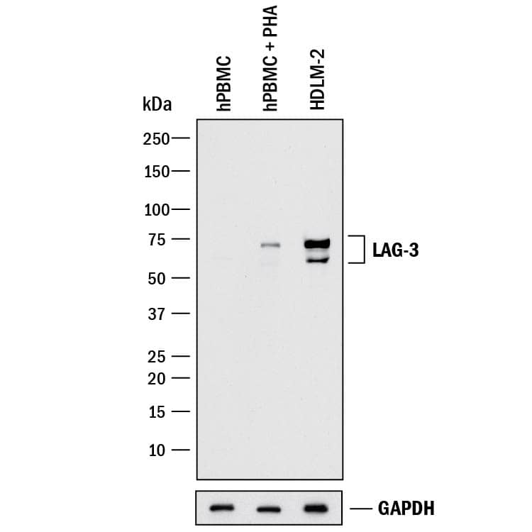 HAF017). Specific bands were detected for LAG-3 at approximately 60-75 kDa (as indicated). GAPDH (Catalog # MAB5718) is shown as a loading control. This experiment was conducted under reducing conditions and using Immunoblot Buffer Group 1." alt="Western blot shows lysates of human peripheral blood mononuclear cells (PBMC) untreated or treated (+) with 1 ug/mL PHA for 5 days and HDLM-2 human Hodgkin's lymphoma cell line. PVDF membrane was probed with 1 µg/mL of Goat Anti-Human LAG-3 Antigen Affinity-purified Polyclonal Antibody (Catalog # AF2319) followed by HRP-conjugated Anti-Goat IgG Secondary Antibody (Catalog # HAF017). Specific bands were detected for LAG-3 at approximately 60-75 kDa (as indicated). GAPDH (Catalog # MAB5718) is shown as a loading control. This experiment was conducted under reducing conditions and using Immunoblot Buffer Group 1." class="big_thumb" />
| Formulation | Catalog # | Availability | Price | |
|---|---|---|---|---|
| Carrier Free | FAB2319C | |||
| Carrier Free | FAB2319F | |||
| Carrier Free | FAB2319P | |||
| Carrier Free | FAB2319A | |||
| Carrier Free | AF2319-SP | |||
 DR647MS) and counterstained with DAPI (blue; Lunaphore Catalog # DR100). Specific staining was localized to the membrane and cytoplasm. Protocol available in COMET™ Panel Builder." alt="LAG-3 was detected in immersion fixed paraffin-embedded sections of human tonsil using Mouse Anti-Human LAG-3 Monoclonal Antibody (NOVUS Catalog # NBP2-80059) at 1:150 at 37 ° Celsius for 4 minutes. Before incubation with the primary antibody, tissue underwent an all-in-one dewaxing and antigen retrieval preprocessing using PreTreatment Module (PT Module) and Dewax and HIER Buffer H (pH 9). Tissue was stained using the Alexa Fluor™ 647 Goat anti-Mouse IgG Secondary Antibody at 1:200 at 37 ° Celsius for 2 minutes. (Yellow; Lunaphore Catalog # DR647MS) and counterstained with DAPI (blue; Lunaphore Catalog # DR100). Specific staining was localized to the membrane and cytoplasm. Protocol available in COMET™ Panel Builder." class="big_thumb" />
 HAF019). Specific bands were detected for LAG-3 at approximately 54 kDa and 75 kDa in mouse liver and mouse heart tissue and 70-80 kDa in EL4.IL-2 and CTLL-2 cell lines (as indicated). This experiment was conducted under reducing conditions and using Immunoblot Buffer Group 1." alt="Western blot shows lysates of mouse liver tissue, mouse heart tissue, EL4.IL-2 mouse lymphoblast cell line, and CTLL-2 mouse cytotoxic T cell line. PVDF membrane was probed with 0.25 µg/mL of Goat Anti-Mouse LAG-3 Antigen Affinity-purified Polyclonal Antibody (Catalog # AF3328) followed by HRP-conjugated Anti-Goat IgG Secondary Antibody (Catalog # HAF019). Specific bands were detected for LAG-3 at approximately 54 kDa and 75 kDa in mouse liver and mouse heart tissue and 70-80 kDa in EL4.IL-2 and CTLL-2 cell lines (as indicated). This experiment was conducted under reducing conditions and using Immunoblot Buffer Group 1." class="big_thumb" />
| Formulation | Catalog # | Availability | Price | |
|---|---|---|---|---|
| Carrier Free | AF3328-SP | |||
 HAF018). Specific bands were detected for LAG-3 at approximately 60-75 kDa (as indicated). GAPDH (Catalog # MAB5718) is shown as a loading control. This experiment was conducted under reducing conditions and using Immunoblot Buffer Group 1." alt="Western blot shows lysates of human peripheral blood mononuclear cells (PBMC) untreated or treated (+) with 1 ug/mL PHA for 5 days and HDLM-2 human Hodgkin's lymphoma cell line . PVDF membrane was probed with 2 µg/mL of Mouse Anti-Human LAG-3 Monoclonal Antibody (Catalog # MAB23193) followed by HRP-conjugated Anti-Mouse IgG Secondary Antibody (Catalog # HAF018). Specific bands were detected for LAG-3 at approximately 60-75 kDa (as indicated). GAPDH (Catalog # MAB5718) is shown as a loading control. This experiment was conducted under reducing conditions and using Immunoblot Buffer Group 1." class="big_thumb" />
 F0101B). Quadrant markers were set based on control antibody staining (Catalog # MAB0041, data not shown). View our protocol for Staining Membrane-associated Proteins." alt="HEK293 human embryonic kidney cell line transfected with either (A) human LAG-3 or (B) irrelevant transfectants and eGFP was stained with Mouse Anti-Human LAG-3 Monoclonal Antibody (Catalog # MAB23196) followed by APC-conjugated Anti-Mouse IgG Secondary Antibody (Catalog # F0101B). Quadrant markers were set based on control antibody staining (Catalog # MAB0041, data not shown). View our protocol for Staining Membrane-associated Proteins." class="big_thumb" />
| Formulation | Catalog # | Availability | Price | |
|---|---|---|---|---|
| Carrier Free | MAB23196-SP | |||
| Carrier Free | FAB23196V-100UG | |||
| Carrier Free | FAB23196U-100UG | |||
| Carrier Free | FAB23196T-100UG | |||
| Carrier Free | FAB23196S-100UG | |||
| Carrier Free | FAB23196R-100UG | |||
| Carrier Free | FAB23196N-100UG | |||
| Carrier Free | FAB23196G-100UG | |||
| Formulation | Catalog # | Availability | Price | |
|---|---|---|---|---|
| BSA Free | NBP3-26841-25ul | |||
| Azide and BSA Free | NBP3-44286 | |||
 F0101B). Quadrant markers were set based on control antibody staining (Catalog # MAB0041, data not shown). View our protocol for Staining Membrane-associated Proteins." alt="HEK293 human embryonic kidney cell line transfected with either (A) human LAG-3 or (B) irrelevant transfectants and eGFP was stained with Mouse Anti-Human LAG-3 Monoclonal Antibody (Catalog # MAB23197) followed by APC-conjugated Anti-Mouse IgG Secondary Antibody (Catalog # F0101B). Quadrant markers were set based on control antibody staining (Catalog # MAB0041, data not shown). View our protocol for Staining Membrane-associated Proteins." class="big_thumb" />
| Formulation | Catalog # | Availability | Price | |
|---|---|---|---|---|
| Carrier Free | MAB23197-SP | |||
| Carrier Free | FAB23197G-100UG | |||
| Carrier Free | FAB23197V-100UG | |||
| Carrier Free | FAB23197U-100UG | |||
| Carrier Free | FAB23197T-100UG | |||
| Carrier Free | FAB23197S-100UG | |||
| Carrier Free | FAB23197R-100UG | |||
| Carrier Free | FAB23197N-100UG | |||
| Formulation | Catalog # | Availability | Price | |
|---|---|---|---|---|
| BSA Free | NBP1-85781-25ul | |||


![Flow Cytometry: LAG-3 Antibody (11E3) [NBP1-97662] - Analysis of 10^6 MCF-7 cells stained using LAG-3 (human) monoclonal antibody (11E3) at a concentration of 50ug/mL.](http://images.novusbio.com/fullsize/LAG-3-Antibody-11E3-Flow-Cytometry-NBP1-97662-img0001.jpg "Flow Cytometry: LAG-3 Antibody (11E3) [NBP1-97662] - Analysis of 10^6 MCF-7 cells stained using LAG-3 (human) monoclonal antibody (11E3) at a concentration of 50ug/mL.")
 F0108) and Mouse Anti-Human CD3e PE-conjugated Monoclonal Antibody (Catalog #
  F0108). Quadrant markers were set based on control antibody staining(Catalog #
 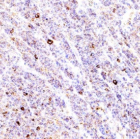 VC004). Before incubation with the primary antibody, tissue was subjected to heat-induced epitope retrieval using Antigen Retrieval Reagent-Basic (
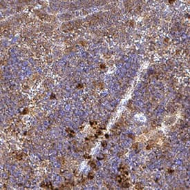 VC004). Before incubation with the primary antibody, tissue was subjected to heat-induced epitope retrieval using Antigen Retrieval Reagent-Basic (
 VC004). Before incubation with the primary antibody, tissue was subjected to heat-induced epitope retrieval using Antigen Retrieval Reagent-Basic (
 VC004). Before incubation with the primary antibody, tissue was subjected to heat-induced epitope retrieval using Antigen Retrieval Reagent-Basic (
![Western Blot: LAG-3 Antibody (BLR027F) [NBP2-76402] - Detection of human LAG3 by WB of HDLM-2, RPMI-8226, Jurkat, MJ, and HEK293T lysate (observed molecular weight of ~ 57 KDa). Antibody: Rabbit anti-LAG3 recombinant monoclonal [BLR027F] (NBP2-76402). Secondary: HRP-conjugated goat anti-rabbit IgG (NB7160). Lower Panel: Rabbit anti-GAPDH (NB300-322).](http://images.novusbio.com/fullsize/LAG-3-Antibody-BLR027F-Western-Blot-NBP2-76402-img0007.jpg "Western Blot: LAG-3 Antibody (BLR027F) [NBP2-76402] - Detection of human LAG3 by WB of HDLM-2, RPMI-8226, Jurkat, MJ, and HEK293T lysate (observed molecular weight of ~ 57 KDa). Antibody: Rabbit anti-LAG3 recombinant monoclonal [BLR027F] (NBP2-76402). Secondary: HRP-conjugated goat anti-rabbit IgG (NB7160). Lower Panel: Rabbit anti-GAPDH (NB300-322).")
![<b>Independent Antibodies Validation. </b>Immunoprecipitation: LAG-3 Antibody (BLR027F) [NBP2-76402] - Detection of human LAG3 by WB of immunoprecipitates from HDLM-2 lysate (observed molecular weight of ~ 57 KDa). Antibodies: Rabbit anti-LAG3 recombinant monoclonal [BLR027F] (NBP2-76402) and another rabbit anti-LAG3 recombinant monoclonal.](http://images.novusbio.com/fullsize/LAG-3-Antibody-BLR027F-Immunoprecipitation-NBP2-76402-img0006.jpg)
![Immunocytochemistry/Immunofluorescence: LAG-3 Antibody (BLR027F) [NBP2-76402] - Detection of human LAG3 in FFPE HDLM-2 cells by ICC. Antibody: Rabbit anti-LAG3 recombinant monoclonal [BLR027F] (NBP2-76402). Secondary: HRP-conjugated goat anti-rabbit IgG. Substate: DAB.](http://images.novusbio.com/fullsize/LAG-3-Antibody-BLR027F-Immunocytochemistry-NBP2-76402-img0002.jpg)
![Immunohistochemistry-Paraffin: LAG-3 Antibody (BLR027F) [NBP2-76402] - Section of human tonsil. Antibody: Rabbit anti-LAG3 recombinant monoclonal antibody [BLR027F] used at 1:250. Secondary: HRP-conjugated goat anti-rabbit IgG.Substrate: Opal™. Counterstain: DAPI (blue).](http://images.novusbio.com/fullsize/LAG-3-Antibody-BLR027F-Immunocytochemistry-Immunofluorescence-NBP2-76402-img0010.jpg)
![Immunohistochemistry-Paraffin: LAG-3 Antibody (BLR027F) [NBP2-76402] - Detection of human LAG3 in FFPE tonsil by IHC. Antibody: Rabbit anti-LAG3 recombinant monoclonal [BLR027F] (NBP2-76402), Secondary: HRP-conjugated goat anti-rabbit IgG. Substate: DAB.](http://images.novusbio.com/fullsize/LAG-3-Antibody-BLR027F-Immunohistochemistry-NBP2-76402-img0001.jpg)
 F0101B) and Mouse Anti-Human CD3e APC-conjugated Monoclonal Antibody (Catalog #
  F0101B) . Quadrant markers were set based on control antibody staining (Catalog #
 ![Western Blot: LAG-3 Antibody (BLR028F) [NBP2-76403] - Detection of human LAG3 by WB of HDLM-2, RPMI-8226, Jurkat, MJ, and HEK293T lysate (observed molecular weight of ~ 57 KDa). Antibody: Rabbit anti-LAG3 recombinant monoclonal [BLR028F] (NBP2-76403). Secondary: HRP-conjugated goat anti-rabbit IgG. Lower Panel: Rabbit anti-GAPDH.](http://images.novusbio.com/fullsize/LAG-3-Antibody-BLR028F-Western-Blot-NBP2-76403-img0003.jpg "Western Blot: LAG-3 Antibody (BLR028F) [NBP2-76403] - Detection of human LAG3 by WB of HDLM-2, RPMI-8226, Jurkat, MJ, and HEK293T lysate (observed molecular weight of ~ 57 KDa). Antibody: Rabbit anti-LAG3 recombinant monoclonal [BLR028F] (NBP2-76403). Secondary: HRP-conjugated goat anti-rabbit IgG. Lower Panel: Rabbit anti-GAPDH.")
![<b>Independent Antibodies Validation. </b>Immunoprecipitation: LAG-3 Antibody (BLR028F) [NBP2-76403] - Detection of human LAG3 by WB of immunoprecipitates from HDLM-2 lysate (observed molecular weight of ~ 57 KDa). Antibodies: Rabbit anti-LAG3 recombinant monoclonal [BLR028F] (NBP2-76403) and rabbit anti-LAG3 recombinant monoclonal [BLR027F] (NBP2-76402).](http://images.novusbio.com/fullsize/LAG-3-Antibody-BLR028F-Immunoprecipitation-NBP2-76403-img0004.jpg)
![Immunocytochemistry: LAG-3 Antibody (BLR028F) [NBP2-76403] - Detection of human LAG3 in FFPE HDLM-2 cells by ICC. Antibody: Rabbit anti-LAG3 recombinant monoclonal [BLR028F] (NBP2-76403) used at 1:200. Secondary: HRP-conjugated goat anti-rabbit IgG. Substrate: DAB.](http://images.novusbio.com/fullsize/LAG-3-Antibody-BLR028F-Immunocytochemistry-NBP2-76403-img0002.jpg)
![Immunohistochemistry-Paraffin: LAG-3 Antibody (BLR028F) [NBP2-76403] - Section of tonsil. Antibody: Rabbit anti-LAG3 recombinant monoclonal antibody [BLR028F] used at 1:200. Secondary: HRP-conjugated goat antirabbit IgG. Substrate: DAB.](http://images.novusbio.com/fullsize/LAG-3-Antibody-BLR028F-Immunohistochemistry-NBP2-76403-img0001.jpg)


 VC001). Before incubation with the primary antibody, tissue was subjected to heat-induced epitope retrieval using Antigen Retrieval Reagent-Basic (
 VC001). Before incubation with the primary antibody, tissue was subjected to heat-induced epitope retrieval using Antigen Retrieval Reagent-Basic (


 VC001). Before incubation with the primary antibody, tissue was subjected to heat-induced epitope retrieval using Antigen Retrieval Reagent-Basic (
![Immunohistochemistry-Paraffin: LAG-3 Antibody [NBP1-85781] - Staining in human tonsil and cerebral cortex tissues using anti-LAG3 antibody. Corresponding LAG3 RNA-seq data are presented for the same tissues.](http://images.novusbio.com/fullsize/LAG-3-Antibody-Immunohistochemistry-Paraffin-NBP1-85781-img0007.jpg "<b>Orthogonal Strategies Validation. </b>Immunohistochemistry-Paraffin: LAG-3 Antibody [NBP1-85781] - Staining in human tonsil and cerebral cortex tissues using anti-LAG3 antibody. Corresponding LAG3 RNA-seq data are presented for the same tissues.")
![<b>Orthogonal Strategies Validation. </b>Immunohistochemistry-Paraffin: LAG-3 Antibody [NBP1-85781] - Staining of human tonsil shows high expression.](http://images.novusbio.com/fullsize/LAG-3-Antibody-Immunohistochemistry-Paraffin-NBP1-85781-img0006.jpg)
![Immunohistochemistry-Paraffin: LAG-3 Antibody [NBP1-85781] - Staining of human cerebral cortex shows low expression as expected.](http://images.novusbio.com/fullsize/LAG-3-Antibody-Immunohistochemistry-Paraffin-NBP1-85781-img0005.jpg)


