1336) and 20 ng/mL Recombinant Human TNF-a (Catalog #
210-TA) for 10 minutes. PVDF membrane was probed with 0.1 µg/mL of Rabbit Anti-Human Phospho-RelA/NF kappa B p65 (S536) Monoclonal Antibody (Catalog # MAB72261) followed by HRP-conjugated Anti-Rabbit IgG Secondary Antibody (Catalog #
HAF008). A specific band was detected for Phospho-RelA/NF kappa B p65 (S536) at approximately 65 kDa (as indicated). This experiment was conducted under reducing conditions and using
Immunoblot Buffer Group 1." class="big_lightbox">
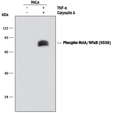
1336) and 20 ng/mL Recombinant Human TNF-a (Catalog #
210-TA) for 10 minutes. PVDF membrane was probed with 0.1 µg/mL of Rabbit Anti-Human Phospho-RelA/NF kappa B p65 (S536) Monoclonal Antibody (Catalog # MAB72261) followed by HRP-conjugated Anti-Rabbit IgG Secondary Antibody (Catalog #
HAF008). A specific band was detected for Phospho-RelA/NF kappa B p65 (S536) at approximately 65 kDa (as indicated). This experiment was conducted under reducing conditions and using
Immunoblot Buffer Group 1." alt="Western blot shows lysates of HeLa human cervical epithelial carcinoma cell line untreated (-) or treated (+) with 100 nM Calyculin A (Catalog #
1336) and 20 ng/mL Recombinant Human TNF-a (Catalog #
210-TA) for 10 minutes. PVDF membrane was probed with 0.1 µg/mL of Rabbit Anti-Human Phospho-RelA/NF kappa B p65 (S536) Monoclonal Antibody (Catalog # MAB72261) followed by HRP-conjugated Anti-Rabbit IgG Secondary Antibody (Catalog #
HAF008). A specific band was detected for Phospho-RelA/NF kappa B p65 (S536) at approximately 65 kDa (as indicated). This experiment was conducted under reducing conditions and using
Immunoblot Buffer Group 1." class="big_thumb" />
1336) and 20 ng/mL Recombinant Human TNF-a (Catalog #
210-TA) for 10 minutes, loaded at 0.2 mg/mL. A specific band was detected for Phospho-RelA/NF kappa B p65 (S536) at approximately 66 kDa (as indicated) using 1.0 µg/mL of Rabbit Anti-Human Phospho-RelA/NF kappa B p65 (S536) Monoclonal Antibody (Catalog # MAB72261). This experiment was conducted under reducing conditions and using the 12-230 kDa separation system.
Non-specific interaction with the 230 kDa Simple Western standard may be seen with this antibody." class="big_lightbox">
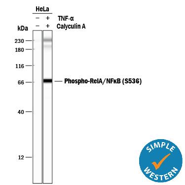 1336) and 20 ng/mL Recombinant Human TNF-a (Catalog # 210-TA) for 10 minutes, loaded at 0.2 mg/mL. A specific band was detected for Phospho-RelA/NF kappa B p65 (S536) at approximately 66 kDa (as indicated) using 1.0 µg/mL of Rabbit Anti-Human Phospho-RelA/NF kappa B p65 (S536) Monoclonal Antibody (Catalog # MAB72261). This experiment was conducted under reducing conditions and using the 12-230 kDa separation system.
Non-specific interaction with the 230 kDa Simple Western standard may be seen with this antibody." class="big_thumb" />
1336) and Recombinant Human TNF-a (Catalog #
210-TA; upper panel) using Rabbit Anti-Human Phospho-RelA/NF kappa B p65 (S536) Monoclonal Antibody (Catalog # MAB72261) at 10 µg/mL for 3 hours at room temperature. Cells were stained using the NorthernLights™ 557-conjugated Anti-Rabbit IgG Secondary Antibody (red; Catalog #
NL004) and counterstained with DAPI (blue). Specific staining was localized to cytoplasm and cell surfaces. View our protocol for
Fluorescent ICC Staining of Cells on Coverslips." class="big_lightbox">
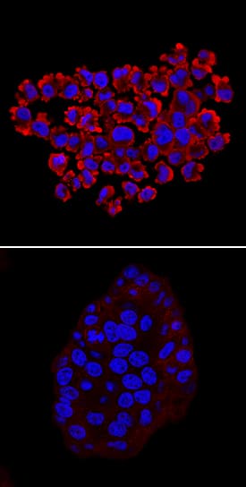
1336) and Recombinant Human TNF-a (Catalog #
210-TA; upper panel) using Rabbit Anti-Human Phospho-RelA/NF kappa B p65 (S536) Monoclonal Antibody (Catalog # MAB72261) at 10 µg/mL for 3 hours at room temperature. Cells were stained using the NorthernLights™ 557-conjugated Anti-Rabbit IgG Secondary Antibody (red; Catalog #
NL004) and counterstained with DAPI (blue). Specific staining was localized to cytoplasm and cell surfaces. View our protocol for
Fluorescent ICC Staining of Cells on Coverslips." class="big_thumb" />
 1336) and 20 ng/mL Recombinant Human TNF-a (Catalog # 210-TA) for 10 minutes. PVDF membrane was probed with 0.1 µg/mL of Rabbit Anti-Human Phospho-RelA/NF kappa B p65 (S536) Monoclonal Antibody (Catalog # MAB72261) followed by HRP-conjugated Anti-Rabbit IgG Secondary Antibody (Catalog # HAF008). A specific band was detected for Phospho-RelA/NF kappa B p65 (S536) at approximately 65 kDa (as indicated). This experiment was conducted under reducing conditions and using Immunoblot Buffer Group 1." alt="Western blot shows lysates of HeLa human cervical epithelial carcinoma cell line untreated (-) or treated (+) with 100 nM Calyculin A (Catalog # 1336) and 20 ng/mL Recombinant Human TNF-a (Catalog # 210-TA) for 10 minutes. PVDF membrane was probed with 0.1 µg/mL of Rabbit Anti-Human Phospho-RelA/NF kappa B p65 (S536) Monoclonal Antibody (Catalog # MAB72261) followed by HRP-conjugated Anti-Rabbit IgG Secondary Antibody (Catalog # HAF008). A specific band was detected for Phospho-RelA/NF kappa B p65 (S536) at approximately 65 kDa (as indicated). This experiment was conducted under reducing conditions and using Immunoblot Buffer Group 1." class="big_thumb" />


![Western Blot: SC65 Antibody (1E12) [H00010609-M01] - Analysis of SC65 expression in transfected 293T cell line by SC65 monoclonal antibody (M01), clone 1E12.Lane 1: SC65 transfected lysate (Predicted MW: 50.381 KDa).Lane 2: Non-transfected lysate.](http://images.novusbio.com/fullsize/SC65-Antibody-1E12-Western-Blot-H00010609-M01-img0002.jpg "Western Blot: SC65 Antibody (1E12) [H00010609-M01] - Analysis of SC65 expression in transfected 293T cell line by SC65 monoclonal antibody (M01), clone 1E12.Lane 1: SC65 transfected lysate (Predicted MW: 50.381 KDa).Lane 2: Non-transfected lysate.")
![Western Blot: SC65 Antibody (1E12) [H00010609-M01] - SC65 monoclonal antibody (M01), clone 1E12. Western Blot analysis of SC65 expression in HepG2 ( Cat # L019V1 ).](http://images.novusbio.com/fullsize/SC65-Antibody-1E12-Western-Blot-H00010609-M01-img0001.jpg)
![Western Blot: SC65 Antibody - BSA Free [NBP3-45441] - Human bladder tissue were subjected to SDS PAGE followed by western blot with (SC65 antibody) at dilution of 1:400](http://images.novusbio.com/fullsize/nbp3-45441_rabbit-sc65-pab-bsa-free-2842025123129.jpg "Western Blot: SC65 Antibody - BSA Free [NBP3-45441] - Human bladder tissue were subjected to SDS PAGE followed by western blot with (SC65 antibody) at dilution of 1:400")
![Western Blot: SC65 Antibody [H00010609-B01P] - Analysis of SC65 expression in transfected 293T cell line by SC65 polyclonal antibody. Lane 1: SC65 transfected lysate(48.07 KDa). Lane 2: Non-transfected lysate.](http://images.novusbio.com/fullsize/SC65-Antibody-Western-Blot-H00010609-B01P-img0001.jpg "Western Blot: SC65 Antibody [H00010609-B01P] - Analysis of SC65 expression in transfected 293T cell line by SC65 polyclonal antibody. Lane 1: SC65 transfected lysate(48.07 KDa). Lane 2: Non-transfected lysate.")
![Immunocytochemistry/Immunofluorescence: YAP1 Antibody - BSA Free [NB110-58358] - Caco-2 cells were fixed in 4% paraformaldehyde for 10 minutes and permeabilized in 0.5% Triton X-100 in PBS for 5 minutes. The cells were incubated with YAP1 Antibody (NB110-58358) at 1 ug/ml overnight at 4C and detected with an anti-rabbit DyLight 488 (Green) at a 1:1000 dilution for 60 minutes. Nuclei were counterstained with DAPI (Blue). Cells were imaged using a 40X objective.](http://images.novusbio.com/fullsize/YAP1-Antibody---BSA-Free-Immunocytochemistry-Immunofluorescence-NB110-58358-img0014.jpg "Immunocytochemistry/Immunofluorescence: YAP1 Antibody - BSA Free [NB110-58358] - Caco-2 cells were fixed in 4% paraformaldehyde for 10 minutes and permeabilized in 0.5% Triton X-100 in PBS for 5 minutes. The cells were incubated with YAP1 Antibody (NB110-58358) at 1 ug/ml overnight at 4C and detected with an anti-rabbit DyLight 488 (Green) at a 1:1000 dilution for 60 minutes. Nuclei were counterstained with DAPI (Blue). Cells were imaged using a 40X objective.")


![Immunohistochemistry-Paraffin: YAP1 Antibody - BSA Free [NB110-58358] - YAP1 was detected in immersion fixed paraffin-embedded sections of human placenta using Rabbit Anti-Human YAP1 polyclonal Antibody (Catalog # NB110-58358) at 1:200 for 1 hour at room temperature followed by incubation with the Anti-Rabbit IgG VisUCyte™ HRP Polymer Antibody (Catalog # VC003). Tissue was stained using DAB (brown) and counterstained with hematoxylin (blue). Specific staining was localized to nuclear and cytoplasm in trophoblast cells.](http://images.novusbio.com/fullsize/YAP1-Antibody---BSA-Free-Immunohistochemistry-Paraffin-NB110-58358-img0010.jpg)
![Western Blot: YAP1 Antibody - BSA Free [NB110-58358] - Analysis in transfected HEK 293 cell lysate using YAP1 antibody. Observed molecular weight 75 kDa.](http://images.novusbio.com/fullsize/YAP1-Antibody---BSA-Free-Western-Blot-NB110-58358-img0003.jpg)


![Simple Western: YAP1 Antibody - BSA Free [NB110-58358] - Simple Western lane view shows a specific band for YAP1 in 0.1 mg/ml of HeLa lysate. Observed molecular weight is 75 kDa. This experiment was performed under reducing conditions using the 12-230kDa separation system.](http://images.novusbio.com/fullsize/YAP1-Antibody---BSA-Free-Simple-Western-NB110-58358-img0007.jpg)
![<b>Genetic Strategies Validation. </b>Western Blot: YAP1 Antibody - BSA Free [NB110-58358] - Western blot shows lysates of HeLa human cervical epithelial carcinoma parental cell line and YAP1 knockout (KO) HeLa cell line. PVDF membrane was probed with 1:1000 of Rabbit Anti-Human YAP1 Polyclonal Antibody (Catalog # NB110-58358) followed by HRP-conjugated Anti-Rabbit IgG Secondary Antibody (Catalog #HAF008). Specific band was detected for YAP1 at approximately 75 kDa (as indicated) in the parental HeLa cell line, but is not detectable in the knockout HeLa cell line. This experiment was conducted under reducing conditions.](http://images.novusbio.com/fullsize/YAP1-Antibody---BSA-Free-Knockout-Validated-NB110-58358-img0008.jpg)
![<b>Genetic Strategies Validation. </b>Simple Western: YAP1 Antibody - BSA Free [NB110-58358] - Western lane view shows lysates of HeLa human cervical epithelial carcinoma parental cell line and YAP1 knockout (KO) HeLa cell line. A specific band was detected for YAP1 at approximately 81 kDa (as indicated) using 50 ug/mL of Rabbit Anti-YAP1 Polyclonal Antibody (Catalog # NB110-58358). This experiment was conducted under reducing conditions and using the 12-230 kDa separation system.](http://images.novusbio.com/fullsize/YAP1-Antibody---BSA-Free-Knockout-Validated-NB110-58358-img0009.jpg)
![Western Blot: YAP1 Antibody - BSA Free [NB110-58358] - Yap deficiency suppresses cell proliferation in vivo & in vitro. (A-F) The Ki67 positive ratio of lens epithelial cells decreased in Yap-deficient mice at different stages (arrowheads indicate Ki67 positive cells). (G) The relative number of Ki67 positive lens epithelial cells (number of Ki67 positive lens epithelial cells / lens epithelium area). The data are shown as mean ± S.E.M. (Student’s t-test, *P<0.05, **P<0.01, n=10). (H-I) Knockdown efficiency of Yap in alpha TN4 cell using siRNA. (J-K) Cell viability & growth assay revealed that proliferation was downregulated in Yap knockdown alpha TN4 cells. The data are shown as mean ± S.E.M. (Two-way RM ANOVA, **P<0.01, n=5). Scale bars: 50 μm. Image collected & cropped by CiteAb from the following publication (//pubmed.ncbi.nlm.nih.gov/31011480), licensed under a CC-BY license. Not internally tested by Novus Biologicals.](http://images.novusbio.com/fullsize/nb110-58358_rabbit-polyclonal-yap1-antibody-31020241535689.jpg)
![Western Blot: YAP1 Antibody - BSA Free [NB110-58358] - Synergistic effect of LW6 & metformin on YAP1. 80 µM LW6, 20 mM metformin (Met) & the combinational treatment metformin plus LW6 increased phosphorylation of YPA1 at serine 127 & decreased cellular YAP1 concentration after treating cells for 24 hours (A). Moreover, this combinational therapy attenuated the nuclear localization of YAP1 compared to Sham treated cells (B). In addition, lysophosphatidic acid (LPA) & the phosphorylation deficient mutant YAP1-S127A stimulate cell migration of 6606PDA cells (C & D). n = 2 per group for A, n = 3 per group for B, n = 7 per group for C, n = 9 per group for D. Bar = 5µm. Arrows point to nuclei. Image collected & cropped by CiteAb from the following publication (//pubmed.ncbi.nlm.nih.gov/31897243), licensed under a CC-BY license. Not internally tested by Novus Biologicals.](http://images.novusbio.com/fullsize/nb110-58358_rabbit-polyclonal-yap1-antibody-310202415371934.jpg)
![Western Blot: YAP1 Antibody - BSA Free [NB110-58358] - Consequences of ORP5 & ORP8 knockdown on downstream MAPK & PI3K/AKT signaling.Protein from BxPC-3, PANC-1, MiaPaCa-2, & MOH parental, single & double ORP knockdowns as well as cells transfected with empty vector control (pLKO.1) were harvested, & 20 μg was subjected to SDS–PAGE & used for Western blotting. EGFR, MAPK, & PI3K signaling were assayed as pEGFR, ppERK, & pAKT levels, respectively. Amplification of YAP-1 was also evaluated. Total ERK, total AKT, & beta -actin levels were used as loading controls. Image collected & cropped by CiteAb from the following publication (//pubmed.ncbi.nlm.nih.gov/31451509), licensed under a CC-BY license. Not internally tested by Novus Biologicals.](http://images.novusbio.com/fullsize/nb110-58358_rabbit-polyclonal-yap1-antibody-310202415291931.jpg)
![Western Blot: YAP1 Antibody - BSA Free [NB110-58358] - Ajuba depletion induces beta -catenin translocation & Cyclin D1 expression in HCC cell lines. a Immunoblotting with specific antibodies against Ajuba, E-cadherin, beta -catenin, Cyclin D1, YAP, TAZ & CYR61 in Ajuba-depleted BEL7402 & HepG2 cells. GAPDH was used as a loading control. The ratios of expression E-Cadherin to their corresponding GAPDH are represented. b Ajuba-depleted HCC cells were fixed for immunofluorescence & stained for beta -catenin protein (green) & DAPI (blue). Representative merged images are also shown for fluorescence signals. Scale bar = 25 μm. c Correlation of Ajuba expression with OS in HCC. Low expression of Ajuba was associated with worse OS compared to high expression of Ajuba. Kaplan-Meier curves & log-rank test were used to evaluate OS. P < 0.05 was considered significant. d, e Representative images & quantification of migration & invasion of Ajuba-depleted (d) or Ajuba-overexpressing (e) HCC cells. Scale bar = 200 μm. HCC, hepatocellular carcinoma. f Expression in response to the overexpression of constructs of Ajuba-Myc was examined by IB, the ratios of expression Ajuba to their corresponding GAPDH are represented. Data are presented as Mean ± SEM from three independent experiments (*p < 0.05, **p < 0.01, ***p < 0.001) Image collected & cropped by CiteAb from the following publication (//pubmed.ncbi.nlm.nih.gov/30041665), licensed under a CC-BY license. Not internally tested by Novus Biologicals.](http://images.novusbio.com/fullsize/nb110-58358_rabbit-polyclonal-yap1-antibody-310202416235517.jpg)
![Immunocytochemistry/ Immunofluorescence: YAP1 Antibody - BSA Free [NB110-58358] - YAP & TAZ expression at high density & during chondrogenic differentiation of human synovial MSCs. (A) YAP & pYAP expression in human synovial membrane-derived (hSM-)MSCs in monolayer at low (L) & high (H) density detected by immunofluorescence staining, shown without (i) & with sytox green nuclear counterstain (ii). (B) YAP & pYAP expression in hSM-MSCs in monolayer at low (L) & high (H) density detected by western blotting with beta -actin as loading control. (C, D) Expression of YAP & TAZ (C) & their target genes CTGF & CYR61 (D) in hSM-MSCs immediately prior to (0 h) or 24 h after plating in micromass culture, determined by quantitative RT-PCR. Data was normalised to GAPDH expression, & is shown as mean ± standard deviation (SD) (three donors) relative to pre-seeding (0 h) control. *P <0.05; **P <0.01; ***P <0.001. (E) Expression of YAP & TAZ in hSM-MSCs after 6 days of treatment with 10 ng/ml TGF-beta 1 or vehicle only in micromass culture to induce chondrogenic differentiation, determined by quantitative RT-PCR. Data was normalised to GAPDH expression, & is shown as mean ± SD (five donors) relative to vehicle-treated control. *P <0.05. (F) Detection of YAP by western blotting during chondrogenic differentiation induced by TGF-beta 1 with detection of beta -actin as loading control. MSC, mesenchymal stromal/stem cell; pYAP, phosphorylated YAP; RT-PCR, reverse transcription PCR; TAZ, transcriptional co-activator with PDZ-binding motif; TGF, transforming growth factor; YAP, Yes-associated protein. Image collected & cropped by CiteAb from the following publication (//pubmed.ncbi.nlm.nih.gov/26025096), licensed under a CC-BY license. Not internally tested by Novus Biologicals.](http://images.novusbio.com/fullsize/nb110-58358_rabbit-polyclonal-yap1-antibody-310202416235539.jpg)
![Immunocytochemistry/ Immunofluorescence: YAP1 Antibody - BSA Free [NB110-58358] - Expression of spcCre is associated with loss of YAP in SOX9+ distal airways.(A–D) Immunostaining of lung sections collected from Yapf/f; spcCre/+ mice at 13.5 days post coitus (dpc). SOX2 expression marks the proximal airway, while the distal airway is distinguished by SOX9 expression (not shown). High levels of spcCre expression were largely confined to the distal airway, where spcCre expression in a given epithelial cell was correlated with loss of YAP immunoreactivity (e.g. arrowheads in D). (E–H) Immunostaining of lung sections collected from Yapf/f; spcCre/+ mice at 14.5 dpc. Only distal airways are shown. YAP was lost mainly in distal airways in Yapf/f; spcCre/+ mice while sporadic loss of YAP was found in the proximal airway. Loss of YAP was most apparent in the more distal part (arrow in H) of the distal airway, while residual YAP could be found in the more proximal part of the distal airway. Together, these results suggest that lung cyst formation in distal airways of Yapf/f; spcCre/+ mice is due to loss of YAP in the distal airway. Scale bar = 25 μm for A–D; E–H.DOI://dx.doi.org/10.7554/eLife.21130.012 Image collected & cropped by CiteAb from the following publication (//pubmed.ncbi.nlm.nih.gov/28323616), licensed under a CC-BY license. Not internally tested by Novus Biologicals.](http://images.novusbio.com/fullsize/nb110-58358_rabbit-polyclonal-yap1-antibody-310202416235526.jpg)
![Immunocytochemistry/ Immunofluorescence: YAP1 Antibody - BSA Free [NB110-58358] - The expression patterns of Yap & GFAP-Cre recombinase in postnatal mouse eyes. (A) Schematic of a transverse section of mouse eye. (B-C) Immunostaining with anti-Yap antibody (green) on frozen eye sections at different ages. Nuclei were counterstained with DAPI (blue). Yap staining was detected in scattered cells within the INL (arrowheads) & GCL of the retina & the lens epithelium (arrows). (D) Cre recombinase (red) was expressed in the lens epithelium & INL, GCL of retina in frozen eye sections of Tomatof/+; GFAP-Cre mice at P14. Nuclei were counterstained with DAPI (blue). LE, lens epithelium; TZ, transitional zone; RPE, retinal pigment epithelium; OS, outer segment; IS, inner segment; ONL, outer nuclear layer; OPL, outer plexiform layer; INL, inner nuclear layer; IPL, inner plexiform layer; GCL, ganglion cell layer. Scale bars: 25 μm (B-C), 100 μm (D). Image collected & cropped by CiteAb from the following publication (//pubmed.ncbi.nlm.nih.gov/31011480), licensed under a CC-BY license. Not internally tested by Novus Biologicals.](http://images.novusbio.com/fullsize/nb110-58358_rabbit-polyclonal-yap1-antibody-310202416235548.jpg)
![Immunocytochemistry/ Immunofluorescence: YAP1 Antibody - BSA Free [NB110-58358] - YAP & phospho-YAP are detected in both the proximal & distal airways during lung development.(A–H) Immunostaining of lung sections collected from wild-type mice at 13.5 days post coitus (dpc). The proximal airway is marked by SOX2 expression, while the distal airway is distinguished by SOX9 expression (not shown). Nuclear YAP can be frequently found in both SOX2+ & SOX9+ domains. Similarly, phospho-YAP at S112 (pYAP) could be detected in both the proximal & distal airways. pYAP levels were, in general, higher in the proximal than distal epithelium but pYAP levels varied significantly from cell to cell in both the proximal & distal airways. Representative cells with higher levels of pYAP (arrowhead) are indicated in (B,F). In many cells, low levels of pYAP were associated with the presence of nuclear YAP. This is consistent with a model in which pYAP is sequestered by 14-3-3 proteins in the cytoplasm & degraded but also indicate a dynamic shuttling & distribution of YAP along the entire airway epithelium. Similar results were obtained for lungs collected at 12.5 dpc. Scale bar = 7.5 μm for A–H.DOI://dx.doi.org/10.7554/eLife.21130.005 Image collected & cropped by CiteAb from the following publication (//pubmed.ncbi.nlm.nih.gov/28323616), licensed under a CC-BY license. Not internally tested by Novus Biologicals.](http://images.novusbio.com/fullsize/nb110-58358_rabbit-polyclonal-yap1-antibody-31020241623554.jpg)
![Immunocytochemistry/ Immunofluorescence: YAP1 Antibody - BSA Free [NB110-58358] - Expression of Nkx2.1Cre is associated with loss of YAP in the upper lobes.(A–D) Immunostaining of lung sections collected from Yapf/f; Nkx2.1Cre/+ mice at 14.5 days post coitus (dpc). SOX2 expression marks the proximal airway, while the distal airway is distinguished by SOX9 expression (not shown). YAP was lost mainly in the upper lobe in Yapf/f; Nkx2.1Cre/+ lung. Loss of YAP was more apparent in the distal airway, while loss of YAP was sporadic in the proximal airway. Lung cyst formation was primarily observed in the distal airway. The boxed region in (B) indicates areas shown in (C,D). Scale bar = 250 μm for A,B; 250 μm for C,D. Sox2 expression was present in sporadic Yap-deficient cells in the transition zone induced by Sox9Cre, spcCre or Nkx2.1Cre. This suggests that Sox2 expression is not controlled by YAP.DOI://dx.doi.org/10.7554/eLife.21130.013 Image collected & cropped by CiteAb from the following publication (//pubmed.ncbi.nlm.nih.gov/28323616), licensed under a CC-BY license. Not internally tested by Novus Biologicals.](http://images.novusbio.com/fullsize/nb110-58358_rabbit-polyclonal-yap1-antibody-310202416235520.jpg)
![Immunocytochemistry/ Immunofluorescence: YAP1 Antibody - BSA Free [NB110-58358] - Active nuclear YAP is distributed throughout the mouse lung epithelium during development.(A–P) Immunostaining of lung sections collected from wild-type mice at 11.5 & 12.5 days post coitus (dpc). The boxed region in (L) indicates areas shown in (N–P). The proximal airway is marked by SOX2 expression, while the distal airway is distinguished by SOX9 expression. Nuclear YAP can be frequently found in both SOX2+ & SOX9+ domains & is not restricted to the junction (the ‘transition zone’) between SOX2+ & SOX9+ domains. Representative cells with nuclear YAP (arrowhead) are indicated in (E,P). YAP immunoreactivity is completely absent in the epithelium (but present in the mesenchyme) of Yapf/f; ShhCre/+ mice (M), demonstrating the specificity of YAP antibodies used in this study. Immunofluorescence & immunohistochemistry yielded the same results (data not shown for immunohistochemistry). (Q–R) Whole-mount immunostaining of wild-type & Yap mutant lungs at 11.5 dpc. Distinct domains of SOX2 were discerned in the absence of YAP. Scale bar = 10 μm for A–J; 25 μm for K, L; 10 μm for N–P; 50 μm for Q, R.DOI://dx.doi.org/10.7554/eLife.21130.004 Image collected & cropped by CiteAb from the following publication (//pubmed.ncbi.nlm.nih.gov/28323616), licensed under a CC-BY license. Not internally tested by Novus Biologicals.](http://images.novusbio.com/fullsize/nb110-58358_rabbit-polyclonal-yap1-antibody-310202416235530.jpg)
![Immunocytochemistry/ Immunofluorescence: YAP1 Antibody - BSA Free [NB110-58358] - The expression patterns of Yap & GFAP-Cre recombinase in postnatal mouse eyes. (A) Schematic of a transverse section of mouse eye. (B-C) Immunostaining with anti-Yap antibody (green) on frozen eye sections at different ages. Nuclei were counterstained with DAPI (blue). Yap staining was detected in scattered cells within the INL (arrowheads) & GCL of the retina & the lens epithelium (arrows). (D) Cre recombinase (red) was expressed in the lens epithelium & INL, GCL of retina in frozen eye sections of Tomatof/+; GFAP-Cre mice at P14. Nuclei were counterstained with DAPI (blue). LE, lens epithelium; TZ, transitional zone; RPE, retinal pigment epithelium; OS, outer segment; IS, inner segment; ONL, outer nuclear layer; OPL, outer plexiform layer; INL, inner nuclear layer; IPL, inner plexiform layer; GCL, ganglion cell layer. Scale bars: 25 μm (B-C), 100 μm (D). Image collected & cropped by CiteAb from the following publication (//pubmed.ncbi.nlm.nih.gov/31011480), licensed under a CC-BY license. Not internally tested by Novus Biologicals.](http://images.novusbio.com/fullsize/nb110-58358_rabbit-polyclonal-yap1-antibody-31020241623552.jpg)


![Western Blot: RelA/NFkB p65 Antibody (112A1021) [NB100-56712] - Analysis of P65 using p65 antibody at 2 ug/mL in 30 ug of A) Ramos, B) Daudi, C) HeLa and D) mouse NIH 3T3 cell lysate.](http://images.novusbio.com/fullsize/RelA-NFkB-p65-Antibody-112A1021-Western-Blot-NB100-56712-img0013.jpg "Western Blot: RelA/NFkB p65 Antibody (112A1021) [NB100-56712] - Analysis of P65 using p65 antibody at 2 ug/mL in 30 ug of A) Ramos, B) Daudi, C) HeLa and D) mouse NIH 3T3 cell lysate.")
![Simple Western: RelA/NFkB p65 Antibody (112A1021) [NB100-56712] - Lane view shows a specific band for NFkB p65 in 1.0 mg/mL of HeLa lysate. This experiment was performed under reducing conditions using the 12-230 kDa separation system.](http://images.novusbio.com/fullsize/RelA-NFkB-p65-Antibody-112A1021-Simple-Western-NB100-56712-img0006.jpg)
![Western Blot: RelA/NFkB p65 Antibody (112A1021) [NB100-56712] - Lysates of HeLa human cervical epithelial carcinoma cell line and Daudi human Burkitt's lymphoma cell line. PVDF membrane was probed with 1 ug/mL mouse anti-RelA/NFkB p65 monoclonal (NB100-56712, Novus Biologicals), followed by 1:2000 dilution of the appropriate HRP-conjugated secondary antibody, donkey anti-mouse IgG (HAF018).](http://images.novusbio.com/fullsize/RelA-NFkB-p65-Antibody-112A1021-Western-Blot-NB100-56712-img0016.jpg)
![Immunohistochemistry-Paraffin: RelA/NFkB p65 Antibody (112A1021) [NB100-56712] - Ovarian cystadenocarcinoma probed with p65 antibody at 5 ug/mL. Human tissue TMA was used for this test. Staining of formalin-fixed tissues is enhanced by boiling tissue sections in 10 mM sodium citrate buffer, pH 6.0 for 10-20 min followed by cooling at RT for 20 min.](http://images.novusbio.com/fullsize/RelA-NFkB-p65-Antibody-112A1021-Immunohistochemistry-Paraffin-NB100-56712-img0009.jpg)
![Flow Cytometry: RelA/NFkB p65 Antibody (112A1021) [NB100-56712] - Intracellular staining of 293 HEK cells using 0.5 ug of p65 antibody. Green histogram represents the isotype control (p65) antibody. This NFkB p65 antibody was used for this test with an anti-mouse IgG PE conjugated secondary antibody.](http://images.novusbio.com/fullsize/RelA-NFkB-p65-Antibody-112A1021-Flow-Cytometry-NB100-56712-img0004.jpg)
![Simple Western: RelA/NFkB p65 Antibody (112A1021) [NB100-56712] - Detection of p65 by Simple Western (JESS) in chemiluminescence in nuclear extract of reconstructed human epidermis lysate (about 600 ug/mL protein concentration).Antibody at 1:20. Simple Western image submitted by a verified customer review.](http://images.novusbio.com/fullsize/RelA-NFkB-p65-Antibody-112A1021-Simple-Western-NB100-56712-img0017.jpg)
![Western Blot: RNF185 Antibody [NBP1-59761] - Human Liver cell lysate, concentration 0.2-1 ug/ml.](http://images.novusbio.com/fullsize/RNF185-Antibody-Western-Blot-NBP1-59761-img0002.jpg "Western Blot: RNF185 Antibody [NBP1-59761] - Human Liver cell lysate, concentration 0.2-1 ug/ml.")
![Immunohistochemistry-Paraffin: RelA/NFkB p65 [p Ser276] Antibody [NB100-82086] - Immunohistochemical analysis of paraffin- embedded human lung carcinoma tissue using NB100-82086](http://images.novusbio.com/fullsize/RelA-NFkB-p65-[p-Ser276]-Antibody-Immunohistochemistry-Paraffin-NB100-82086-img0009.jpg "Immunohistochemistry-Paraffin: RelA/NFkB p65 [p Ser276] Antibody [NB100-82086] - Immunohistochemical analysis of paraffin- embedded human lung carcinoma tissue using NB100-82086")
![Immunocytochemistry/Immunofluorescence: RelA/NFkB p65 [p Ser276] Antibody [NB100-82086] - Staining of methanol-fixed Hela cells.](http://images.novusbio.com/fullsize/RelA-NFkB-p65-[p-Ser276]-Antibody-Immunocytochemistry-Immunofluorescence-NB100-82086-img0014.jpg)
![Immunohistochemistry-Paraffin: RelA/NFkB p65 [p Ser276] Antibody [NB100-82086] - Human breast carcinoma tissue.](http://images.novusbio.com/fullsize/RelA-NFkB-p65-[p-Ser276]-Antibody-Immunohistochemistry-Paraffin-NB100-82086-img0010.jpg)
![Western Blot: RelA/NFkB p65 [p Ser276] Antibody [NB100-82086] - Western Blot analysis of various cells using NB100-82086 diluted at 1:1000](http://images.novusbio.com/fullsize/nb100-82086_rabbit-polyclonal-rela-nfkb-p65-p-ser276-antibody-23420241545358.jpg)
![Immunohistochemistry-Paraffin: RelA/NFkB p65 [p Ser276] Antibody [NB100-82086] - Analysis of paraffin- embedded human breast carcinoma tissue using NB100-82086 incubated with blocking peptide (right)](http://images.novusbio.com/fullsize/nb100-82086_rabbit-polyclonal-rela-nfkb-p65-p-ser276-antibody-103202523464543.jpg)
![Immunocytochemistry/Immunofluorescence: RelA/NFkB p65 [p Ser276] Antibody [NB100-82086] - Immunofluorescence staining of methanol-fixed MEF cells untreated or treated with TNF using NB100-82086](http://images.novusbio.com/fullsize/nb100-82086_rabbit-polyclonal-rela-nfkb-p65-p-ser276-antibody-11320251555922.jpg)
![Western Blot: RelA/NFkB p65 [p Ser276] Antibody [NB100-82086] - Lane 1: HEK293. Lane 2: Lentivirus mediated NFkB overexpression. We image submitted by a verified customer review.](http://images.novusbio.com/fullsize/antibody/nb100-82086_rabbit-polyclonal-rela-nfkb-p65-p-ser276-antibody-western-blot-1132025204015..jpg)
![Western Blot: C11orf65 Antibody [NBP1-91439] - MCF-7 whole cell lysates, concentration 0.2-1 ug/ml.](http://images.novusbio.com/fullsize/C11orf65-Antibody-Western-Blot-NBP1-91439-img0002.jpg "Western Blot: C11orf65 Antibody [NBP1-91439] - MCF-7 whole cell lysates, concentration 0.2-1 ug/ml.")
![Western Blot: RelA/NFkB p65 Antibody [NB100-2176] - Human glomerular endothelial cell lysates, time-response treatment with 10ng/ml TNF-alpha. This image was submitted via customer Review.](http://images.novusbio.com/fullsize/RelA-NFkB-p65-Antibody-Western-Blot-NB100-2176-img0006.jpg "<b>Biological Strategies Validation. </b>Western Blot: RelA/NFkB p65 Antibody [NB100-2176] - Human glomerular endothelial cell lysates, time-response treatment with 10ng/ml TNF-alpha. This image was submitted via customer Review.")
![<b>Biological Strategies Validation. </b>Immunohistochemistry-Paraffin: RelA/NFkB p65 Antibody [NB100-2176] - Staining of formalin-fixed, paraffin-embedded human DLBCL showing nuclear expression of NFkB p65 in the tumor cells.](http://images.novusbio.com/fullsize/RelA-NFkB-p65-Antibody-Immunohistochemistry-Paraffin-NB100-2176-img0004.jpg)
![Western Blot: RelA/NFkB p65 Antibody [NB100-2176] - Analysis of NFkB p65 expression in A431 (A) and HeLa (B) whole cell lysates.](http://images.novusbio.com/fullsize/RelA-NFkB-p65-Antibody-Western-Blot-NB100-2176-img0005.jpg)
![Immunohistochemistry-Paraffin: Rabbit Polyclonal RelA/NFkB p65 Antibody [NB100-2176] - RelA/NFkB p65 IHC (brown) in the mice liver tissue; bars, 200 μm. Image from a verified customer review.](http://images.novusbio.com/fullsize/antibody/nb100-2176_rabbit-polyclonal-rela-nfkb-p65-antibody-immunohistochemistry-paraffin-1111202410307..jpg)
![Immunocytochemistry/Immunofluorescence: Rabbit Polyclonal RelA/NFkB p65 Antibody [NB100-2176] - Mice hepatocytes stained for RelA/NFkB p65. Image from a verified customer review.](http://images.novusbio.com/fullsize/antibody/nb100-2176_rabbit-polyclonal-rela-nfkb-p65-antibody-immunocytochemistry-immunofluorescence-2511202410544..png)
 1336) and 20 ng/mL Recombinant Human TNF-a (Catalog #
  1336) and Recombinant Human TNF-a (Catalog #
 ![Western Blot: Neuroplastin Antibody [NBP1-62423] - HepG2 cell lysate, Antibody Titration: 5.0ug/ml](http://images.novusbio.com/fullsize/Neuroplastin-Antibody-Western-Blot-NBP1-62423-img0001.jpg "Western Blot: Neuroplastin Antibody [NBP1-62423] - HepG2 cell lysate, Antibody Titration: 5.0ug/ml")
![Immunohistochemistry-Paraffin: Neuroplastin Antibody [NBP1-62423] - Human Muscle Tissue, Skeletal muscle cells (Indicated with Arrows) 4-8ug/ml.](http://images.novusbio.com/fullsize/Neuroplastin-Antibody-Immunohistochemistry-Paraffin-NBP1-62423-img0003.jpg)


