 CTS008) and counterstained with hematoxylin (blue). Specific staining was localized to membranes in blood vessels in CNS. View our protocol for Chromogenic IHC Staining of Frozen Tissue Sections." alt="Laminin a4 was detected in immersion fixed frozen sections of mouse embryo (13 d.p.c.) using Goat Anti-Mouse Laminin a4 Antigen Affinity-purified Polyclonal Antibody (Catalog # AF3837) at 3 µg/mL overnight at 4 °C. Tissue was stained using the Anti-Goat HRP-DAB Cell & Tissue Staining Kit (brown; Catalog # CTS008) and counterstained with hematoxylin (blue). Specific staining was localized to membranes in blood vessels in CNS. View our protocol for Chromogenic IHC Staining of Frozen Tissue Sections." class="big_thumb" />
| Formulation | Catalog # | Availability | Price | |
|---|---|---|---|---|
| Carrier Free | AF3837-SP | |||
 HAF016). A specific band was detected for Laminin a4 at approximately 200-220 kDa (as indicated). This experiment was conducted under reducing conditions and using Immunoblot Buffer Group 1." alt="Western blot shows lysates of human placenta tissue. PVDF membrane was probed with 2 µg/mL of Sheep Anti-Human Laminin a4 Antigen Affinity-purified Polyclonal Antibody (Catalog # AF7340) followed by HRP-conjugated Anti-Sheep IgG Secondary Antibody (Catalog # HAF016). A specific band was detected for Laminin a4 at approximately 200-220 kDa (as indicated). This experiment was conducted under reducing conditions and using Immunoblot Buffer Group 1." class="big_thumb" />
| Formulation | Catalog # | Availability | Price | |
|---|---|---|---|---|
| Carrier Free | AF7340-SP | |||
| Formulation | Catalog # | Availability | Price | |
|---|---|---|---|---|
| Azide and BSA Free | NBP3-44408 | |||
| BSA Free | NBP2-42392-25ul | |||
| Formulation | Catalog # | Availability | Price | |
|---|---|---|---|---|
| Azide and BSA Free | NBP3-44409 | |||
| BSA Free | NBP2-42393-25ul | |||
| Formulation | Catalog # | Availability | Price | |
|---|---|---|---|---|
| Azide and BSA Free | NBP2-71113 | |||
 CTS017) and counterstained with hematoxylin (blue). Specific staining was localized to endothelial cells in gastric pits. View our protocol for Chromogenic IHC Staining of Frozen Tissue Sections." alt="Laminin a4 was detected in perfusion fixed frozen sections of mouse stomach using Rat Anti-Mouse Laminin a4 Monoclonal Antibody (Catalog # MAB3837) at 25 µg/mL overnight at 4 °C. Tissue was stained using the Anti-Rat HRP-DAB Cell & Tissue Staining Kit (brown; Catalog # CTS017) and counterstained with hematoxylin (blue). Specific staining was localized to endothelial cells in gastric pits. View our protocol for Chromogenic IHC Staining of Frozen Tissue Sections." class="big_thumb" />
| Formulation | Catalog # | Availability | Price | |
|---|---|---|---|---|
| Carrier Free | MAB3837-SP | |||
 CCM017) using Mouse Anti-Human Laminin a4 Monoclonal Antibody (Catalog # MAB7340) at 10 µg/mL for 3 hours at room temperature. Cells were stained using the NorthernLights™ 557-conjugated Anti-Mouse IgG Secondary Antibody (red; Catalog # NL007) and counterstained with DAPI (blue). Specific staining was localized to cytoplasm. View our protocol for Fluorescent ICC Staining of Cells on Coverslips." alt="Laminin a4 was detected in immersion fixed T98G human glioblastoma cells cultured with (upper panel) or without (lower panel) StemXVivo EMT Inducing Media Supplement (Catalog # CCM017) using Mouse Anti-Human Laminin a4 Monoclonal Antibody (Catalog # MAB7340) at 10 µg/mL for 3 hours at room temperature. Cells were stained using the NorthernLights™ 557-conjugated Anti-Mouse IgG Secondary Antibody (red; Catalog # NL007) and counterstained with DAPI (blue). Specific staining was localized to cytoplasm. View our protocol for Fluorescent ICC Staining of Cells on Coverslips." class="big_thumb" />
| Formulation | Catalog # | Availability | Price | |
|---|---|---|---|---|
| Carrier Free | MAB7340-SP | |||
| Formulation | Catalog # | Availability | Price | |
|---|---|---|---|---|
| BSA Free | NBP1-87716-25ul | |||


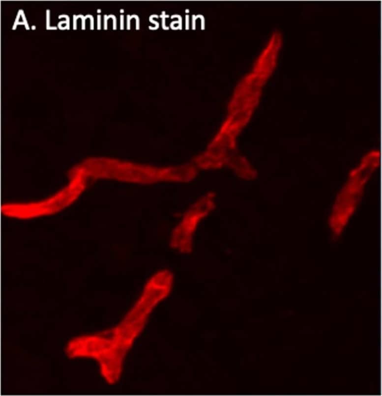


 CCM017) , using Sheep Anti-Human Laminin a4 Antigen Affinity-purified Polyclonal Antibody (Catalog # AF7340) at 10 µg/mL for 3 hours at room temperature. Cells were stained using the NorthernLights™ 557-conjugated Anti-Sheep IgG Secondary Antibody (red; Catalog #
  VC006). Before incubation with the primary antibody, tissue was subjected to heat-induced epitope retrieval using Antigen Retrieval Reagent-Basic (Catalog #
  HAF016). This experiment was conducted under reducing conditions and using the 66-440 kDa separation system." class="big_thumb" />
 7340-A4) supports adhesion in the HT1080 human fibrosarcoma cell line in a dose-dependent manner (orange line), as measured by Calcein AM. Adhesion elicited by Recombinant Human Laminin a4 (5 µg/mL) is neutralized (green line) by increasing concentrations of Sheep Anti-Human Laminin a4 Antigen Affinity-purified Polyclonal Antibody (Catalog # AF7340). The ND50 is typically 1.5-7.5 µg/mL." class="big_thumb" />


![Western Blot: Laminin alpha 4 Antibody (CL3183) [NBP2-42392] - Analysis of purified human recombinant Laminin-411, Laminin-421, Laminin-511, Laminin-121, Laminin-221 and Laminin-332.](http://images.novusbio.com/fullsize/Laminin-alpha-4-Antibody-CL3183-Western-Blot-NBP2-42392-img0020.jpg "Western Blot: Laminin alpha 4 Antibody (CL3183) [NBP2-42392] - Analysis of purified human recombinant Laminin-411, Laminin-421, Laminin-511, Laminin-121, Laminin-221 and Laminin-332.")
![Immunohistochemistry-Paraffin: Laminin alpha 4 Antibody (CL3183) [NBP2-42392] - Staining of rat rectum shows immunoreactivity in the basement membrane of glandular epithelium.](http://images.novusbio.com/fullsize/Laminin-alpha-4-Antibody-CL3183-Immunohistochemistry-Paraffin-NBP2-42392-img0031.jpg)
![Western Blot: Laminin alpha 4 Antibody (CL3183) [NBP2-42392] - Analysis in human placenta tissue.](http://images.novusbio.com/fullsize/Laminin-alpha-4-Antibody-CL3183-Western-Blot-NBP2-42392-img0019.jpg)
![Immunohistochemistry-Paraffin: Laminin alpha 4 Antibody (CL3183) [NBP2-42392] - Staining of human colon shows strong immunoreactivity in basement membrane of glandular epithelium.](http://images.novusbio.com/fullsize/Laminin-alpha-4-Antibody-CL3183-Immunohistochemistry-Paraffin-NBP2-42392-img0007.jpg)
![Immunohistochemistry-Paraffin: Laminin alpha 4 Antibody (CL3183) [NBP2-42392] - Staining of human pancreas shows strong immunoreactivity in basement membrane of glandular epithelium.](http://images.novusbio.com/fullsize/Laminin-alpha-4-Antibody-CL3183-Immunohistochemistry-Paraffin-NBP2-42392-img0009.jpg)
![Immunohistochemistry-Paraffin: Laminin alpha 4 Antibody (CL3183) [NBP2-42392] - Staining of human lymph node shows absence of immunoreactivity in lymphoid tissue (negative control).](http://images.novusbio.com/fullsize/Laminin-alpha-4-Antibody-CL3183-Immunohistochemistry-Paraffin-NBP2-42392-img0011.jpg)
![Immunohistochemistry-Paraffin: Laminin alpha 4 Antibody (CL3183) [NBP2-42392] - Staining of mouse stomach shows positivity in the basement membrane of glandular epithelium.](http://images.novusbio.com/fullsize/Laminin-alpha-4-Antibody-CL3183-Immunohistochemistry-Paraffin-NBP2-42392-img0017.jpg)
![Immunohistochemistry-Paraffin: Laminin alpha 4 Antibody (CL3183) [NBP2-42392] - Staining of rat heart muscle shows membranous positivity in cardiomyocytes.](http://images.novusbio.com/fullsize/Laminin-alpha-4-Antibody-CL3183-Immunohistochemistry-Paraffin-NBP2-42392-img0022.jpg)
![Immunohistochemistry-Paraffin: Laminin alpha 4 Antibody (CL3183) [NBP2-42392] - Staining of rat epididymis shows strong immunoreactivity in lamina propria.](http://images.novusbio.com/fullsize/Laminin-alpha-4-Antibody-CL3183-Immunohistochemistry-Paraffin-NBP2-42392-img0024.jpg)
![Immunohistochemistry-Paraffin: Laminin alpha 4 Antibody (CL3183) [NBP2-42392] - Staining of mouse epididymis shows immunoreactivity in smooth muscle in the lamina propria.](http://images.novusbio.com/fullsize/Laminin-alpha-4-Antibody-CL3183-Immunohistochemistry-Paraffin-NBP2-42392-img0025.jpg)
![Immunohistochemistry-Paraffin: Laminin alpha 4 Antibody (CL3183) [NBP2-42392] - Staining of rat stomach shows immunoreactivity in the basement membrane of glandular epithelium.](http://images.novusbio.com/fullsize/Laminin-alpha-4-Antibody-CL3183-Immunohistochemistry-Paraffin-NBP2-42392-img0027.jpg)
![Immunohistochemistry-Paraffin: Laminin alpha 4 Antibody (CL3183) [NBP2-42392] - Staining of human placenta shows strong positivity in basement membrane and endothelium.](http://images.novusbio.com/fullsize/Laminin-alpha-4-Antibody-CL3183-Immunohistochemistry-Paraffin-NBP2-42392-img0028.jpg)
![Immunohistochemistry-Paraffin: Laminin alpha 4 Antibody (CL3183) [NBP2-42392] - Staining of human prostate shows moderate membranous positivity in smooth muscle cells.](http://images.novusbio.com/fullsize/Laminin-alpha-4-Antibody-CL3183-Immunohistochemistry-Paraffin-NBP2-42392-img0029.jpg)
![Immunohistochemistry-Paraffin: Laminin alpha 4 Antibody (CL3183) [NBP2-42392] - Staining of rat kidney shows basement membrane immunoreactivity in renal tubules and glomeruli.](http://images.novusbio.com/fullsize/Laminin-alpha-4-Antibody-CL3183-Immunohistochemistry-Paraffin-NBP2-42392-img0030.jpg)
![Immunohistochemistry-Paraffin: Laminin alpha 4 Antibody (CL3185) [NBP2-42393] - Analysis in human smooth muscle and lymph node tissues using NBP2-42393 antibody. Corresponding LAMA4 RNA-seq data are presented for the same tissues.](http://images.novusbio.com/fullsize/Laminin-alpha-4-Antibody-CL3185-Immunohistochemistry-Paraffin-NBP2-42393-img0014.jpg "<b>Orthogonal Strategies Validation. </b>Immunohistochemistry-Paraffin: Laminin alpha 4 Antibody (CL3185) [NBP2-42393] - Analysis in human smooth muscle and lymph node tissues using NBP2-42393 antibody. Corresponding LAMA4 RNA-seq data are presented for the same tissues.")
![<b>Orthogonal Strategies Validation. </b>Western Blot: Laminin alpha 4 Antibody (CL3185) [NBP2-42393] - Lane 1: Marker [kDa] 460, 268, 238, 171, 117, 71, 55, 41, 31 Lane 2: Purified human recombinant protein Laminin-411 Lane 3: Purified human recombinant protein Laminin-421 Lane 4: Purified human recombinant protein Laminin-511 Lane 5: Purified human recombinant protein Laminin-121 Lane 6: Purified human recombinant protein Laminin-221 Lane 7: Purified human recombinant protein Laminin-332](http://images.novusbio.com/fullsize/Laminin-alpha-4-Antibody-CL3185-Western-Blot-NBP2-42393-img0001.jpg)
![Immunohistochemistry-Paraffin: Laminin alpha 4 Antibody (CL3185) [NBP2-42393] - Staining of human rectum shows strong immunoreactivity in basement membrane of glandular epithelium.](http://images.novusbio.com/fullsize/Laminin-alpha-4-Antibody-CL3185-Immunohistochemistry-Paraffin-NBP2-42393-img0008.jpg)
![Immunohistochemistry-Paraffin: Laminin alpha 4 Antibody (CL3185) [NBP2-42393] - Staining of human placenta shows strong positivity in basement membrane of trophoblast and in endothelium.](http://images.novusbio.com/fullsize/Laminin-alpha-4-Antibody-CL3185-Immunohistochemistry-Paraffin-NBP2-42393-img0009.jpg)
![Immunohistochemistry-Paraffin: Laminin alpha 4 Antibody (CL3185) [NBP2-42393] - Staining of human prostate shows moderate membranous positivity in smooth muscle cells.](http://images.novusbio.com/fullsize/Laminin-alpha-4-Antibody-CL3185-Immunohistochemistry-Paraffin-NBP2-42393-img0010.jpg)
![Immunohistochemistry-Paraffin: Laminin alpha 4 Antibody (CL3185) [NBP2-42393] - Staining of human lymph node shows absence of immunoreactivity in lymphoid tissue (negative control).](http://images.novusbio.com/fullsize/Laminin-alpha-4-Antibody-CL3185-Immunohistochemistry-Paraffin-NBP2-42393-img0011.jpg)
![Immunohistochemistry-Paraffin: Laminin alpha 4 Antibody (CL3185) [NBP2-42393] - Staining of mouse heart muscle shows membranous positivity in cardiomyocytes.](http://images.novusbio.com/fullsize/Laminin-alpha-4-Antibody-CL3185-Immunohistochemistry-Paraffin-NBP2-42393-img0006.jpg)
![Immunohistochemistry-Paraffin: Laminin alpha 4 Antibody (CL3185) [NBP2-42393] - Staining of mouse stomach shows strong positivity in basement membrane of glandular epithelium and in smooth muscle layer.](http://images.novusbio.com/fullsize/Laminin-alpha-4-Antibody-CL3185-Immunohistochemistry-Paraffin-NBP2-42393-img0007.jpg)
![Immunohistochemistry-Paraffin: Laminin alpha 4 Antibody (CL3185) [NBP2-42393] - Staining of mouse epididymis shows immunoreactivity in smooth muscle in the lamina propria.](http://images.novusbio.com/fullsize/Laminin-alpha-4-Antibody-CL3185-Immunohistochemistry-Paraffin-NBP2-42393-img0012.jpg)
![Western Blot: Laminin alpha 4 Antibody (5G8) [NBP2-45582] - Analysis of HEK293T cells were transiently transfected with the pCMV6-ENTRY control (Left lane) or pCMV6-ENTRY Laminin alpha 4.](http://images.novusbio.com/fullsize/Laminin-alpha-4-Antibody-5G8-Western-Blot-NBP2-45582-img0009.jpg "Western Blot: Laminin alpha 4 Antibody (5G8) [NBP2-45582] - Analysis of HEK293T cells were transiently transfected with the pCMV6-ENTRY control (Left lane) or pCMV6-ENTRY Laminin alpha 4.")
![Immunohistochemistry: Laminin alpha 4 Antibody (5G8) [NBP2-45582] - Analysis of Human lymph node tissue.](http://images.novusbio.com/fullsize/Laminin-alpha-4-Antibody-5G8-Immunohistochemistry-NBP2-45582-img0008.jpg)
![Western Blot: Laminin alpha 4 Antibody (5G8) [NBP2-45582] - Analysis of Carcinoma of Human prostate tissue.](http://images.novusbio.com/fullsize/Laminin-alpha-4-Antibody-5G8-Western-Blot-NBP2-45582-img0007.jpg)
![Immunohistochemistry: Laminin alpha 4 Antibody (5G8) [NBP2-45582] - Analysis of Adenocarcinoma Human colon tissue.](http://images.novusbio.com/fullsize/Laminin-alpha-4-Antibody-5G8-Immunohistochemistry-NBP2-45582-img0001.jpg)
![Immunohistochemistry: Laminin alpha 4 Antibody (5G8) [NBP2-45582] - Analysis of Human Kidney tissue.](http://images.novusbio.com/fullsize/Laminin-alpha-4-Antibody-5G8-Immunohistochemistry-NBP2-45582-img0002.jpg)
![Immunohistochemistry: Laminin alpha 4 Antibody (5G8) [NBP2-45582] - Analysis of Carcinoma of Human liver tissue.](http://images.novusbio.com/fullsize/Laminin-alpha-4-Antibody-5G8-Immunohistochemistry-NBP2-45582-img0003.jpg)
![Immunohistochemistry: Laminin alpha 4 Antibody (5G8) [NBP2-45582] - Analysis of Carcinoma of Human lung tissue.](http://images.novusbio.com/fullsize/Laminin-alpha-4-Antibody-5G8-Immunohistochemistry-NBP2-45582-img0004.jpg)
![Immunohistochemistry: Laminin alpha 4 Antibody (5G8) [NBP2-45582] - Analysis of Human pancreas tissue.](http://images.novusbio.com/fullsize/Laminin-alpha-4-Antibody-5G8-Immunohistochemistry-NBP2-45582-img0005.jpg)
![Immunohistochemistry: Laminin alpha 4 Antibody (5G8) [NBP2-45582] - Analysis of Human prostate tissue.](http://images.novusbio.com/fullsize/Laminin-alpha-4-Antibody-5G8-Immunohistochemistry-NBP2-45582-img0006.jpg)
![Immunohistochemistry-Paraffin: Laminin alpha 4 Antibody [NBP1-87716] - Staining of human smooth muscle shows high expression.](http://images.novusbio.com/fullsize/Laminin-alpha-4-Antibody-Immunohistochemistry-Paraffin-NBP1-87716-img0006.jpg "Immunohistochemistry-Paraffin: Laminin alpha 4 Antibody [NBP1-87716] - Staining of human smooth muscle shows high expression.")
![Immunohistochemistry-Paraffin: Laminin alpha 4 Antibody [NBP1-87716] - Staining of human smooth muscle shows cytoplasmic positivity in smooth muscle cells.](http://images.novusbio.com/fullsize/Laminin-alpha-4-Antibody-Immunohistochemistry-Paraffin-NBP1-87716-img0004.jpg)
![Immunohistochemistry-Paraffin: Laminin alpha 4 Antibody [NBP1-87716] - Staining of human skeletal muscle shows low expression as expected.](http://images.novusbio.com/fullsize/Laminin-alpha-4-Antibody-Immunohistochemistry-Paraffin-NBP1-87716-img0005.jpg)
![<b>Orthogonal Strategies Validation. </b>Immunohistochemistry-Paraffin: Laminin alpha 4 Antibody [NBP1-87716] - Staining in human smooth muscle and skeletal muscle tissues using anti-LAMA4 antibody. Corresponding LAMA4 RNA-seq data are presented for the same tissues.](http://images.novusbio.com/fullsize/Laminin-alpha-4-Antibody-Immunohistochemistry-Paraffin-NBP1-87716-img0007.jpg)
![ELISA: Human Laminin alpha 4 ELISA Kit (Colorimetric) [NBP2-68048] - Samples were spiked with high concentrations of Human Laminin alpha 4 and diluted with Reference Standard & Sample Diluent to produce samples with values within the range of the assay.](http://images.novusbio.com/fullsize/nbp2-68048_human-laminin-alpha-4-elisa-kit-colorimetric-132202416215348.jpg "ELISA: Human Laminin alpha 4 ELISA Kit (Colorimetric) [NBP2-68048] - Samples were spiked with high concentrations of Human Laminin alpha 4 and diluted with Reference Standard & Sample Diluent to produce samples with values within the range of the assay.")
![ELISA: Human Laminin alpha 4 ELISA Kit (Colorimetric) [NBP2-68048] - Standard Curve Reference](http://images.novusbio.com/fullsize/nbp2-68048_human-laminin-alpha-4-elisa-kit-colorimetric-432024983528.jpg)
![Immunohistochemistry-Paraffin: Laminin alpha 4 Antibody [NBP1-32159] - Mouse lung. Laminin alpha 4 antibody [C3], C-term diluted at 1:500. Antigen Retrieval: Trilogy™ (EDTA based, pH 8.0) buffer, 15min.](http://images.novusbio.com/fullsize/Laminin-alpha-4-Antibody-Immunohistochemistry-Paraffin-NBP1-32159-img0004.jpg "Immunohistochemistry-Paraffin: Laminin alpha 4 Antibody [NBP1-32159] - Mouse lung. Laminin alpha 4 antibody [C3], C-term diluted at 1:500. Antigen Retrieval: Trilogy™ (EDTA based, pH 8.0) buffer, 15min.")
![Western Blot: Laminin alpha 4 Antibody [H00003910-D01P] - Analysis of LAMA4 expression in transfected 293T cell line by LAMA4 polyclonal antibody.Lane 1: LAMA4 transfected lysate(12.80 KDa).Lane 2: Non-transfected lysate.](http://images.novusbio.com/fullsize/Laminin-alpha-4-Antibody-Western-Blot-H00003910-D01P-img0002.jpg "Western Blot: Laminin alpha 4 Antibody [H00003910-D01P] - Analysis of LAMA4 expression in transfected 293T cell line by LAMA4 polyclonal antibody.Lane 1: LAMA4 transfected lysate(12.80 KDa).Lane 2: Non-transfected lysate.")


