HAF018). A specific band was detected for PTEN at approximately 54 kDa (as indicated). This experiment was conducted under reducing conditions and using Western Blot Buffer Group 1.**For Western blot, we recommend the use of Rabbit Anti-Human/Mouse/Rat PTEN Affinitiy-purified Polyclonal Ab (
AF847)." class="big_lightbox">
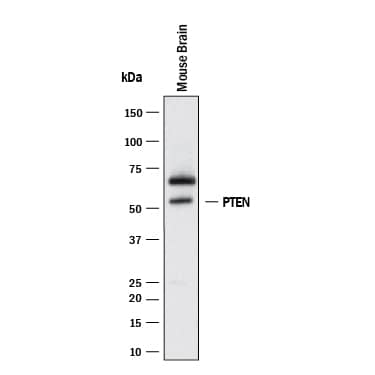
HAF018). A specific band was detected for PTEN at approximately 54 kDa (as indicated). This experiment was conducted under reducing conditions and using Western Blot Buffer Group 1.**For Western blot, we recommend the use of Rabbit Anti-Human/Mouse/Rat PTEN Affinitiy-purified Polyclonal Ab (
AF847)." alt="Western blot shows lysates of mouse brain tissue. PVDF membrane was probed with 2 µg/mL of Mouse Anti-Human/Mouse PTEN Monoclonal Antibody (Catalog # MAB847) followed by HRP-conjugated Anti-Mouse IgG Secondary Antibody (
HAF018). A specific band was detected for PTEN at approximately 54 kDa (as indicated). This experiment was conducted under reducing conditions and using Western Blot Buffer Group 1.**For Western blot, we recommend the use of Rabbit Anti-Human/Mouse/Rat PTEN Affinitiy-purified Polyclonal Ab (
AF847)." class="big_thumb" />
CTS002) and counterstained with hematoxylin (blue). Lower panel shows a lack of labeling if primary antibodies are omitted and tissue is stained only with secondary antibody followed by incubation with detection reagents. View our protocol for
Chromogenic IHC Staining of Paraffin-embedded Tissue Sections." class="big_lightbox">
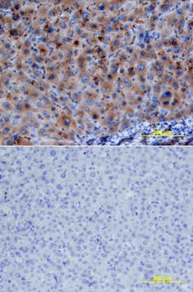
CTS002) and counterstained with hematoxylin (blue). Lower panel shows a lack of labeling if primary antibodies are omitted and tissue is stained only with secondary antibody followed by incubation with detection reagents. View our protocol for
Chromogenic IHC Staining of Paraffin-embedded Tissue Sections." class="big_thumb" />
MAB002, open histogram), followed by Allophycocyanin-conjugated Anti-Mouse IgG F(ab')2Secondary Antibody (
F0101B). To facilitate intracellular staining, cells were fixed with Flow Cytometry Fixation Buffer (
FC004) and permeabilized with Flow Cytometry Permeabilization/Wash Buffer I (
FC005). Staining was performed using our protocol for Staining Intracellular Molecules. " class="big_lightbox">
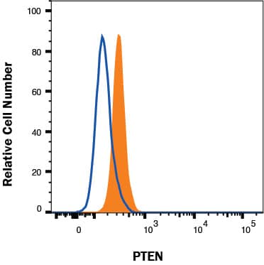
MAB002, open histogram), followed by Allophycocyanin-conjugated Anti-Mouse IgG F(ab')2Secondary Antibody (
F0101B). To facilitate intracellular staining, cells were fixed with Flow Cytometry Fixation Buffer (
FC004) and permeabilized with Flow Cytometry Permeabilization/Wash Buffer I (
FC005). Staining was performed using our protocol for Staining Intracellular Molecules. " class="big_thumb" />
MAB002, open histogram), followed by Phycoerythrin-conjugated Anti-Mouse IgG F(ab')
2Secondary Antibody (Catalog #
F0102B). To facilitate intracellular staining, cells were fixed with paraformaldehyde and permeabilized with saponin." class="big_lightbox">

MAB002, open histogram), followed by Phycoerythrin-conjugated Anti-Mouse IgG F(ab')
2Secondary Antibody (Catalog #
F0102B). To facilitate intracellular staining, cells were fixed with paraformaldehyde and permeabilized with saponin." class="big_thumb" />
HAF018). A specific band was detected for PTEN at approximately 55 kDa (as indicated) in the parental HeLa cell line, but is not detectable in knockout HeLa cell line. GAPDH (
MAB5718) is shown as a loading control. This experiment was conducted under reducing conditions and using Western Blot Buffer Group 1." class="big_lightbox">
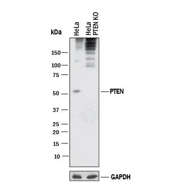
HAF018). A specific band was detected for PTEN at approximately 55 kDa (as indicated) in the parental HeLa cell line, but is not detectable in knockout HeLa cell line. GAPDH (
MAB5718) is shown as a loading control. This experiment was conducted under reducing conditions and using Western Blot Buffer Group 1." class="big_thumb" />
 HAF008). For additional reference, recombinant human PTEN (5 ng) was included. A specific band for PTEN was detected at approximately 54 kDa (as indicated). This experiment was conducted under reducing conditions and using Immunoblot Buffer Group 4." alt="Western blot shows lysates of mouse and rat brain tissue, A431 human epithelial carcinoma cell line, and MRC-5 human embryonic lung fibroblast cell line. PVDF membrane was probed with 0.1 µg/mL Rabbit Anti-Human/Mouse/Rat PTEN Antigen Affinity-purified Polyclonal Antibody (Catalog # AF847) followed by HRP-conjugated Anti-Rabbit IgG Secondary Antibody (Catalog # HAF008). For additional reference, recombinant human PTEN (5 ng) was included. A specific band for PTEN was detected at approximately 54 kDa (as indicated). This experiment was conducted under reducing conditions and using Immunoblot Buffer Group 4." class="big_thumb" />
 HAF018). A specific band was detected for PTEN at approximately 54 kDa (as indicated). This experiment was conducted under reducing conditions and using Western Blot Buffer Group 1.**For Western blot, we recommend the use of Rabbit Anti-Human/Mouse/Rat PTEN Affinitiy-purified Polyclonal Ab (AF847)." alt="Western blot shows lysates of mouse brain tissue. PVDF membrane was probed with 2 µg/mL of Mouse Anti-Human/Mouse PTEN Monoclonal Antibody (Catalog # MAB847) followed by HRP-conjugated Anti-Mouse IgG Secondary Antibody (HAF018). A specific band was detected for PTEN at approximately 54 kDa (as indicated). This experiment was conducted under reducing conditions and using Western Blot Buffer Group 1.**For Western blot, we recommend the use of Rabbit Anti-Human/Mouse/Rat PTEN Affinitiy-purified Polyclonal Ab (AF847)." class="big_thumb" />
 CTS013). Tissue was stained using the Anti-Goat HRP-DAB Cell & Tissue Staining Kit (brown; Catalog # CTS008) and counterstained with hematoxylin (blue). Specific staining was localized to cytoplasm of hepatocytes. View our protocol for Chromogenic IHC Staining of Paraffin-embedded Tissue Sections." alt="PTEN was detected in immersion fixed paraffin-embedded sections of human liver using Goat Anti- Human PTEN Antigen Affinity-purified Polyclonal Antibody (Catalog # AF6655) at 10 µg/mL overnight at 4 °C. Before incubation with the primary antibody, tissue was subjected to heat-induced epitope retrieval using Antigen Retrieval Reagent-Basic (Catalog # CTS013). Tissue was stained using the Anti-Goat HRP-DAB Cell & Tissue Staining Kit (brown; Catalog # CTS008) and counterstained with hematoxylin (blue). Specific staining was localized to cytoplasm of hepatocytes. View our protocol for Chromogenic IHC Staining of Paraffin-embedded Tissue Sections." class="big_thumb" />
 MAB847) immunopreciptate of MRC-5 human embryonic lung fibroblast cell line, mouse and rat brain tissue untreated (-) or treated (+) with 300 U/mL CIP for 1 hour. PVDF membrane was probed with 1 µg/mL of Rabbit Anti-Human/Mouse/ Rat Phospho-PTEN (S380) Antigen Affinity-purified Polyclonal Antibody (Catalog # AF2838), followed by HRP-conjugated Anti-Rabbit IgG Secondary Antibody (Catalog # HAF008). A specific band was detected for Phospho-PTEN (S380) at approximately 54 kDa (as indicated). This experiment was conducted under reducing conditions and using Immunoblot Buffer Group 1." alt="Western blot shows Human/Mouse/Rat PTEN Monoclonal Antibody (Catalog # MAB847) immunopreciptate of MRC-5 human embryonic lung fibroblast cell line, mouse and rat brain tissue untreated (-) or treated (+) with 300 U/mL CIP for 1 hour. PVDF membrane was probed with 1 µg/mL of Rabbit Anti-Human/Mouse/ Rat Phospho-PTEN (S380) Antigen Affinity-purified Polyclonal Antibody (Catalog # AF2838), followed by HRP-conjugated Anti-Rabbit IgG Secondary Antibody (Catalog # HAF008). A specific band was detected for Phospho-PTEN (S380) at approximately 54 kDa (as indicated). This experiment was conducted under reducing conditions and using Immunoblot Buffer Group 1." class="big_thumb" />


 VC003). Before incubation with the primary antibody, tissue was subjected to heat-induced epitope retrieval using Antigen Retrieval Reagent-Basic (
 VC003). Before incubation with the primary antibody, tissue was subjected to heat-induced epitope retrieval using Antigen Retrieval Reagent-Basic (
 AB-105-C, open histogram), followed by Phycoerythrin-conjugated Anti-Rabbit IgG Secondary Antibody (Catalog #
 HAF008). A specific band was detected for PTEN at approximately 55 kDa (as indicated) in the parental HeLa cell line, but is not detectable in knockout HeLa cell line. GAPDH (Catalog #
 CTS002) and counterstained with hematoxylin (blue). Lower panel shows a lack of labeling if primary antibodies are omitted and tissue is stained only with secondary antibody followed by incubation with detection reagents. View our protocol for
  MAB002, open histogram), followed by Allophycocyanin-conjugated Anti-Mouse IgG F(ab')2Secondary Antibody (
 MAB002, open histogram), followed by Phycoerythrin-conjugated Anti-Mouse IgG F(ab')2Secondary Antibody (Catalog #
  HAF018). A specific band was detected for PTEN at approximately 55 kDa (as indicated) in the parental HeLa cell line, but is not detectable in knockout HeLa cell line. GAPDH (
 NL001) and counterstained with DAPI (blue). Specific staining was localized to cytoplasm. View our protocol for
![Western Blot: PTEN Antibody (7O6F4) [NBP3-15666] - Analysis of extracts of C6 cells, using PTEN antibody (NBP3-15666) at 1:1000 dilution. Secondary antibody: HRP Goat Anti-Rabbit IgG (H+L) at 1:10000 dilution. Lysates/proteins: 25ug per lane. Blocking buffer: 3% nonfat dry milk in TBST. Detection: ECL Basic Kit. Exposure time: 60s.](http://images.novusbio.com/fullsize/PTEN-Antibody-7O6F4-Western-Blot-NBP3-15666-img0005.jpg "Western Blot: PTEN Antibody (7O6F4) [NBP3-15666] - Analysis of extracts of C6 cells, using PTEN antibody (NBP3-15666) at 1:1000 dilution. Secondary antibody: HRP Goat Anti-Rabbit IgG (H+L) at 1:10000 dilution. Lysates/proteins: 25ug per lane. Blocking buffer: 3% nonfat dry milk in TBST. Detection: ECL Basic Kit. Exposure time: 60s.")
![Western Blot: PTEN Antibody (7O6F4) [NBP3-15666] - Analysis of extracts of various cell lines, using PTEN antibody (NBP3-15666) at 1:1000 dilution. Secondary antibody: HRP Goat Anti-Rabbit IgG (H+L) at 1:10000 dilution. Lysates/proteins: 25ug per lane. Blocking buffer: 3% nonfat dry milk in TBST. Detection: ECL Basic Kit. Exposure time: 1s.](http://images.novusbio.com/fullsize/PTEN-Antibody-7O6F4-Western-Blot-NBP3-15666-img0004.jpg)
![<b>Genetic Strategies Validation. </b>Western Blot: PTEN Antibody (7O6F4) [NBP3-15666] - Analysis of extracts from normal (control) and PTEN knockout (KO) HeLa cells, using PTEN antibody (NBP3-15666) at 1:1000 dilution. Secondary antibody: HRP Goat Anti-Rabbit IgG (H+L) at 1:10000 dilution. Lysates/proteins: 25ug per lane. Blocking buffer: 3% nonfat dry milk in TBST. Detection: ECL Basic Kit. Exposure time: 60s.](http://images.novusbio.com/fullsize/PTEN-Antibody-7O6F4-Knockout-Validated-NBP3-15666-img0001.jpg)
![Immunoprecipitation: PTEN Antibody (7O6F4) [NBP3-15666] - Analysis of 300ug extracts of NIH/3T3 cells using 3ug PTEN antibody (NBP3-15666). Western blot was performed from the immunoprecipitate using PTEN antibody (NBP3-15666) at a dilition of 1:1000.](http://images.novusbio.com/fullsize/PTEN-Antibody-7O6F4-Immunoprecipitation-NBP3-15666-img0006.jpg)
![Immunocytochemistry/ Immunofluorescence: PTEN Antibody (7O6F4) [PTEN] - Confocal imaging of NIH/3T3 cells using [KO Validated] PTEN Rabbit mAb followed by a further incubation with Cy3 Goat Anti-Rabbit IgG (H+L) . The cells were counterstained with alpha-Tubulin Mouse mAb followed by incubation with ABflo 488-conjugated Goat Anti-Mouse IgG (H+L) Ab (Green). DAPI was used for nuclear staining (Blue). Objective: 100x.](http://images.novusbio.com/fullsize/nbp3-15666_rabbit-pten-mab-7o6f4-52202517361229.jpg)


![Western Blot: PTEN Antibody [NB100-2229] - Detection of Human and Mouse PTEN on HeLa whole cell lysate using NB100-2229. PTEN was also IPed by rabbit anti-PTEN antibodies NB100-2227, NB100-2228 and another PTEN antibody.](http://images.novusbio.com/fullsize/PTEN-Antibody-Western-Blot-NB100-2229-img0001.jpg "<b>Independent Antibodies Validation and Biological Strategies Validation.</b>Western Blot: PTEN Antibody [NB100-2229] - Detection of Human and Mouse PTEN on HeLa whole cell lysate using NB100-2229. PTEN was also IPed by rabbit anti-PTEN antibodies NB100-2227, NB100-2228 and another PTEN antibody.")
![<b>Independent Antibodies Validation and Biological Strategies Validation.</b>Immunohistochemistry-Paraffin: PTEN Antibody [NB100-2229] - Section of human breast carcinoma. Antibody: Affinity purified rabbit anti- PTEN used at a dilution of 1:200 (1ug/ml). Detection: DAB](http://images.novusbio.com/fullsize/PTEN-Antibody-Immunohistochemistry-NB100-2229-img0008.jpg)
![Western Blot: PTEN [p Thr366, p Ser370] Antibody [NBP3-12919] - Various whole cell extracts (30 ug) were separated by 10% SDS-PAGE, and the membrane was blotted with PTEN (phospho Thr366/Ser370) antibody (NBP3-12919) diluted at 1:500. The HRP-conjugated anti-rabbit IgG antibody (NBP2-19301) was used to detect the primary antibody.](http://images.novusbio.com/fullsize/PTEN-[p-Thr366--p-Ser370]-Antibody-Western-Blot-NBP3-12919-img0004.jpg "Western Blot: PTEN [p Thr366, p Ser370] Antibody [NBP3-12919] - Various whole cell extracts (30 ug) were separated by 10% SDS-PAGE, and the membrane was blotted with PTEN (phospho Thr366/Ser370) antibody (NBP3-12919) diluted at 1:500. The HRP-conjugated anti-rabbit IgG antibody (NBP2-19301) was used to detect the primary antibody.")
![Western Blot: PTEN [p Thr366, p Ser370] Antibody [NBP3-12919] - Various whole cell extracts (30 ug) were separated by 10% SDS-PAGE, and the membrane was blotted with PTEN (phospho Thr366/Ser370) antibody (NBP3-12919) diluted at 1:500. The HRP-conjugated anti-rabbit IgG antibody (NBP2-19301) was used to detect the primary antibody.](http://images.novusbio.com/fullsize/PTEN-[p-Thr366--p-Ser370]-Antibody-Western-Blot-NBP3-12919-img0003.jpg)
![Western Blot: PTEN [p Thr366, p Ser370] Antibody [NBP3-12919] - Untreated (-) and treated (+) A431 whole cell extracts (30 ug) were separated by 10% SDS-PAGE, and the membrane was blotted with PTEN antibody (NBP3-12919) diluted at 1:500. The HRP-conjugated anti-rabbit IgG antibody was used to detect the primary antibody.](http://images.novusbio.com/fullsize/nbp3-12919_rabbit-pten-p-thr-366-p-ser-370-pab-9102024204126.jpg)
![Immunocytochemistry/ Immunofluorescence: PTEN [p Thr366, p Ser370] Antibody [NBP3-12919] - PTEN antibody detects PTEN protein at cytoplasm and nucleus by immunofluorescent analysis.</br>Sample: HeLa cells were fixed in 4% paraformaldehyde at RT for 15 min.</br>Green: PTEN stained by PTEN antibody (NBP3-12919) diluted at 1:500.</br>Red: alpha Tubulin, a cytoskeleton marker, stained by alpha Tubulin antibody [GT114] diluted at 1:1000.</br>Blue: Fluoroshield with DAPI .</br>Scale bar= 10um.](http://images.novusbio.com/fullsize/nbp3-12919_rabbit-pten-p-thr-366-p-ser-370-pab-91020242001095.jpg)
![Immunohistochemistry: PTEN Antibody (JE51-96) [NBP3-32873] - Immunohistochemical analysis of paraffin-embedded mouse brain tissue with Rabbit anti-PTEN antibody (NBP3-32873) at 1/500 dilution.<br/><br/> The section was pre-treated using heat mediated antigen retrieval with sodium citrate buffer (pH 6.0) for 2 minutes. The tissues were blocked in 1% BSA for 20 minutes at room temperature, washed with ddH2O and PBS, and then probed with the primary antibody (NBP3-32873) at 1/500 dilution for 1 hour at room temperature. The detection was performed using an HRP conjugated compact polymer system. DAB was used as the chromogen. Tissues were counterstained with hematoxylin and mounted with DPX.](http://images.novusbio.com/fullsize/nbp3-32873_rabbit-pten-mab-je51-96-29820241492644.jpg "Immunohistochemistry: PTEN Antibody (JE51-96) [NBP3-32873] - Immunohistochemical analysis of paraffin-embedded mouse brain tissue with Rabbit anti-PTEN antibody (NBP3-32873) at 1/500 dilution.<br/><br/> The section was pre-treated using heat mediated antigen retrieval with sodium citrate buffer (pH 6.0) for 2 minutes. The tissues were blocked in 1% BSA for 20 minutes at room temperature, washed with ddH2O and PBS, and then probed with the primary antibody (NBP3-32873) at 1/500 dilution for 1 hour at room temperature. The detection was performed using an HRP conjugated compact polymer system. DAB was used as the chromogen. Tissues were counterstained with hematoxylin and mounted with DPX.")
![Flow Cytometry: PTEN Antibody (JE51-96) [NBP3-32873] - Flow cytometric analysis of A431 cells labeling PTEN.<br/><br/> Cells were fixed and permeabilized. Then stained with the primary antibody (NBP3-32873, 1μg/mL) (red) compared with Rabbit IgG Isotype Control (green). After incubation of the primary antibody at +4℃ for an hour, the cells were stained with a iFluor™ 488 conjugate-Goat anti-Rabbit IgG Secondary antibody at 1/1,000 dilution for 30 minutes at +4℃. Unlabelled sample was used as a control (cells without incubation with primary antibody; black).](http://images.novusbio.com/fullsize/nbp3-32873_rabbit-pten-mab-je51-96-29202416372137.jpg)
![Western blot analysis on different lysates with NBP3-32873 at 1/1,000 dilution. Lane 1: HeLa cell lysate Lane 2: MCF7 cell lysate Lane 3: MDA-MB-468 cell lysate (negative) Lane 4: A431 cell lysate Lane 5: NIH/3T3 cell lysate Lane 6: C6 cell lysate Lane 7: PC-12 cell lysate Lysates/proteins at 20 µg/Lane. Predicted band size: 47 kDa Observed band size: 55 kDa Exposure time: 2 minutes 18 seconds; 4-20% SDS-PAGE gel. Proteins were transferred to a PVDF membrane and blocked with 5% NFDM/TBST for 1 hour at room temperature. NBP3-32873 at 1/1,000 dilution was used in 5% NFDM/TBST at room temperature for 2 hours. Goat Anti-Rabbit IgG [HRP] Secondary Antibody at 1/50,000 dilution was used for 1 hour at room temperature.](http://images.novusbio.com/fullsize/nbp3-32873_-western-blot-639204598085869092.jpg)


