396-HB) for five minutes to induce tyrosine phosphorylation of ErbB3. Serial dilutions of lysates were analyzed by R&D Systems’ Phospho-ErbB3 DuoSet IC ELISA (Catalog # DYC1769) and by IP-Western blot (inset). For IP-Westerns, IPs were done using anti-ErbB3 monoclonal antibody and anti-mouse IgG agarose. Immunoblots were incubated with biotinylated anti-phosphotyrosine monoclonal antibody (Cat #
BAM1676) to detect phospho-ErbB3. Bands were visualized with Streptavidin-HRP (Cat #
DY998) followed by ECL detection. ErbB3 was observed at its predicted size (inset; 185 kDa, arrow) in Western blot analysis." class="big_lightbox">
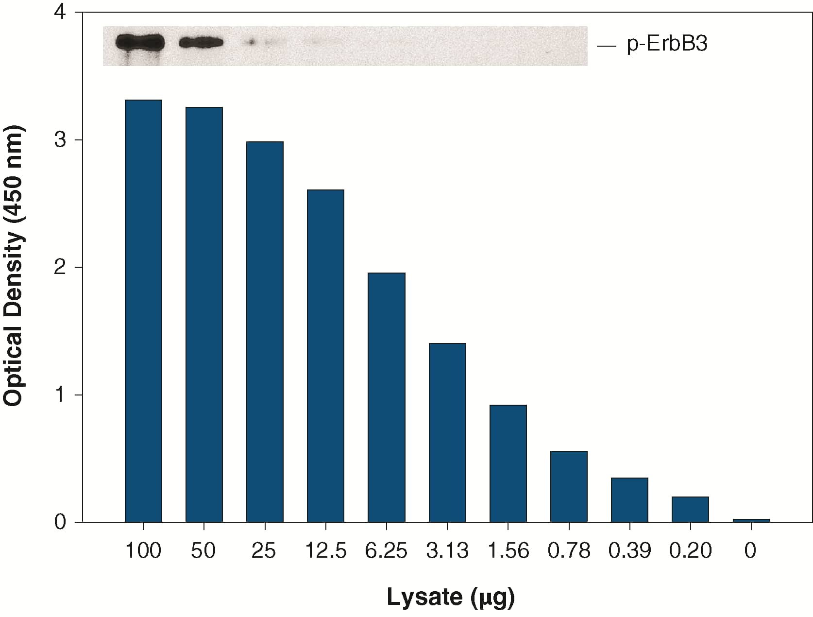
396-HB) for five minutes to induce tyrosine phosphorylation of ErbB3. Serial dilutions of lysates were analyzed by R&D Systems’ Phospho-ErbB3 DuoSet IC ELISA (Catalog # DYC1769) and by IP-Western blot (inset). For IP-Westerns, IPs were done using anti-ErbB3 monoclonal antibody and anti-mouse IgG agarose. Immunoblots were incubated with biotinylated anti-phosphotyrosine monoclonal antibody (Cat #
BAM1676) to detect phospho-ErbB3. Bands were visualized with Streptavidin-HRP (Cat #
DY998) followed by ECL detection. ErbB3 was observed at its predicted size (inset; 185 kDa, arrow) in Western blot analysis." alt="The human breast cancer cell line, MDA-MB-453, was treated with 100 ng/mL recombinant HRG-beta 1 (Catalog #
396-HB) for five minutes to induce tyrosine phosphorylation of ErbB3. Serial dilutions of lysates were analyzed by R&D Systems’ Phospho-ErbB3 DuoSet IC ELISA (Catalog # DYC1769) and by IP-Western blot (inset). For IP-Westerns, IPs were done using anti-ErbB3 monoclonal antibody and anti-mouse IgG agarose. Immunoblots were incubated with biotinylated anti-phosphotyrosine monoclonal antibody (Cat #
BAM1676) to detect phospho-ErbB3. Bands were visualized with Streptavidin-HRP (Cat #
DY998) followed by ECL detection. ErbB3 was observed at its predicted size (inset; 185 kDa, arrow) in Western blot analysis." class="big_thumb" />
396-HB) for five minutes. Cell lysates (100 μg) were analyzed by R&D Systems’ Phospho-ErbB3 DuoSet IC ELISA (Catalog # DYC1769) and by IP-Western blot (inset). IP-Western blots for phospho-ErbB3 (p-ErbB3) were done as described in Figure 1. Blots were stripped and total ErbB3 (ErbB3) was detected using biotinylated anti-ErbB3 polyclonal antibody (Catalog #
BAF234). " class="big_lightbox">
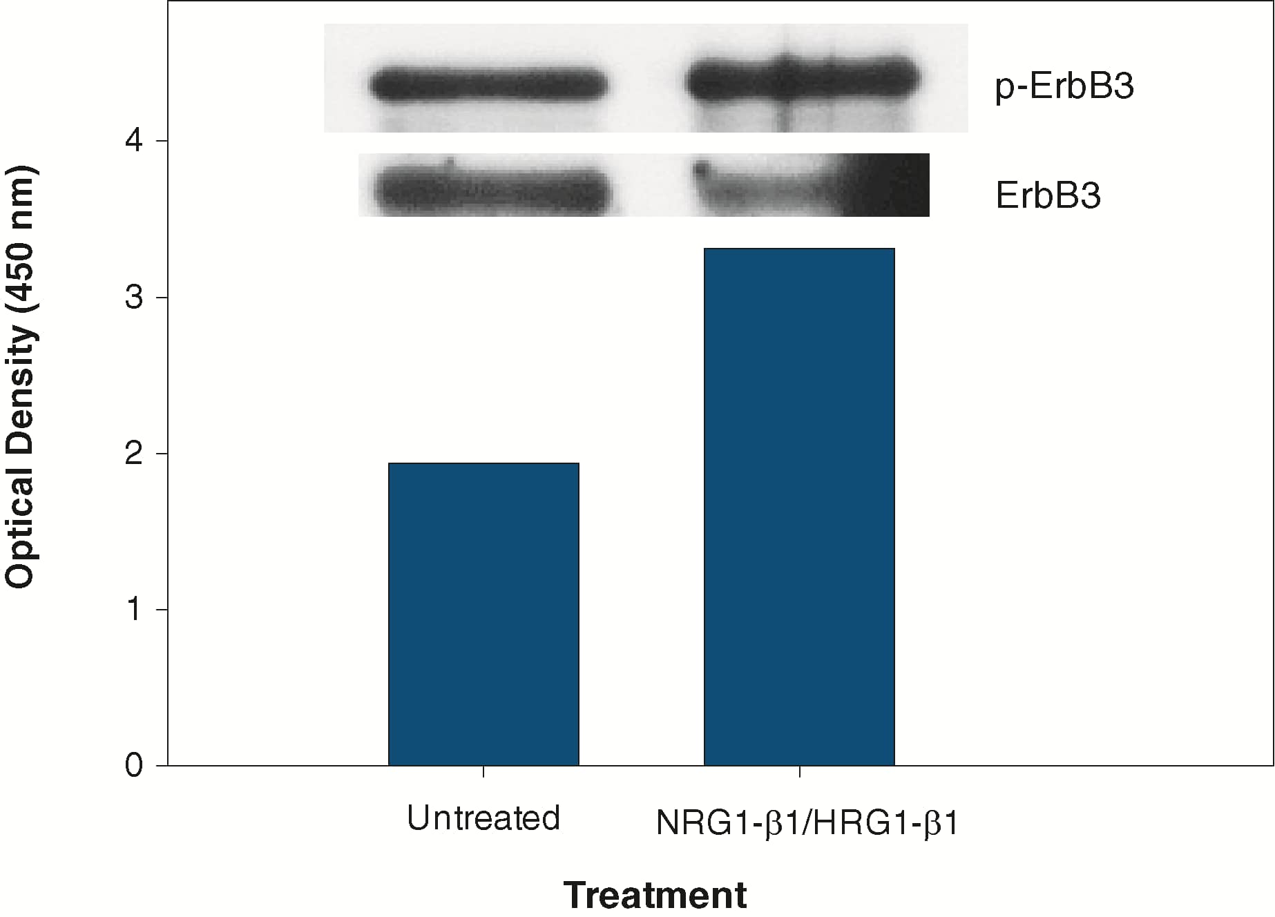
396-HB) for five minutes. Cell lysates (100 μg) were analyzed by R&D Systems’ Phospho-ErbB3 DuoSet IC ELISA (Catalog # DYC1769) and by IP-Western blot (inset). IP-Western blots for phospho-ErbB3 (p-ErbB3) were done as described in Figure 1. Blots were stripped and total ErbB3 (ErbB3) was detected using biotinylated anti-ErbB3 polyclonal antibody (Catalog #
BAF234). " class="big_thumb" />
396-HB) for five minutes. The indicated amounts of ErbB3 (Catalog # 348-ER), ErbB2 (Catalog #
1129-ER) or EGF R (Catalog #
1095-ER) recombinant extracellular domains were added to 5 μg lysate and analyzed using R&D Systems’ Human Phospho-ErbB3 DuoSet IC ELISA (Catalog # DYC1769). Competition was observed only with recombinant ErbB3. " class="big_lightbox">
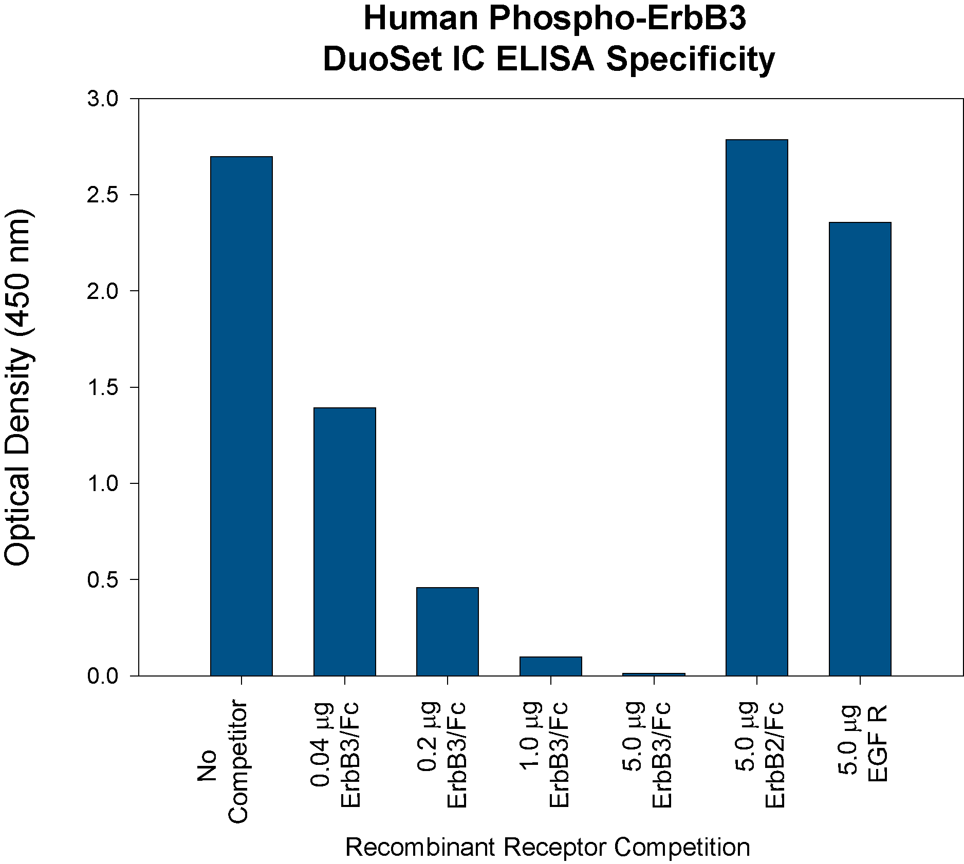
396-HB) for five minutes. The indicated amounts of ErbB3 (Catalog # 348-ER), ErbB2 (Catalog #
1129-ER) or EGF R (Catalog #
1095-ER) recombinant extracellular domains were added to 5 μg lysate and analyzed using R&D Systems’ Human Phospho-ErbB3 DuoSet IC ELISA (Catalog # DYC1769). Competition was observed only with recombinant ErbB3. " class="big_thumb" />
 377-HB) stimulates proliferation in the MCF-17 human breast cancer cell line in a dose-dependent manner (orange line). Proliferation elicited by Recombinant Human NRG1-1 beta 1/HRG1-1 beta 1 (10 ng/mL) is neutralized (green line) by increasing concentrations of Human ErbB3/Her3 Monoclonal Antibody (Catalog # MAB3481). The ND50 is typically 0.0075-0.03 µg/mL." alt="Recombinant Human NRG1-1 beta 1/HRG1-1 beta 1 (Catalog # 377-HB) stimulates proliferation in the MCF-17 human breast cancer cell line in a dose-dependent manner (orange line). Proliferation elicited by Recombinant Human NRG1-1 beta 1/HRG1-1 beta 1 (10 ng/mL) is neutralized (green line) by increasing concentrations of Human ErbB3/Her3 Monoclonal Antibody (Catalog # MAB3481). The ND50 is typically 0.0075-0.03 µg/mL." class="big_thumb" />
 HAF019). A specific band was detected for ErbB3/Her3 at approximately 180kDa (as indicated). This experiment was conducted under reducing conditions and using Immunoblot Buffer Group 8." alt="Western blot shows lysates of MCF-7 human breast cancer cell line. PVDF Membrane was probed with 1 µg/mL of Goat Anti-Human ErbB3/Her3 Antigen Affinity-purified Polyclonal Antibody (Catalog # AF234) followed by HRP-conjugated Anti-Goat IgG Secondary Antibody (Catalog # HAF019). A specific band was detected for ErbB3/Her3 at approximately 180kDa (as indicated). This experiment was conducted under reducing conditions and using Immunoblot Buffer Group 8." class="big_thumb" />
 5-001-A, open histogram), followed by Allophycocyanin-conjugated Anti-Sheep IgG Secondary Antibody (Catalog # F0127)." alt="C2C12 mouse myoblast cell line was stained with Sheep Anti-Mouse ErbB3/Her3 Antigen Affinity-purified Polyclonal Antibody (Catalog # AF4518, filled histogram) or control antibody (Catalog # 5-001-A, open histogram), followed by Allophycocyanin-conjugated Anti-Sheep IgG Secondary Antibody (Catalog # F0127)." class="big_thumb" />
 396-HB) for 5 minutes. PVDF membrane was probed with 1 µg/mL of Human Phospho-ErbB3/Her3 (Y1262) Antigen Affinity-purified Polyclonal Antibody (Catalog # AF5817), followed by HRP-conjugated Anti-Rabbit IgG Secondary Antibody (Catalog # HAF008). A specific band was detected for Phospho-ErbB3/Her3 (Y1262) at approximately 185 kDa (as indicated). This experiment was conducted under reducing conditions and using Immunoblot Buffer Group 1." alt="Western blot shows lysates of MDA-MB-453 human breast cancer cell line untreated (-) or treated (+) with 300 ng/mL Recombinant Human NRG1-beta /HRG1-beta (Catalog # 396-HB) for 5 minutes. PVDF membrane was probed with 1 µg/mL of Human Phospho-ErbB3/Her3 (Y1262) Antigen Affinity-purified Polyclonal Antibody (Catalog # AF5817), followed by HRP-conjugated Anti-Rabbit IgG Secondary Antibody (Catalog # HAF008). A specific band was detected for Phospho-ErbB3/Her3 (Y1262) at approximately 185 kDa (as indicated). This experiment was conducted under reducing conditions and using Immunoblot Buffer Group 1." class="big_thumb" />
 396-HB) for five minutes to induce tyrosine phosphorylation of ErbB3. Serial dilutions of lysates were analyzed by R&D Systems’ Phospho-ErbB3 DuoSet IC ELISA (Catalog # DYC1769) and by IP-Western blot (inset). For IP-Westerns, IPs were done using anti-ErbB3 monoclonal antibody and anti-mouse IgG agarose. Immunoblots were incubated with biotinylated anti-phosphotyrosine monoclonal antibody (Cat # BAM1676) to detect phospho-ErbB3. Bands were visualized with Streptavidin-HRP (Cat # DY998) followed by ECL detection. ErbB3 was observed at its predicted size (inset; 185 kDa, arrow) in Western blot analysis." alt="The human breast cancer cell line, MDA-MB-453, was treated with 100 ng/mL recombinant HRG-beta 1 (Catalog # 396-HB) for five minutes to induce tyrosine phosphorylation of ErbB3. Serial dilutions of lysates were analyzed by R&D Systems’ Phospho-ErbB3 DuoSet IC ELISA (Catalog # DYC1769) and by IP-Western blot (inset). For IP-Westerns, IPs were done using anti-ErbB3 monoclonal antibody and anti-mouse IgG agarose. Immunoblots were incubated with biotinylated anti-phosphotyrosine monoclonal antibody (Cat # BAM1676) to detect phospho-ErbB3. Bands were visualized with Streptavidin-HRP (Cat # DY998) followed by ECL detection. ErbB3 was observed at its predicted size (inset; 185 kDa, arrow) in Western blot analysis." class="big_thumb" />
 HAF007). A specific band was detected for ErbB3/Her3 at approximately 185 kDa (as indicated). This experiment was conducted under reducing conditions and using Immunoblot Buffer Group 1." alt="Western blot shows lysates of MDA-MB-453 human breast cancer cell line and MCF-7 human breast cancer cell line. PVDF membrane was probed with 1 µg/mL of Human ErbB3/Her3 Monoclonal Antibody (Catalog # MAB3482) followed by HRP-conjugated Anti-Mouse IgG Secondary Antibody (Catalog # HAF007). A specific band was detected for ErbB3/Her3 at approximately 185 kDa (as indicated). This experiment was conducted under reducing conditions and using Immunoblot Buffer Group 1." class="big_thumb" />


 348-RB) was serially diluted and captured by Mouse Anti-Human ErbB3/Her3 Monoclonal Antibody (Catalog # MAB3481) coated on a Clear Polystyrene Microplate (Catalog #
  377-HB) stimulates proliferation in the MCF-7 human breast cancer cell line in a dose-dependent manner (orange line). Proliferation elicited by Recombinant Human NRG1-beta 1/HRG1-beta 1 Extracellular Domain (10 ng/mL) is neutralized (green line) by increasing concentrations of Goat Anti-Human ErbB3/Her3 Antigen Affinity-purified Polyclonal Antibody (Catalog # AF234). The ND50 is typically 0.125-0.5 µg/mL." class="big_thumb" />
![Immunohistochemistry-Paraffin: ErbB3/Her3 Antibody (OTI3E10) [NBP2-45481] - Analysis of Human colon tissue. (Heat-induced epitope retrieval by 1 mM EDTA in 10mM Tris, pH8.5, 120C for 3min)](http://images.novusbio.com/fullsize/ErbB3-Her3-Antibody-OTI3E10-Immunohistochemistry-Paraffin-NBP2-45481-img0001.jpg "Immunohistochemistry-Paraffin: ErbB3/Her3 Antibody (OTI3E10) [NBP2-45481] - Analysis of Human colon tissue. (Heat-induced epitope retrieval by 1 mM EDTA in 10mM Tris, pH8.5, 120C for 3min)")
![Flow Cytometry: ErbB3/Her3 Antibody (OTI3E10) [NBP2-45481] - HEK293T cells transfected with either ErbB3/Her3 Human Tagged ORF Clone overexpress plasmid (Red), compared to an IgG isotype control, (Green) or empty vector control plasmid (Blue) were immunostained by anti-ERBB3 antibody , and then analyzed by flow cytometry (1:100).](http://images.novusbio.com/fullsize/ErbB3-Her3-Antibody-OTI3E10-Flow-Cytometry-NBP2-45481-img0009.jpg)
![Immunohistochemistry: ErbB3/Her3 Antibody (OTI3E10) [NBP2-45481] - Analysis of Human Kidney tissue. (Heat-induced epitope retrieval by 1 mM EDTA in 10mM Tris, pH8.5, 120C for 3min)](http://images.novusbio.com/fullsize/ErbB3-Her3-Antibody-OTI3E10-Immunohistochemistry-NBP2-45481-img0002.jpg)
![Immunohistochemistry: ErbB3/Her3 Antibody (OTI3E10) [NBP2-45481] - Analysis of Human liver tissue. (Heat-induced epitope retrieval by 1 mM EDTA in 10mM Tris, pH8.5, 120C for 3min)](http://images.novusbio.com/fullsize/ErbB3-Her3-Antibody-OTI3E10-Immunohistochemistry-NBP2-45481-img0003.jpg)
![Immunohistochemistry: ErbB3/Her3 Antibody (OTI3E10) [NBP2-45481] - Analysis of Carcinoma of Human liver tissue.(Heat-induced epitope retrieval by 1 mM EDTA in 10mM Tris, pH8.5, 120C for 3min)](http://images.novusbio.com/fullsize/ErbB3-Her3-Antibody-OTI3E10-Immunohistochemistry-NBP2-45481-img0004.jpg)
![Immunohistochemistry: ErbB3/Her3 Antibody (OTI3E10) [NBP2-45481] - Analysis of Carcinoma of Human lung tissue.(Heat-induced epitope retrieval by 1 mM EDTA in 10mM Tris, pH8.5, 120C for 3min)](http://images.novusbio.com/fullsize/ErbB3-Her3-Antibody-OTI3E10-Immunohistochemistry-NBP2-45481-img0005.jpg)
![Immunohistochemistry: ErbB3/Her3 Antibody (OTI3E10) [NBP2-45481] - Analysis of Adenocarcinoma of Human ovary tissue. (Heat-induced epitope retrieval by 1 mM EDTA in 10mM Tris, pH8.5, 120C for 3min)](http://images.novusbio.com/fullsize/ErbB3-Her3-Antibody-OTI3E10-Immunohistochemistry-NBP2-45481-img0006.jpg)
![Immunohistochemistry: ErbB3/Her3 Antibody (OTI3E10) [NBP2-45481] - Analysis of Carcinoma of Human prostate tissue. (Heat-induced epitope retrieval by 1 mM EDTA in 10mM Tris, pH8.5, 120C for 3min)](http://images.novusbio.com/fullsize/ErbB3-Her3-Antibody-OTI3E10-Immunohistochemistry-NBP2-45481-img0008.jpg)
![Immunohistochemistry: ErbB3/Her3 Antibody (OTI3E10) [NBP2-45481] - Analysis of Human thyroid tissue. (Heat-induced epitope retrieval by 1 mM EDTA in 10mM Tris, pH8.5, 120C for 3min)](http://images.novusbio.com/fullsize/ErbB3-Her3-Antibody-OTI3E10-Immunohistochemistry-NBP2-45481-img0007.jpg)
 NL010) and counterstained with DAPI (blue). Specific staining was localized to cytoplasm. View our protocol for
  NL010) and counterstained with DAPI (blue). View our protocol for
 396-HB) for five minutes. Cell lysates (100 μg) were analyzed by R&D Systems’ Phospho-ErbB3 DuoSet IC ELISA (Catalog # DYC1769) and by IP-Western blot (inset). IP-Western blots for phospho-ErbB3 (p-ErbB3) were done as described in Figure 1. Blots were stripped and total ErbB3 (ErbB3) was detected using biotinylated anti-ErbB3 polyclonal antibody (Catalog #
  396-HB) for five minutes. The indicated amounts of ErbB3 (Catalog # 348-ER), ErbB2 (Catalog #

![N/A ErbB3/Her3 [Biotin]](http://images.novusbio.com/fullsize/elisa/DATA_ErbB3_DY348_ELISA_1947.jpg "N/A ErbB3/Her3 [Biotin]")
![ELISA ErbB3/Her3 [Biotin]](http://images.novusbio.com/fullsize/dyc234-2_human-total-erbb3-her3-duoset-ic-elisa-2-plate-elisa-169202091343.jpg "ELISA ErbB3/Her3 [Biotin]")


