HAF008). A specific band was detected for TL1A/TNFSF15 at approximately 22 kDa (as indicated). This experiment was conducted under reducing conditions and using
Immunoblot Buffer Group 1." class="big_lightbox">
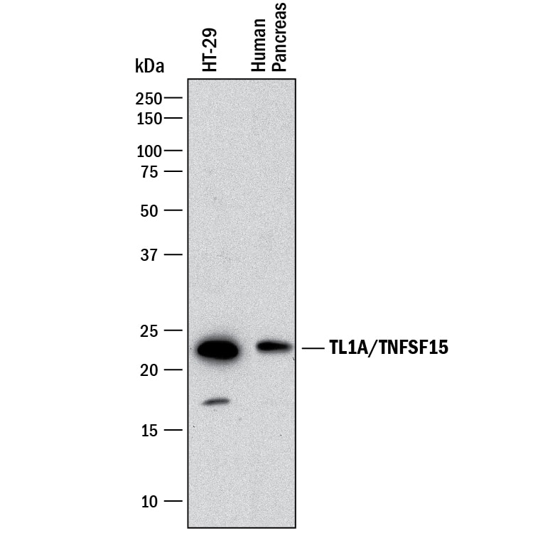
HAF008). A specific band was detected for TL1A/TNFSF15 at approximately 22 kDa (as indicated). This experiment was conducted under reducing conditions and using
Immunoblot Buffer Group 1." alt="Western blot shows lysates of HT-29 human colon adenocarcinoma cell line and human pancreas tissue. PVDF membrane was probed with 1 µg/mL of Rabbit Anti-Human TL1A/TNFSF15 Monoclonal Antibody (Catalog # MAB74422) followed by HRP-conjugated Anti-Rabbit IgG Secondary Antibody (
HAF008). A specific band was detected for TL1A/TNFSF15 at approximately 22 kDa (as indicated). This experiment was conducted under reducing conditions and using
Immunoblot Buffer Group 1." class="big_thumb" />
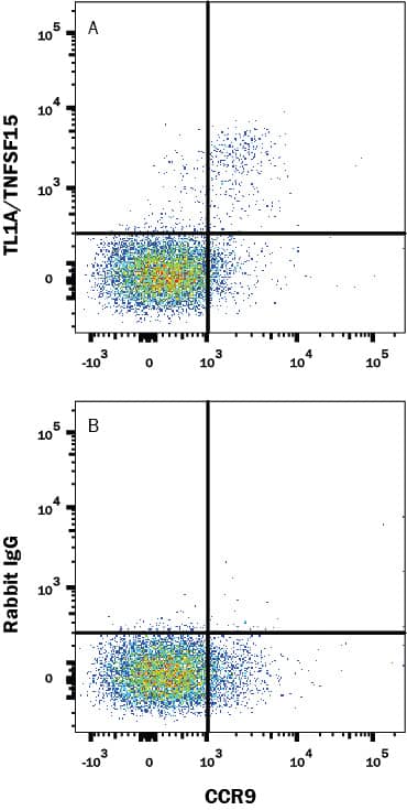
MAB1050) followed by Goat anti-Rabbit IgG PE-conjugated Secondary Antibody (
F0110) and Mouse anti-Human CCR9 APC-conjugated Monoclonal Antibody (
FAB1791A). Cells were gated on CD4+ lymphocytes using Mouse anti-Human CD4 FITC-conjugated Monoclonal Antibody (
FAB3791F). View our protocol for
Staining Membrane-associated Proteins." class="big_lightbox">
MAB1050) followed by Goat anti-Rabbit IgG PE-conjugated Secondary Antibody (
F0110) and Mouse anti-Human CCR9 APC-conjugated Monoclonal Antibody (
FAB1791A). Cells were gated on CD4+ lymphocytes using Mouse anti-Human CD4 FITC-conjugated Monoclonal Antibody (
FAB3791F). View our protocol for
Staining Membrane-associated Proteins." class="big_thumb" />
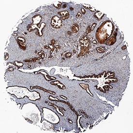
VC003). Before incubation with the primary antibody, tissue was subjected to heat-induced epitope retrieval using Antigen Retrieval Reagent-Basic (
CTS013). Tissue was stained using DAB (brown) and counterstained with hematoxylin (blue). Specific staining was localized to cytoplasm in epithelial cells. View our protocol for
IHC Staining with VisUCyte HRP Polymer Detection Reagents." class="big_lightbox">
VC003). Before incubation with the primary antibody, tissue was subjected to heat-induced epitope retrieval using Antigen Retrieval Reagent-Basic (
CTS013). Tissue was stained using DAB (brown) and counterstained with hematoxylin (blue). Specific staining was localized to cytoplasm in epithelial cells. View our protocol for
IHC Staining with VisUCyte HRP Polymer Detection Reagents." class="big_thumb" />
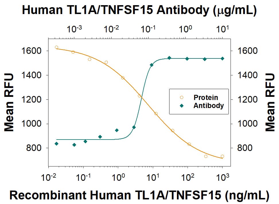
1319-TL) induces apoptosis in the TF-1 human erythroleukemic cell line in a dose-dependent manner (orange line), as measured by Resazurin (
AR002). Apoptosis elicited by Recombinant Human TL1A/TNFSF15 (80 ng/mL) is neutralized (green line) by increasing concentrations of Rabbit Anti-Human TL1A/TNFSF15 Monoclonal Antibody (Catalog # MAB74422). The ND
50 is typically 0.04‑0.2 µg/mL." class="big_lightbox">
1319-TL) induces apoptosis in the TF-1 human erythroleukemic cell line in a dose-dependent manner (orange line), as measured by Resazurin (
AR002). Apoptosis elicited by Recombinant Human TL1A/TNFSF15 (80 ng/mL) is neutralized (green line) by increasing concentrations of Rabbit Anti-Human TL1A/TNFSF15 Monoclonal Antibody (Catalog # MAB74422). The ND
50 is typically 0.04‑0.2 µg/mL." class="big_thumb" />
 HAF008). A specific band was detected for TL1A/TNFSF15 at approximately 22 kDa (as indicated). This experiment was conducted under reducing conditions and using Immunoblot Buffer Group 1." alt="Western blot shows lysates of HT-29 human colon adenocarcinoma cell line and human pancreas tissue. PVDF membrane was probed with 1 µg/mL of Rabbit Anti-Human TL1A/TNFSF15 Monoclonal Antibody (Catalog # MAB74422) followed by HRP-conjugated Anti-Rabbit IgG Secondary Antibody (HAF008). A specific band was detected for TL1A/TNFSF15 at approximately 22 kDa (as indicated). This experiment was conducted under reducing conditions and using Immunoblot Buffer Group 1." class="big_thumb" />
 VC004). Tissue was stained using DAB (brown) and counterstained with hematoxylin (blue). Specific staining was localized to epithelial cells. View our protocol for IHC Staining with VisUCyte HRP Polymer Detection Reagents." alt="TL1A/TNFSF15 was detected in immersion fixed paraffin-embedded sections of human prostate using Goat Anti-Human TL1A/TNFSF15 Antigen Affinity-purified Polyclonal Antibody (Catalog # AF744) at 3 µg/mL for 1 hour at room temperature followed by incubation with the Anti-Goat IgG VisUCyte™ HRP Polymer Antibody (Catalog # VC004). Tissue was stained using DAB (brown) and counterstained with hematoxylin (blue). Specific staining was localized to epithelial cells. View our protocol for IHC Staining with VisUCyte HRP Polymer Detection Reagents." class="big_thumb" />
 HAF008). A specific band was detected for TL1A/TNFSF15 at approximately 22 kDa (as indicated). This experiment was conducted under reducing conditions and using Immunoblot Buffer Group 1." alt="Western blot shows lysates of human prostate tissue, human lung tissue, and HUVEC human umbilical vein endothelial cells. PVDF membrane was probed with 1 µg/mL of Rabbit Anti-Human TL1A/ TNFSF15 Monoclonal Antibody (Catalog # MAB74421) followed by HRP-conjugated Anti-Rabbit IgG Secondary Antibody (Catalog # HAF008). A specific band was detected for TL1A/TNFSF15 at approximately 22 kDa (as indicated). This experiment was conducted under reducing conditions and using Immunoblot Buffer Group 1." class="big_thumb" />


 MAB1050) followed by Goat anti-Rabbit IgG PE-conjugated Secondary Antibody (
 VC003). Before incubation with the primary antibody, tissue was subjected to heat-induced epitope retrieval using Antigen Retrieval Reagent-Basic (
 1319-TL) induces apoptosis in the TF-1 human erythroleukemic cell line in a dose-dependent manner (orange line), as measured by Resazurin (
 AB-105-C, open histogram), followed by Allophycocyanin-conjugated Anti-Rabbit IgG Secondary Antibody (Catalog #
 ![SDS-Page: Recombinant Human TL1A/TNFSF15 Protein [NBP2-35067]](http://images.novusbio.com/fullsize/Recombinant-Human-TL1A-TNFSF15-Protein-SDS-Page-NBP2-35067-img0002.jpg "SDS-Page: Recombinant Human TL1A/TNFSF15 Protein [NBP2-35067]")
![Immunohistochemistry-Paraffin: TL1A/TNFSF15 Antibody (VEGI/2052R) [NBP3-07481] - Formalin-fixed, paraffin-embedded human Colon Carcinoma stained with TL1A/TNFSF15 Rabbit Recombinant Monoclonal Antibody (TL1A/TNFSF15 /2052R).](http://images.novusbio.com/fullsize/TL1A-TNFSF15-Antibody-VEGI-2052R-Immunohistochemistry-Paraffin-NBP3-07481-img0004.jpg "Immunohistochemistry-Paraffin: TL1A/TNFSF15 Antibody (VEGI/2052R) [NBP3-07481] - Formalin-fixed, paraffin-embedded human Colon Carcinoma stained with TL1A/TNFSF15 Rabbit Recombinant Monoclonal Antibody (TL1A/TNFSF15 /2052R).")
![Immunohistochemistry-Paraffin: TL1A/TNFSF15 Antibody (VEGI/2052R) [NBP3-07481] - Formalin-fixed, paraffin-embedded human Spleen stained with TL1A/TNFSF15 Rabbit Recombinant Monoclonal Antibody (TL1A/TNFSF15 /2052R).](http://images.novusbio.com/fullsize/TL1A-TNFSF15-Antibody-VEGI-2052R-Immunohistochemistry-Paraffin-NBP3-07481-img0002.jpg)
![Immunohistochemistry-Paraffin: TL1A/TNFSF15 Antibody (VEGI/2052R) [NBP3-07481] - Formalin-fixed, paraffin-embedded human Parathyroid Mass stained with TL1A/TNFSF15 Rabbit Recombinant Monoclonal Antibody (TL1A/TNFSF15 /2052R).](http://images.novusbio.com/fullsize/TL1A-TNFSF15-Antibody-VEGI-2052R-Immunohistochemistry-Paraffin-NBP3-07481-img0003.jpg)
![Protein Array: TL1A/TNFSF15 Antibody (VEGI/2052R) [NBP3-07481] - Analysis of Protein Array containing more than 19,000 full-length human proteins using TL1A/TNFSF15 Rabbit Recombinant Monoclonal Antibody (TL1A/TNFSF15/2052R).](http://images.novusbio.com/fullsize/TL1A-TNFSF15-Antibody-VEGI-2052R-Protein-Array-NBP3-07481-img0001.jpg)
![Western Blot: TL1A/TNFSF15 Antibody (2116C) [MAB7442] - Analysis of human prostate, kidney, and lung tissue lysates with 1 ug/ml of hTL1A Monoclonal Antibody followed by HRP-conjugated Anti-Rabbit IgG Antibody. A specific band was detected for hTL1A at approximately 22 kDa (as indicated). This experiment was conducted under reducing conditions and using Immunoblot Buffer Group 1.](http://images.novusbio.com/fullsize/TL1A-TNFSF15-Antibody-2116C-Western-Blot-MAB7442-img0001.jpg "Western Blot: TL1A/TNFSF15 Antibody (2116C) [MAB7442] - Analysis of human prostate, kidney, and lung tissue lysates with 1 ug/ml of hTL1A Monoclonal Antibody followed by HRP-conjugated Anti-Rabbit IgG Antibody. A specific band was detected for hTL1A at approximately 22 kDa (as indicated). This experiment was conducted under reducing conditions and using Immunoblot Buffer Group 1.")
 VC001). Tissue was stained using DAB (brown) and counterstained with hematoxylin (blue). Specific staining was localized to cytoplasm of epithelial cells. View our protocol for
  1319-TL) induces apoptosis in the TF-1 human erythroleukemic cell line in a dose-dependent manner (orange line), as measured by Resazurin (Catalog #
 ![Immunohistochemistry-Paraffin: TL1A/TNFSF15 Antibody (VEGI/1283) [NBP2-53388] - Formalin-fixed, paraffin-embedded human Parathyroid Mass stained with VEGI Monoclonal Antibody (VEGI /1283).](http://images.novusbio.com/fullsize/TL1A-TNFSF15-Antibody-VEGI-1283-Immunohistochemistry-Paraffin-NBP2-53388-img0002.jpg "Immunohistochemistry-Paraffin: TL1A/TNFSF15 Antibody (VEGI/1283) [NBP2-53388] - Formalin-fixed, paraffin-embedded human Parathyroid Mass stained with VEGI Monoclonal Antibody (VEGI /1283).")
![Immunohistochemistry-Paraffin: TL1A/TNFSF15 Antibody (VEGI/1283) [NBP2-53388] - Formalin-fixed, paraffin-embedded human Colon Carcinoma stained with VEGI Monoclonal Antibody (VEGI /1283).](http://images.novusbio.com/fullsize/TL1A-TNFSF15-Antibody-VEGI-1283-Immunohistochemistry-Paraffin-NBP2-53388-img0001.jpg)
![N/A TL1A/TNFSF15 [Biotin]](http://images.novusbio.com/fullsize/elisa/DATA_TL1A_DY131905_ELISA_2352.jpg "N/A TL1A/TNFSF15 [Biotin]")


