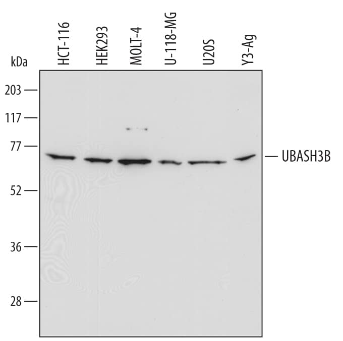
 HAF016). A specific band was detected for UBASH3B/STS1 at approximately 72 kDa (as indicated). This experiment was conducted under reducing conditions and using Immunoblot Buffer Group 1." alt="Western blot shows lysates of HCT-116 human colorectal carcinoma cell line, HEK293 human embryonic kidney cell line, MOLT-4 human acute lymphoblastic leukemia cell line, U-118-MG human glioblastoma/astrocytoma cell line, U2OS human osteosarcoma cell line, and Y3-Ag rat myeloid cell line. PVDF Membrane was probed with 2 µg/mL of Human/Rat UBASH3B/STS1 Antigen Affinity-purified Polyclonal Antibody (Catalog # AF6696) followed by HRP-conjugated Anti-Sheep IgG Secondary Antibody (Catalog # HAF016). A specific band was detected for UBASH3B/STS1 at approximately 72 kDa (as indicated). This experiment was conducted under reducing conditions and using Immunoblot Buffer Group 1." class="big_thumb" />
| Formulation | Catalog # | Availability | Price | |
|---|---|---|---|---|
| Carrier Free | AF6696-SP | |||
| Formulation | Catalog # | Availability | Price | |
|---|---|---|---|---|
| BSA Free | NBP2-34053-25ul | |||
| Formulation | Catalog # | Availability | Price | |
|---|---|---|---|---|
| BSA Free | NBP1-92456-25ul | |||


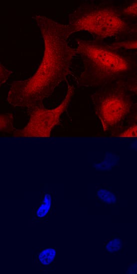 NL010) and counterstained with DAPI (blue, lower panel). Specific staining was localized to nuclei and cytoplasm. View our protocol for
 ![Immunocytochemistry/Immunofluorescence: UBASH3B/STS1/Tula-2 Antibody (3G7) [H00084959-M01] - Analysis of monoclonal antibody to UBASH3B on HeLa cell . Antibody concentration 10 ug/ml.](http://images.novusbio.com/fullsize/UBASH3B-STS1-Tula-2-Antibody-3G7-Immunocytochemistry-Immunofluorescence-H00084959-M01-img0002.jpg "Immunocytochemistry/Immunofluorescence: UBASH3B/STS1/Tula-2 Antibody (3G7) [H00084959-M01] - Analysis of monoclonal antibody to UBASH3B on HeLa cell . Antibody concentration 10 ug/ml.")
![Immunohistochemistry-Paraffin: UBASH3B/STS1/Tula-2 Antibody (3G7) [H00084959-M01] - Analysis of monoclonal antibody to UBASH3B on formalin-fixed paraffin-embedded human colon. Antibody concentration 3 ug/ml.](http://images.novusbio.com/fullsize/UBASH3B-STS1-Tula-2-Antibody-3G7-Immunohistochemistry-Paraffin-H00084959-M01-img0004.jpg)
![Sandwich ELISA: UBASH3B/STS1/Tula-2 Antibody (3G7) [H00084959-M01] - Detection limit for recombinant GST tagged UBASH3B is 1 ng/ml as a capture antibody.](http://images.novusbio.com/fullsize/UBASH3B-STS1-Tula-2-Antibody-3G7-Sandwich-ELISA-H00084959-M01-img0005.jpg)
![Western Blot: UBASH3B/STS1/Tula-2 Antibody [NBP1-32592] - HeLa whole cell and membrane extracts (30 ug) were separated by 7.5% SDS-PAGE, and the membrane was blotted with UBASH3B antibody diluted at 1:1000. HRP-conjugated anti-rabbit IgG antibody was used to detect the primary antibody.](http://images.novusbio.com/fullsize/UBASH3B-STS1-Tula-2-Antibody-Western-Blot-NBP1-32592-img0008.jpg "Western Blot: UBASH3B/STS1/Tula-2 Antibody [NBP1-32592] - HeLa whole cell and membrane extracts (30 ug) were separated by 7.5% SDS-PAGE, and the membrane was blotted with UBASH3B antibody diluted at 1:1000. HRP-conjugated anti-rabbit IgG antibody was used to detect the primary antibody.")
![Immunohistochemistry-Paraffin: UBASH3B/STS1/Tula-2 Antibody [NBP1-32592] - SW480 xenograft , using Steroid sulfatase antibody at 1:100 dilution. Antigen Retrieval: Trilogy™ (EDTA based, pH 8.0) buffer, 15min.](http://images.novusbio.com/fullsize/UBASH3B-STS1-Tula-2-Antibody-Immunohistochemistry-Paraffin-NBP1-32592-img0005.jpg)
![Western Blot: UBASH3B/STS1/Tula-2 Antibody [NBP1-32592] - Human tissue extract (30 ug) was separated by 7.5% SDS-PAGE, and the membrane was blotted with Steroid sulfatase antibody diluted at 1:500. HRP-conjugated anti-rabbit IgG antibody was used to detect the primary antibody.](http://images.novusbio.com/fullsize/UBASH3B-STS1-Tula-2-Antibody-Western-Blot-NBP1-32592-img0007.jpg)
![Immunohistochemistry-Paraffin: UBASH3B/STS1/Tula-2 Antibody [NBP2-34053] - Staining of human lymph node shows strong cytoplasmic positivity in germinal and non germinal center cells.](http://images.novusbio.com/fullsize/UBASH3B-STS1-Tula-2-Antibody-Immunohistochemistry-Paraffin-NBP2-34053-img0006.jpg "Immunohistochemistry-Paraffin: UBASH3B/STS1/Tula-2 Antibody [NBP2-34053] - Staining of human lymph node shows strong cytoplasmic positivity in germinal and non germinal center cells.")

![Western Blot: UBASH3B/STS1/Tula-2 Antibody [NBP2-20523] - Sample (30 ug of whole cell lysate) A: Jurkat B: THP-1 7. 5% SDS PAGE gel, diluted at 1:5000.](http://images.novusbio.com/fullsize/STS-1-Tula-2-Antibody-Western-Blot-NBP2-20523-img0001.jpg "Western Blot: UBASH3B/STS1/Tula-2 Antibody [NBP2-20523] - Sample (30 ug of whole cell lysate) A: Jurkat B: THP-1 7. 5% SDS PAGE gel, diluted at 1:5000.")
![Immunohistochemistry-Paraffin: UBASH3B/STS1/Tula-2 Antibody [NBP2-20523] - Human hepatoma, using Sts1 antibody at 1:500 dilution. Antigen Retrieval: Trilogy™ (EDTA based, pH 8.0) buffer, 15min.](http://images.novusbio.com/fullsize/STS-1-Tula-2-Antibody-Immunohistochemistry-Paraffin-NBP2-20523-img0002.jpg)

![Western Blot: UBASH3B/STS1/Tula-2 Antibody [NBP2-20524] - Sample (30 ug of whole cell lysate) A: A431 B: Jurkat 7. 5% SDS PAGE gel, diluted at 1:1000.](http://images.novusbio.com/fullsize/UBASH3B-STS1-Tula-2-Antibody-Western-Blot-NBP2-20524-img0002.jpg "Western Blot: UBASH3B/STS1/Tula-2 Antibody [NBP2-20524] - Sample (30 ug of whole cell lysate) A: A431 B: Jurkat 7. 5% SDS PAGE gel, diluted at 1:1000.")

