220-BB) for 20 minutes. PVDF membrane was probed with 1 µg/mL of Rabbit Anti-Human/Mouse Phospho-SHP-2 (Y542) Antigen Affinity-purified Polyclonal Antibody (Catalog # AF3790), followed by HRP-conjugated Anti-Rabbit IgG Secondary Antibody (Catalog #
HAF008). A specific band was detected for Phospho-SHP-2 (Y542) at approximately 72 kDa (as indicated). The lysates were also probed for total SHP-2 with Human/Mouse/Rat SHP-2 Antigen Affinity-purified Polyclonal Antibody (Catalog #
AF1894). This experiment was conducted under reducing conditions and using
Immunoblot Buffer Group 1." class="big_lightbox">

220-BB) for 20 minutes. PVDF membrane was probed with 1 µg/mL of Rabbit Anti-Human/Mouse Phospho-SHP-2 (Y542) Antigen Affinity-purified Polyclonal Antibody (Catalog # AF3790), followed by HRP-conjugated Anti-Rabbit IgG Secondary Antibody (Catalog #
HAF008). A specific band was detected for Phospho-SHP-2 (Y542) at approximately 72 kDa (as indicated). The lysates were also probed for total SHP-2 with Human/Mouse/Rat SHP-2 Antigen Affinity-purified Polyclonal Antibody (Catalog #
AF1894). This experiment was conducted under reducing conditions and using
Immunoblot Buffer Group 1." alt="Western blot shows lysates of NIH-3T3 mouse embryonic fibroblast cell line untreated (-) or treated (+) with 50 ng/mL Human PDGF-BB (Catalog #
220-BB) for 20 minutes. PVDF membrane was probed with 1 µg/mL of Rabbit Anti-Human/Mouse Phospho-SHP-2 (Y542) Antigen Affinity-purified Polyclonal Antibody (Catalog # AF3790), followed by HRP-conjugated Anti-Rabbit IgG Secondary Antibody (Catalog #
HAF008). A specific band was detected for Phospho-SHP-2 (Y542) at approximately 72 kDa (as indicated). The lysates were also probed for total SHP-2 with Human/Mouse/Rat SHP-2 Antigen Affinity-purified Polyclonal Antibody (Catalog #
AF1894). This experiment was conducted under reducing conditions and using
Immunoblot Buffer Group 1." class="big_thumb" />
NL004) and counterstained with DAPI (blue). Specific staining was localized to cytoplasm. Staining was performed using our protocol for Fluorescent ICC Staining of Non-adherent Cells." class="big_lightbox">
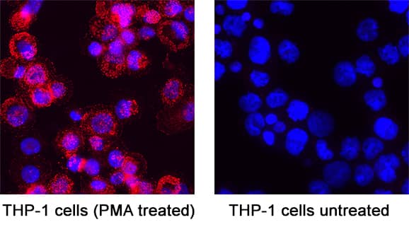
NL004) and counterstained with DAPI (blue). Specific staining was localized to cytoplasm. Staining was performed using our protocol for Fluorescent ICC Staining of Non-adherent Cells." class="big_thumb" />
220-BB) for 20 minutes, loaded at 0.2 mg/mL. A specific band was detected for Phospho-SHP-2 (Y542) at approximately 72 kDa (as indicated) using 10 µg/mL of Rabbit Anti-Human/Mouse Phospho-SHP-2 (Y542) Antigen Affinity-purified Polyclonal Antibody (Catalog # AF3790). This experiment was conducted under reducing conditions and using the 12-230 kDa separation system.*Non-specific interaction with the 230 kDa Simple Western standard may be seen with this antibody." class="big_lightbox">
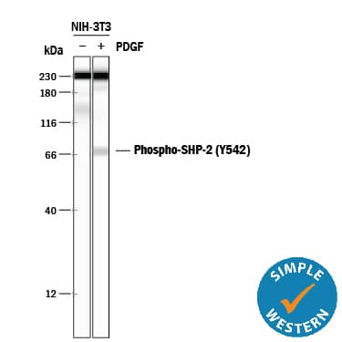
220-BB) for 20 minutes, loaded at 0.2 mg/mL. A specific band was detected for Phospho-SHP-2 (Y542) at approximately 72 kDa (as indicated) using 10 µg/mL of Rabbit Anti-Human/Mouse Phospho-SHP-2 (Y542) Antigen Affinity-purified Polyclonal Antibody (Catalog # AF3790). This experiment was conducted under reducing conditions and using the 12-230 kDa separation system.*Non-specific interaction with the 230 kDa Simple Western standard may be seen with this antibody." class="big_thumb" />
 220-BB) for 20 minutes. PVDF membrane was probed with 1 µg/mL of Rabbit Anti-Human/Mouse Phospho-SHP-2 (Y542) Antigen Affinity-purified Polyclonal Antibody (Catalog # AF3790), followed by HRP-conjugated Anti-Rabbit IgG Secondary Antibody (Catalog # HAF008). A specific band was detected for Phospho-SHP-2 (Y542) at approximately 72 kDa (as indicated). The lysates were also probed for total SHP-2 with Human/Mouse/Rat SHP-2 Antigen Affinity-purified Polyclonal Antibody (Catalog # AF1894). This experiment was conducted under reducing conditions and using Immunoblot Buffer Group 1." alt="Western blot shows lysates of NIH-3T3 mouse embryonic fibroblast cell line untreated (-) or treated (+) with 50 ng/mL Human PDGF-BB (Catalog # 220-BB) for 20 minutes. PVDF membrane was probed with 1 µg/mL of Rabbit Anti-Human/Mouse Phospho-SHP-2 (Y542) Antigen Affinity-purified Polyclonal Antibody (Catalog # AF3790), followed by HRP-conjugated Anti-Rabbit IgG Secondary Antibody (Catalog # HAF008). A specific band was detected for Phospho-SHP-2 (Y542) at approximately 72 kDa (as indicated). The lysates were also probed for total SHP-2 with Human/Mouse/Rat SHP-2 Antigen Affinity-purified Polyclonal Antibody (Catalog # AF1894). This experiment was conducted under reducing conditions and using Immunoblot Buffer Group 1." class="big_thumb" />
 HAF005). For additional reference, Recombinant Human Active SHP-1 aa 205-595 (Catalog # 1878-SH) and Recombinant Human SHP-2 (Catalog # 1894-SH) (2 ng/lane) were included. A specific band for SHP-2 was detected at approximately 80 kDa (as indicated). This experiment was conducted under reducing conditions and using Immunoblot Buffer Group 2." alt="Western blot shows lysates of HepG2 human hepatocellular carcinoma cell line, TS1 mouse helper T cell line, and PC-12 rat adrenal pheochromocytoma cell line. PVDF membrane was probed with 2 µg/mL Rat Anti-Human/Mouse/Rat SHP-2 Monoclonal Antibody (Catalog # MAB1894) followed by HRP-conjugated Anti-Rat IgG Secondary Antibody (Catalog # HAF005). For additional reference, Recombinant Human Active SHP-1 aa 205-595 (Catalog # 1878-SH) and Recombinant Human SHP-2 (Catalog # 1894-SH) (2 ng/lane) were included. A specific band for SHP-2 was detected at approximately 80 kDa (as indicated). This experiment was conducted under reducing conditions and using Immunoblot Buffer Group 2." class="big_thumb" />
 HAF109). For additional reference, Recombinant Human Active SHP-1 aa 205-595 (Catalog # 1878-SH) and Recombinant Human SHP-2 (Catalog # 1894-SH) (2 ng/lane) were included. A specific band for SHP-2 was detected at approximately 72 kDa (as indicated). This experiment was conducted under reducing conditions and using Immunoblot Buffer Group 1." alt="Western blot shows lysates of HepG2 human hepatocellular carcinoma cell line, HT-29 human colon adenocarcinoma cell line, Raji human Burkitt's lymphoma cell line, and NR-6 mouse fibroblast cell line. PVDF membrane was probed with 0.1 µg/mL Goat Anti-Human/Mouse/Rat SHP-2 Antigen Affinity-purified Polyclonal Antibody (Catalog # AF1894) followed by HRP-conjugated Anti-Goat IgG Secondary Antibody (Catalog # HAF109). For additional reference, Recombinant Human Active SHP-1 aa 205-595 (Catalog # 1878-SH) and Recombinant Human SHP-2 (Catalog # 1894-SH) (2 ng/lane) were included. A specific band for SHP-2 was detected at approximately 72 kDa (as indicated). This experiment was conducted under reducing conditions and using Immunoblot Buffer Group 1." class="big_thumb" />
 HAF017). A specific band was detected for SHP-2 at approximately 72 kDa (as indicated). This experiment was conducted under reducing conditions and using Immunoblot Buffer Group 1." alt="Western blot shows lysates of HeLa human cervical epithelial carcinoma cell line, HEK293T human embryonic kidney cell line, mouse brain tissue, and rat brain tissue. PVDF membrane was probed with 1 µg/mL of Goat Anti-Human/Mouse/Rat SHP-2 Antigen Affinity-purified Polyclonal Antibody (Catalog # AF10121) followed by HRP-conjugated Anti-Goat IgG Secondary Antibody (Catalog # HAF017). A specific band was detected for SHP-2 at approximately 72 kDa (as indicated). This experiment was conducted under reducing conditions and using Immunoblot Buffer Group 1." class="big_thumb" />


 NL004) and counterstained with DAPI (blue). Specific staining was localized to cytoplasm. Staining was performed using our protocol for Fluorescent ICC Staining of Non-adherent Cells." class="big_thumb" />
 220-BB) for 20 minutes, loaded at 0.2 mg/mL. A specific band was detected for Phospho-SHP-2 (Y542) at approximately 72 kDa (as indicated) using 10 µg/mL of Rabbit Anti-Human/Mouse Phospho-SHP-2 (Y542) Antigen Affinity-purified Polyclonal Antibody (Catalog # AF3790). This experiment was conducted under reducing conditions and using the 12-230 kDa separation system.*Non-specific interaction with the 230 kDa Simple Western standard may be seen with this antibody." class="big_thumb" />
![Western Blot: SHP-2/PTPN11 Antibody (6D9) [NBP2-37454] - Analysis using PTPN11 mAb against human PTPN11 recombinant protein. (Expected MW is 33.4 kDa)](http://images.novusbio.com/fullsize/SHP-2-PTPN11-Antibody-6D9-Western-Blot-NBP2-37454-img0002.jpg "Western Blot: SHP-2/PTPN11 Antibody (6D9) [NBP2-37454] - Analysis using PTPN11 mAb against human PTPN11 recombinant protein. (Expected MW is 33.4 kDa)")
![Immunocytochemistry/Immunofluorescence: SHP-2/PTPN11 Antibody (6D9) [NBP2-37454] - Analysis of HeLa cells using PTPN11 mouse mAb (green). Blue: DRAQ5 fluorescent DNA dye.](http://images.novusbio.com/fullsize/SHP-2-PTPN11-Antibody-6D9-Immunocytochemistry-Immunofluorescence-NBP2-37454-img0003.jpg)
![Immunohistochemistry-Paraffin: SHP-2/PTPN11 Antibody (6D9) [NBP2-37454] - Analysis of breast cancer tissues using PTPN11 mouse mAb with DAB staining.](http://images.novusbio.com/fullsize/SHP-2-PTPN11-Antibody-6D9-Immunohistochemistry-NBP2-37454-img0005.jpg)
![Flow Cytometry: SHP-2/PTPN11 Antibody (6D9) [NBP2-37454] - An intracellular stain was performed on HeLa cells with SHP-2/PTPN11 Antibody (6D9) NBP2-37454F and a matched isotype control (orange). Cells were fixed with 4% PFA and then permeabilized with 0.1% saponin. Cells were incubated in an antibody dilution of 10 ug/mL for 30 minutes at room temperature. Both antibodies were conjugated to FITC.](http://images.novusbio.com/fullsize/SHP-2-PTPN11-Antibody-6D9-Flow-Cytometry-NBP2-37454-img0006.jpg)
![Flow Cytometry: SHP-2/PTPN11 Antibody (6D9) [NBP2-37454] - Analysis of HepG2 cells using PTPN11 mouse mAb (green) and negative control (red).](http://images.novusbio.com/fullsize/SHP-2-PTPN11-Antibody-6D9-Flow-Cytometry-NBP2-37454-img0004.jpg)
![ELISA: SHP-2/PTPN11 Antibody (6D9) [NBP2-37454] - Red: Control Antigen (100ng); Purple: Antigen (10ng); Green: Antigen (50ng); Blue: Antigen (100ng);](http://images.novusbio.com/fullsize/SHP-2-PTPN11-Antibody-6D9-ELISA-NBP2-37454-img0001.jpg)
![Immunohistochemistry-Paraffin: SHP-2/PTPN11 Antibody [NBP1-33064] - Paraffin-embedded BT483 xenograft.SHP2 antibody dilution: 1:500.](http://images.novusbio.com/fullsize/SHP-2-PTPN11-Antibody-Immunohistochemistry-Paraffin-NBP1-33064-img0011.jpg "Immunohistochemistry-Paraffin: SHP-2/PTPN11 Antibody [NBP1-33064] - Paraffin-embedded BT483 xenograft.SHP2 antibody dilution: 1:500.")
![Western Blot: SHP-2/PTPN11 Antibody [NBP1-33064] - Sample (50 ug of whole cell lysate) A: Mouse brain 7. 5% SDS PAGE, antibody diluted at 1:500.](http://images.novusbio.com/fullsize/SHP-2-PTPN11-Antibody-Western-Blot-NBP1-33064-img0008.jpg)
![Western Blot: SHP-2/PTPN11 Antibody [NBP1-33064] - 50 ug rat brain lysate/extract 7. 5 % SDS-PAGE gel, antibody dilution 1:1000.](http://images.novusbio.com/fullsize/SHP-2-PTPN11-Antibody-Western-Blot-NBP1-33064-img0009.jpg)
![Immunohistochemistry-Paraffin: SHP-2/PTPN11 Antibody [NBP1-33064] - Paraffin-embedded A549 xenograft, using antibody at 1:500 dilution.](http://images.novusbio.com/fullsize/SHP-2-PTPN11-Antibody-Immunohistochemistry-Paraffin-NBP1-33064-img0007.jpg)
![<b>Genetic Strategies Validation. </b>Western Blot: SHP-2/PTPN11 Antibody [NBP1-33064] - Wild-type (WT) and SHP2 knockout (KO) 293T cell extracts (30 ug) were separated by 7.5% SDS-PAGE, and the membrane was blotted with SHP2 antibody diluted at 1:500. The HRP-conjugated anti-rabbit IgG antibody was used to detect the primary antibody.](http://images.novusbio.com/fullsize/nbp1-33064_rabbit-polyclonal-shp-2-ptpn11-antibody-1610202419414556.jpg)
 HAF005). A specific band was detected for SHP-2 at approximately 80 kDa (as indicated) in the parental HEK293T cell line, but is not detectable in knockout HEK293T cell line. GAPDH (Catalog #
  HAF017). A specific band was detected for SHP-2 at approximately 80 kDa (as indicated) in the parental HEK293T cell line, but is not detectable in knockout HEK293T cell line. GAPDH (Catalog #
 ![Western Blot: SHP-2/PTPN11 Antibody [NBP3-03486] - Analysis of extracts from normal (control) and PTPN11 knockout (KO) 293T cells, using SHP-2/PTPN11 antibody at 1:1000 dilution. Secondary antibody: HRP Goat Anti-Rabbit IgG (H+L) at 1:10000 dilution. Lysates/proteins: 25ug per lane. Blocking buffer: 3% nonfat dry milk in TBST.](http://images.novusbio.com/fullsize/SHP-2-PTPN11-Antibody-Western-Blot-NBP3-03486-img0002.jpg "<b>Genetic Strategies Validation. </b>Western Blot: SHP-2/PTPN11 Antibody [NBP3-03486] - Analysis of extracts from normal (control) and PTPN11 knockout (KO) 293T cells, using SHP-2/PTPN11 antibody at 1:1000 dilution. Secondary antibody: HRP Goat Anti-Rabbit IgG (H+L) at 1:10000 dilution. Lysates/proteins: 25ug per lane. Blocking buffer: 3% nonfat dry milk in TBST.")
![<b>Genetic Strategies Validation. </b>Western Blot: SHP-2/PTPN11 Antibody [NBP3-03486] - Analysis of extracts of various cell lines, using SHP-2/PTPN11 antibody at 1:1000 dilution. Secondary antibody: HRP Goat Anti-Rabbit IgG (H+L) at 1:10000 dilution. Lysates/proteins: 25ug per lane. Blocking buffer: 3% nonfat dry milk in TBST.](http://images.novusbio.com/fullsize/SHP-2-PTPN11-Antibody-Western-Blot-NBP3-03486-img0003.jpg)
![Immunoprecipitation: SHP-2/PTPN11 Antibody [NBP3-03486] - Immunoprecipitation analysis of 300ug extracts of MCF7 cells using 3ug SHP-2/PTPN11 antibody (NBP3-03486). Western blot was performed from the immunoprecipitate using SHP-2/PTPN11 antibody (NBP3-03486) at a dilution of 1:2000.](http://images.novusbio.com/fullsize/SHP-2-PTPN11-Antibody-Immunoprecipitation-NBP3-03486-img0004.jpg)
![Immunocytochemistry/ Immunofluorescence: SHP-2/PTPN11 Antibody - Azide and BSA Free [SHP-2/PTPN11] - Immunofluorescence analysis of C6 cells using [KO Validated] SHP-2/PTPN11 Rabbit pAb at dilution of 1:100. Blue: DAPI for nuclear staining.](http://images.novusbio.com/fullsize/nbp3-03486_rabbit-polyclonal-shp-2-ptpn11-antibody-5220251634524.jpg)
![Western Blot: SHP-2/PTPN11 Antibody [NB100-806] - Human Muscle lysate (RIPA buffer, 35 ug total protein per lane). Antibody at 2 ug/mL. Detected by chemiluminescence.](http://images.novusbio.com/fullsize/SHP-2-PTPN11-Antibody-Western-Blot-NB100-806-img0001.jpg "Western Blot: SHP-2/PTPN11 Antibody [NB100-806] - Human Muscle lysate (RIPA buffer, 35 ug total protein per lane). Antibody at 2 ug/mL. Detected by chemiluminescence.")
![Immunocytochemistry/Immunofluorescence: SHP-2/PTPN11 Antibody [NB100-806] - Analysis of paraformaldehyde fixed HeLa cells, permeabilized with 0.15% Triton. Primary incubation 1hr (10 ug/mL) followed by Alexa Fluor 488 secondary antibody (2 ug/mL), showing nuclear and cytoplasmic staining. The nuclear stain is DAPI (blue). Negative control: Unimmunized goat IgG (10 ug/mL) followed by Alexa Fluor 488 secondary antibody (2 ug/mL).](http://images.novusbio.com/fullsize/SHP-2-PTPN11-Antibody-Immunocytochemistry-Immunofluorescence-NB100-806-img0002.jpg)
![Flow Cytometry: SHP-2/PTPN11 Antibody [NB100-806] - Analysis of paraformaldehyde fixed A431 cells (blue line), permeabilized with 0.5% Triton. Primary incubation 1hr (10 ug/mL) followed by Alexa Fluor 488 secondary antibody (1 ug/mL). IgG control: Unimmunized goat IgG (black line) followed by Alexa Fluor 488 secondary antibody.](http://images.novusbio.com/fullsize/SHP-2-PTPN11-Antibody-Flow-Cytometry-NB100-806-img0003.jpg)
![Western Blot: PTPN11/SHP2 Antibody (3F2) [NBP2-03063] Analysis of extracts (35ug) from 9 different cell lines by using anti-PTPN11/SHP2 monoclonal antibody.](http://images.novusbio.com/fullsize/PTPN11-SHP2-Antibody-3F2-Western-Blot-NBP2-03063-img0004.jpg "Western Blot: PTPN11/SHP2 Antibody (3F2) [NBP2-03063] Analysis of extracts (35ug) from 9 different cell lines by using anti-PTPN11/SHP2 monoclonal antibody.")
![Flow Cytometry: PTPN11/SHP2 Antibody (3F2) [NBP2-03063] - Analysis of Jurkat cells, using anti-PTPN11/SHP2 antibody, (Red), compared to a nonspecific negative control antibody (Blue).](http://images.novusbio.com/fullsize/PTPN11-SHP2-Antibody-3F2-Flow-Cytometry-NBP2-03063-img0002.jpg)
![Western Blot: PTPN11/SHP2 Antibody (3F2) [NBP2-03063] - HEK293T cells were transfected with the pCMV6-ENTRY control (Left lane) or pCMV6-ENTRY PTPN11/SHP2 (Right lane) cDNA for 48 hrs and lysed. Equivalent amounts of cell lysates (5 ug per lane) were separated by SDS-PAGE and immunoblotted with anti-PTPN11/SHP2.](http://images.novusbio.com/fullsize/PTPN11-SHP2-Antibody-3F2-Western-Blot-NBP2-03063-img0003.jpg)
![Flow Cytometry: PTPN11/SHP2 Antibody (3F2) [NBP2-03063] - Analysis of Hela cells, using anti-PTPN11/SHP2 antibody, (Red), compared to a nonspecific negative control antibody (Blue).](http://images.novusbio.com/fullsize/PTPN11-SHP2-Antibody-3F2-Flow-Cytometry-NBP2-03063-img0001.jpg)
![Immunocytochemistry/Immunofluorescence: SHP-2/PTPN11 Antibody [NBP2-42920] - Analysis of Denque virus 2-infected BHK-21, using NS1 (Dengue virus) antibody at 1:2000 dilution.](http://images.novusbio.com/fullsize/SHP-2-PTPN11-Antibody-Immunocytochemistry-Immunofluorescence-NBP2-42920-img0001.jpg "Immunocytochemistry/Immunofluorescence: SHP-2/PTPN11 Antibody [NBP2-42920] - Analysis of Denque virus 2-infected BHK-21, using NS1 (Dengue virus) antibody at 1:2000 dilution.")


![Western Blot: SHP-2/PTPN11 Antibody [H00005781-B02P] - Analysis of PTPN11 expression in transfected 293T cell line by PTPN11 polyclonal antibody. Lane 1: PTPN11 transfected lysate(50.6 KDa). Lane 2: Non-transfected lysate.](http://images.novusbio.com/fullsize/SHP-2-PTPN11-Antibody-Western-Blot-H00005781-B02P-img0007.jpg "Western Blot: SHP-2/PTPN11 Antibody [H00005781-B02P] - Analysis of PTPN11 expression in transfected 293T cell line by PTPN11 polyclonal antibody. Lane 1: PTPN11 transfected lysate(50.6 KDa). Lane 2: Non-transfected lysate.")
![Proximity Ligation Assay: SHP-2/PTPN11 Antibody [H00005781-B02P] - Analysis of protein-protein interactions between FLT1 and PTPN11. Huh7 cells were stained with anti-FLT1 rabbit purified polyclonal 1:600 and anti-PTPN11 mouse purified polyclonal antibody 1:100. Each red dot represents the detection of protein-protein in](http://images.novusbio.com/fullsize/SHP-2-PTPN11-Antibody-Proximity-Ligation-Assay-H00005781-B02P-img0005.jpg)
![Proximity Ligation Assay: SHP-2/PTPN11 Antibody [H00005781-B02P] - Analysis of protein-protein interactions between CRKL and PTPN11. Mahlavu cells were stained with anti-CRKL rabbit purified polyclonal 1:600 and anti-PTPN11 mouse purified polyclonal antibody 1:100. Each red dot represents the detection of protein-protein](http://images.novusbio.com/fullsize/SHP-2-PTPN11-Antibody-Proximity-Ligation-Assay-H00005781-B02P-img0006.jpg)


