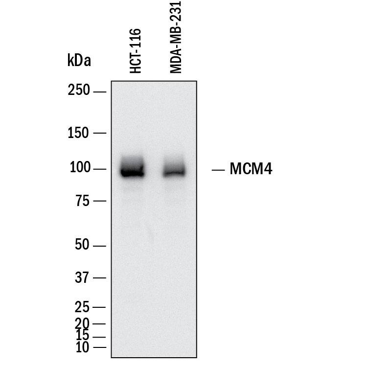
HAF018). A specific band was detected for MCM4 at approximately 96 kDa (as indicated). This experiment was conducted under reducing conditions and using
Western Blot Buffer Group 1." class="big_lightbox">
HAF018). A specific band was detected for MCM4 at approximately 96 kDa (as indicated). This experiment was conducted under reducing conditions and using
Western Blot Buffer Group 1." alt="Western Blot shows lysates of HCT‑116 human colorectal carcinoma cell line and MDA‑MB‑231 human breast cancer cell line. PVDF membrane was probed with 0.5 µg/ml of Mouse Anti-Human MCM4 Monoclonal Antibody (Catalog # MAB11718) followed by HRP-conjugated Anti-Mouse IgG Secondary Antibody (Catalog #
HAF018). A specific band was detected for MCM4 at approximately 96 kDa (as indicated). This experiment was conducted under reducing conditions and using
Western Blot Buffer Group 1." class="big_thumb" />
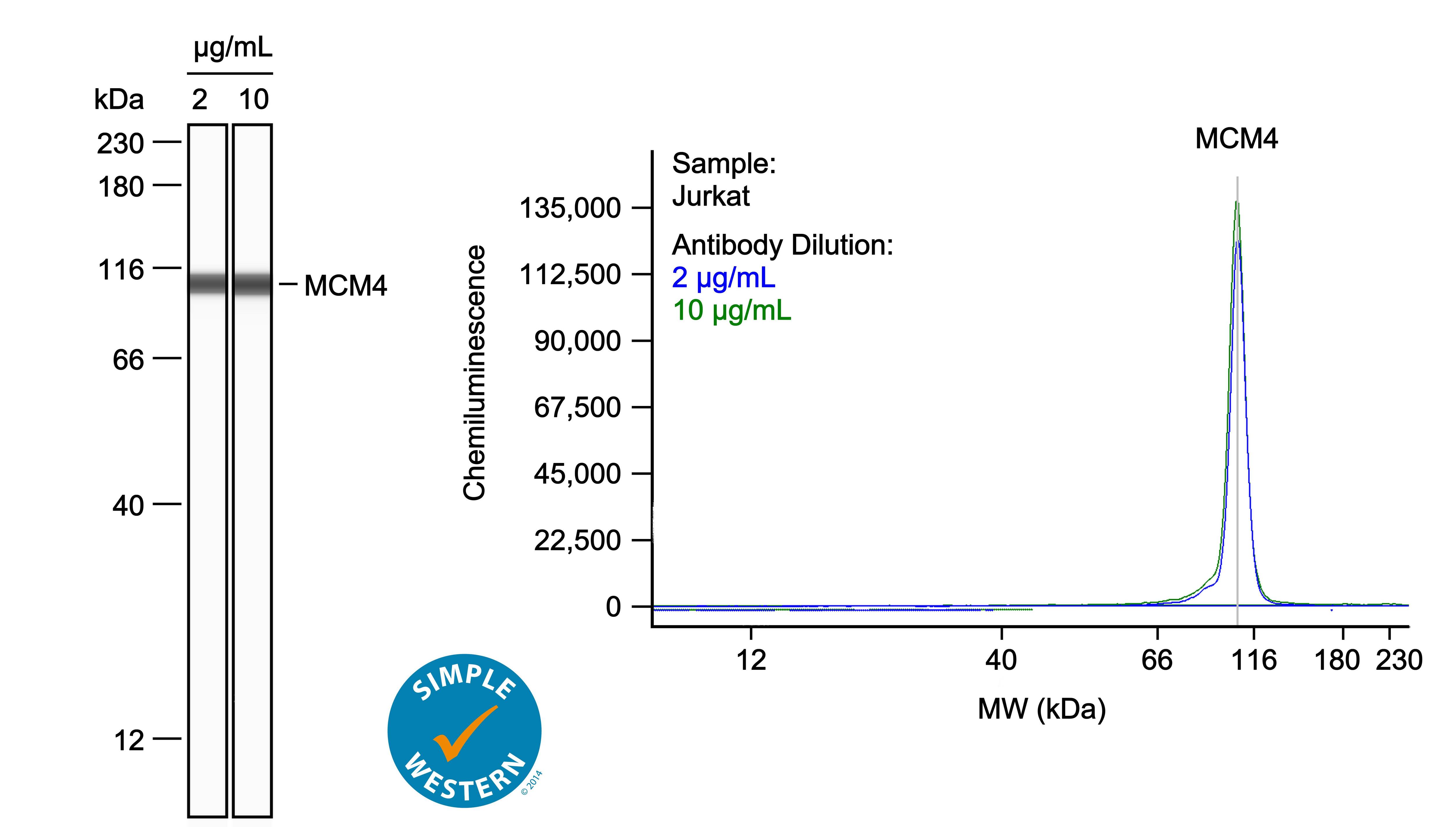
042-205). This experiment was conducted under reducing conditions and using the 12-230kDa separation system.
Right: Simple Western electropherogram showing the same Mouse Anti-Human MCM4 Monoclonal Antibody (Catalog # MAB11718) tested at 2 µg/ml (blue line) and 10 µg/ml (green line) in the Jurkat human acute T cell leukemia cell line." class="big_lightbox">
042-205). This experiment was conducted under reducing conditions and using the 12-230kDa separation system.
Right: Simple Western electropherogram showing the same Mouse Anti-Human MCM4 Monoclonal Antibody (Catalog # MAB11718) tested at 2 µg/ml (blue line) and 10 µg/ml (green line) in the Jurkat human acute T cell leukemia cell line." class="big_thumb" />
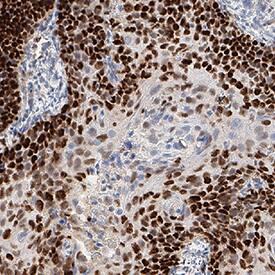
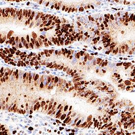
VC001) or the HRP-conjugated Anti-Mouse IgG Secondary Antibody (Catalog #
HAF007). Before incubation with the primary antibody, tissue was subjected to heat-induced epitope retrieval using VisUCyte Antigen Retrieval Reagent-Basic (Catalog #
VCTS021). Tissue was stained using DAB (brown) and counterstained with hematoxylin (blue). Specific staining was localized to the nucleus. View our protocol for
Chromogenic IHC Staining of Paraffin-embedded Tissue Sections." class="big_lightbox">
VC001) or the HRP-conjugated Anti-Mouse IgG Secondary Antibody (Catalog #
HAF007). Before incubation with the primary antibody, tissue was subjected to heat-induced epitope retrieval using VisUCyte Antigen Retrieval Reagent-Basic (Catalog #
VCTS021). Tissue was stained using DAB (brown) and counterstained with hematoxylin (blue). Specific staining was localized to the nucleus. View our protocol for
Chromogenic IHC Staining of Paraffin-embedded Tissue Sections." class="big_thumb" />
VC001) or the HRP-conjugated Anti-Mouse IgG Secondary Antibody (Catalog #
HAF007). Before incubation with the primary antibody, tissue was subjected to heat-induced epitope retrieval using VisUCyte Antigen Retrieval Reagent-Basic (Catalog #
VCTS021). Tissue was stained using DAB (brown) and counterstained with hematoxylin (blue). Specific staining was localized to the nucleus. View our protocol for
Chromogenic IHC Staining of Paraffin-embedded Tissue Sections." class="big_lightbox">
VC001) or the HRP-conjugated Anti-Mouse IgG Secondary Antibody (Catalog #
HAF007). Before incubation with the primary antibody, tissue was subjected to heat-induced epitope retrieval using VisUCyte Antigen Retrieval Reagent-Basic (Catalog #
VCTS021). Tissue was stained using DAB (brown) and counterstained with hematoxylin (blue). Specific staining was localized to the nucleus. View our protocol for
Chromogenic IHC Staining of Paraffin-embedded Tissue Sections." class="big_thumb" />
VC001) or the HRP-conjugated Anti-Mouse IgG Secondary Antibody (Catalog #
HAF007). Before incubation with the primary antibody, tissue was subjected to heat-induced epitope retrieval using VisUCyte Antigen Retrieval Reagent-Basic (Catalog #
VCTS021). Tissue was stained using DAB (brown) and counterstained with hematoxylin (blue). Specific staining was localized to the nucleus. View our protocol for
Chromogenic IHC Staining of Paraffin-embedded Tissue Sections." class="big_lightbox">
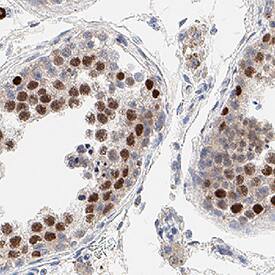
VC001) or the HRP-conjugated Anti-Mouse IgG Secondary Antibody (Catalog #
HAF007). Before incubation with the primary antibody, tissue was subjected to heat-induced epitope retrieval using VisUCyte Antigen Retrieval Reagent-Basic (Catalog #
VCTS021). Tissue was stained using DAB (brown) and counterstained with hematoxylin (blue). Specific staining was localized to the nucleus. View our protocol for
Chromogenic IHC Staining of Paraffin-embedded Tissue Sections." class="big_thumb" />
 HAF018). A specific band was detected for MCM4 at approximately 96 kDa (as indicated). This experiment was conducted under reducing conditions and using Western Blot Buffer Group 1." alt="Western Blot shows lysates of HCT‑116 human colorectal carcinoma cell line and MDA‑MB‑231 human breast cancer cell line. PVDF membrane was probed with 0.5 µg/ml of Mouse Anti-Human MCM4 Monoclonal Antibody (Catalog # MAB11718) followed by HRP-conjugated Anti-Mouse IgG Secondary Antibody (Catalog # HAF018). A specific band was detected for MCM4 at approximately 96 kDa (as indicated). This experiment was conducted under reducing conditions and using Western Blot Buffer Group 1." class="big_thumb" />


 042-205). This experiment was conducted under reducing conditions and using the 12-230kDa separation system. Right: Simple Western electropherogram showing the same Mouse Anti-Human MCM4 Monoclonal Antibody (Catalog # MAB11718) tested at 2 µg/ml (blue line) and 10 µg/ml (green line) in the Jurkat human acute T cell leukemia cell line." class="big_thumb" />
 VC001) or the HRP-conjugated Anti-Mouse IgG Secondary Antibody (Catalog #
  VC001) or the HRP-conjugated Anti-Mouse IgG Secondary Antibody (Catalog #
  VC001) or the HRP-conjugated Anti-Mouse IgG Secondary Antibody (Catalog #
  NL007) and counterstained with DAPI (blue). Specific staining was localized to the nucleus. View our protocol for
  NL007) and counterstained with DAPI (blue). Specific staining was localized to the nucleus. View our protocol for
 ![Western Blot: MCM4 Antibody [NBP1-33105] - Various whole cell extracts (30 ug) were separated by 7.5% SDS-PAGE, and the membrane was blotted with MCM4 antibody diluted at 1:1000.](http://images.novusbio.com/fullsize/MCM4-Antibody-Western-Blot-NBP1-33105-img0007.jpg "Western Blot: MCM4 Antibody [NBP1-33105] - Various whole cell extracts (30 ug) were separated by 7.5% SDS-PAGE, and the membrane was blotted with MCM4 antibody diluted at 1:1000.")
![Immunocytochemistry/Immunofluorescence: MCM4 Antibody [NBP1-33105] - Paraformaldehyde-fixed HeLa, using antibody at 1:500 dilution.](http://images.novusbio.com/fullsize/MCM4-Antibody-Immunocytochemistry-Immunofluorescence-NBP1-33105-img0003.jpg)
![Immunohistochemistry-Paraffin: MCM4 Antibody [NBP1-33105] - Paraffin-embedded A549 Xenograft, using antibody at 1:100 dilution.](http://images.novusbio.com/fullsize/MCM4-Antibody-Immunohistochemistry-Paraffin-NBP1-33105-img0004.jpg)
![Western Blot: MCM4 Antibody [NBP1-33105] - MCM4 antibody detects MCM4 protein by Western blot analysis. A. 30 ug Jurkat whole cell lysate/extract. B. 30 ug Raji whole cell lysate/extract. C. 30 ug NCI-H929 whole cell lysate/extract. 7.5 % SDS-PAGE. MCM4 antibody dilution: 1:5000.](http://images.novusbio.com/fullsize/MCM4-Antibody-Western-Blot-NBP1-33105-img0006.jpg)
![Immunoprecipitation: MCM4 Antibody [NBP1-33105] - MCM4 antibody immunoprecipitates MCM4 protein in IP experiments. IP samples: Jurkat whole cell extract. A. 40 ug Jurkat whole cell extract. B. Control with 4 ug of preimmune Rabbit IgG. C. Immunoprecipitation of MCM4 protein by 4 ug MCM4 antibody. 7.5 % SDS-PAGE. The immunoprecipitated MCM4 protein was detected by MCM4 antibody diluted at 1:500.](http://images.novusbio.com/fullsize/MCM4-Antibody-Immunoprecipitation-NBP1-33105-img0008.jpg)
![Immunohistochemistry: MCM4 Antibody [NBP1-33105] - Immunohistochemical analysis of paraffin-embedded zebrafish tissue, using MCM4 antibody (NBP1-33105) at 1:300 dilution.](http://images.novusbio.com/fullsize/nbp1-33105_rabbit-polyclonal-mcm4-antibody-91020242041276.jpg)
![Chromatin Immunoprecipitation (ChIP): MCM4 Antibody [NBP1-33105] - Cross-linked ChIP was performed with HeLa chromatin extract and 5 ug of either control rabbit IgG or anti-MCM4 antibody. The precipitated DNA was detected by PCR with primer set targeting to Myc promoter.](http://images.novusbio.com/fullsize/nbp1-33105_rabbit-polyclonal-mcm4-antibody-910202420134039.jpg)
![Western Blot: MCM4 Antibody [NBP1-33105] - Sample (30 ug of whole cell lysate) <br /> A: zebrafish eye <br /> 7.5% SDS PAGE <br /> NBP1-33105 diluted at 1:1000 <br />](http://images.novusbio.com/fullsize/nbp1-33105_rabbit-polyclonal-mcm4-antibody-91020242010442.jpg)
![Immunohistochemistry-Paraffin: MCM4 Antibody [NBP1-85728] - Analysis in human testis and pancreas tissues. Corresponding MCM4 RNA-seq data are presented for the same tissues.](http://images.novusbio.com/fullsize/MCM4-Antibody-Immunohistochemistry-Paraffin-NBP1-85728-img0012.jpg "<b>Orthogonal Strategies Validation. </b>Immunohistochemistry-Paraffin: MCM4 Antibody [NBP1-85728] - Analysis in human testis and pancreas tissues. Corresponding MCM4 RNA-seq data are presented for the same tissues.")
![<b>Orthogonal Strategies Validation. </b>Immunohistochemistry-Paraffin: MCM4 Antibody [NBP1-85728] - Staining of human skin shows strong nuclear positivity in squamous epithelial cells.](http://images.novusbio.com/fullsize/MCM4-Antibody-Immunohistochemistry-Paraffin-NBP1-85728-img0016.jpg)
![Immunohistochemistry-Paraffin: MCM4 Antibody [NBP1-85728] - Staining of human pancreas shows low positivity in glandular cells as expected.](http://images.novusbio.com/fullsize/MCM4-Antibody-Immunohistochemistry-Paraffin-NBP1-85728-img0013.jpg)
![Immunohistochemistry-Paraffin: MCM4 Antibody [NBP1-85728] - Staining of human testis shows strong nuclear positivity in cells in seminiferous ducts.](http://images.novusbio.com/fullsize/MCM4-Antibody-Immunohistochemistry-Paraffin-NBP1-85728-img0014.jpg)
![Immunohistochemistry-Paraffin: MCM4 Antibody [NBP1-85728] - Staining of human tonsil shows strong nuclear positivity in germinal center cells.](http://images.novusbio.com/fullsize/MCM4-Antibody-Immunohistochemistry-Paraffin-NBP1-85728-img0015.jpg)


![Western Blot: MCM4 Antibody [H00004173-B01P] - Analysis of MCM4 expression in human placenta.](http://images.novusbio.com/fullsize/MCM4-Antibody-Western-Blot-H00004173-B01P-img0004.jpg "Western Blot: MCM4 Antibody [H00004173-B01P] - Analysis of MCM4 expression in human placenta.")
![Immunocytochemistry/Immunofluorescence: MCM4 Antibody [H00004173-B01P] - Analysis of purified antibody to MCM4 on HeLa cell. (antibody concentration 10 ug/ml)](http://images.novusbio.com/fullsize/MCM4-Antibody-Immunocytochemistry-Immunofluorescence-H00004173-B01P-img0001.jpg)
![Immunohistochemistry-Paraffin: MCM4 Antibody [H00004173-B01P] - Analysis of purified antibody to MCM4 on formalin-fixed paraffin-embedded human tonsil. (antibody concentration 3 ug/ml)](http://images.novusbio.com/fullsize/MCM4-Antibody-Immunohistochemistry-Paraffin-H00004173-B01P-img0002.jpg)
![Western Blot: MCM4 Antibody [H00004173-B01P] - Analysis of MCM4 expression in HeLa.](http://images.novusbio.com/fullsize/MCM4-Antibody-Western-Blot-H00004173-B01P-img0003.jpg)
![Western Blot: MCM4 Antibody [H00004173-B01P] - Analysis of MCM4 expression in transfected 293T cell line by MCM4 polyclonal antibody. Lane 1: MCM4 transfected lysate(94.93 KDa). Lane 2: Non-transfected lysate.](http://images.novusbio.com/fullsize/MCM4-Antibody-Western-Blot-H00004173-B01P-img0005.jpg)
![Western Blot: MCM4 Antibody [NB100-290] - RIPA extract (50 ug) from HeLa cells. Antibody used at 0.2 ug/ml.](http://images.novusbio.com/fullsize/MCM4-Antibody-Western-Blot-NB100-290-img0002.jpg "Western Blot: MCM4 Antibody [NB100-290] - RIPA extract (50 ug) from HeLa cells. Antibody used at 0.2 ug/ml.")
![Immunohistochemistry: MCM4 Antibody [NB100-290] - Sample: FFPE section of human testicular seminoma (left) and mouse squamous cell carcinoma (right). Antibody: Affinity purified rabbit anti-MCM4 used at a dilution of 1:1,000 (1ug/ml). Detection: DAB](http://images.novusbio.com/fullsize/MCM4-Antibody-Immunohistochemistry-NB100-290-img0009.jpg)
![Western Blot: MCM4 Antibody [NBP2-58370] - Analysis in U2OS cells transfected with control siRNA, target specific siRNA probe #1 and #2. Remaining relative intensity is presented.](http://images.novusbio.com/fullsize/MCM4-Antibody-Western-Blot-NBP2-58370-img0004.jpg "<b>Genetic Strategies Validation. </b>Western Blot: MCM4 Antibody [NBP2-58370] - Analysis in U2OS cells transfected with control siRNA, target specific siRNA probe #1 and #2. Remaining relative intensity is presented.")
![<b>Genetic Strategies Validation. </b>Immunocytochemistry/Immunofluorescence: MCM4 Antibody [NBP2-58370] - Staining of human cell line U-2 OS shows localization to nucleoplasm.](http://images.novusbio.com/fullsize/MCM4-Antibody-Immunocytochemistry-Immunofluorescence-NBP2-58370-img0001.jpg)
![Immunohistochemistry: MCM4 Antibody (JE38-76) [NBP3-32578] - Immunohistochemical analysis of paraffin-embedded human tonsil tissue with Rabbit anti-MCM4 antibody (NBP3-32578) at 1/1,000 dilution.<br/><br/> The section was pre-treated using heat mediated antigen retrieval with sodium citrate buffer (pH 6.0) for 2 minutes. The tissues were blocked in 1% BSA for 20 minutes at room temperature, washed with ddH2O and PBS, and then probed with the primary antibody (NBP3-32578) at 1/1,000 dilution for 1 hour at room temperature. The detection was performed using an HRP conjugated compact polymer system. DAB was used as the chromogen. Tissues were counterstained with hematoxylin and mounted with DPX.](http://images.novusbio.com/fullsize/nbp3-32578_rabbit-mcm4-mab-je38-76-298202414161853.jpg "Immunohistochemistry: MCM4 Antibody (JE38-76) [NBP3-32578] - Immunohistochemical analysis of paraffin-embedded human tonsil tissue with Rabbit anti-MCM4 antibody (NBP3-32578) at 1/1,000 dilution.<br/><br/> The section was pre-treated using heat mediated antigen retrieval with sodium citrate buffer (pH 6.0) for 2 minutes. The tissues were blocked in 1% BSA for 20 minutes at room temperature, washed with ddH2O and PBS, and then probed with the primary antibody (NBP3-32578) at 1/1,000 dilution for 1 hour at room temperature. The detection was performed using an HRP conjugated compact polymer system. DAB was used as the chromogen. Tissues were counterstained with hematoxylin and mounted with DPX.")
![Western blot analysis on different lysates with NBP3-32578 at 1/1,000 dilution. Lane 1: HeLa cell lysate Lane 2: Jurkat cell lysate Lane 3: 293T cell lysate Lane 4: HL-60 cell lysate Lysates/proteins at 40 µg/Lane. Predicted band size: 97 kDa Observed band size: 97 kDa Exposure time: 1 minute; 4-20% SDS-PAGE gel. Proteins were transferred to a PVDF membrane and blocked with 5% NFDM/TBST for 1 hour at room temperature. NBP3-32578 at 1/1,000 dilution was used in 5% NFDM/TBST at room temperature for 2 hours. Goat Anti-Rabbit IgG [HRP] Secondary Antibody at 1:100,000 dilution was used for 1 hour at room temperature.](http://images.novusbio.com/fullsize/nbp3-32578_-western-blot-639204595237201669.jpg)


![Immunohistochemistry:[NBP3-29310]-Detection of human MCM4 by immunohistochemistry. Sample: FFPE section of human lung small cell carcinoma. Antibody: Affinity purified rabbit anti-MCM4.](http://images.novusbio.com/fullsize/nbp3-29310_rabbit-mcm4-pab-231202511585557.jpg "Immunohistochemistry:[NBP3-29310]-Detection of human MCM4 by immunohistochemistry. Sample: FFPE section of human lung small cell carcinoma. Antibody: Affinity purified rabbit anti-MCM4.")
![Western Blot: MCM4 Antibody (5N0X3) [NBP3-16791] - Western blot analysis of extracts of various cell lines, using MCM4 Rabbit mAb (NBP3-16791) at 1:1000 dilution. Secondary antibody: HRP Goat Anti-Rabbit IgG (H+L) at 1:10000 dilution. Lysates/proteins: 25ug per lane. Blocking buffer: 3% nonfat dry milk in TBST. Detection: ECL Basic Kit. Exposure time: 30s.](http://images.novusbio.com/fullsize/MCM4-Antibody-5N0X3-Western-Blot-NBP3-16791-img0001.jpg "Western Blot: MCM4 Antibody (5N0X3) [NBP3-16791] - Western blot analysis of extracts of various cell lines, using MCM4 Rabbit mAb (NBP3-16791) at 1:1000 dilution. Secondary antibody: HRP Goat Anti-Rabbit IgG (H+L) at 1:10000 dilution. Lysates/proteins: 25ug per lane. Blocking buffer: 3% nonfat dry milk in TBST. Detection: ECL Basic Kit. Exposure time: 30s.")
![Immunohistochemistry:[NBP3-29264]-Detection of human MCM4 by immunohistochemistry. Sample: FFPE section of human seminoma. Antibody: Affinity purified goat anti-MCM4.](http://images.novusbio.com/fullsize/nbp3-29264_goat-mcm4-pab-23120251252833.jpg "Immunohistochemistry:[NBP3-29264]-Detection of human MCM4 by immunohistochemistry. Sample: FFPE section of human seminoma. Antibody: Affinity purified goat anti-MCM4.")


