DR555RB) and counterstained with DAPI (blue; Lunaphore Catalog #
DR100). Specific staining was localized to the membrane. Protocol available in
COMET™ Panel Builder." class="big_lightbox">
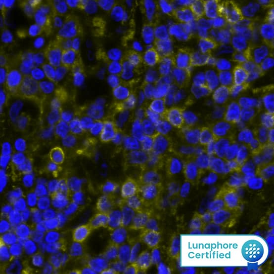
DR555RB) and counterstained with DAPI (blue; Lunaphore Catalog #
DR100). Specific staining was localized to the membrane. Protocol available in
COMET™ Panel Builder." alt="IgM was detected in immersion fixed paraffin-embedded sections of human Non-Hodgkins Lymphoma using Rabbit Anti-Human IgM, Monoclonal Antibody (Catalog #MAB9435) at 0.2ug/mL at 37° Celsius for 4 minutes. Before incubation with the primary antibody, tissue underwent an all-in-one dewaxing and antigen retrieval preprocessing using PreTreatment Module (PT Module) and Dewax and HIER Buffer H (pH 9; Epredia Catalog # TA-999-DHBH). Tissue was stained using the Alexa Fluor™ Plus 555 Goat anti-Rabbit IgG Secondary Antibody at 1:100 at 37 ° Celsius for 2 minutes. (Yellow; Lunaphore Catalog #
DR555RB) and counterstained with DAPI (blue; Lunaphore Catalog #
DR100). Specific staining was localized to the membrane. Protocol available in
COMET™ Panel Builder." class="big_thumb" />
DR555RB) and counterstained with DAPI (blue; Lunaphore Catalog #
DR100). Specific staining was localized to the membrane. Protocol available in
COMET™ Panel Builder." class="big_lightbox">
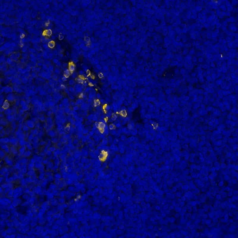
DR555RB) and counterstained with DAPI (blue; Lunaphore Catalog #
DR100). Specific staining was localized to the membrane. Protocol available in
COMET™ Panel Builder." class="big_thumb" />
DR555RB) and counterstained with DAPI (blue; Lunaphore Catalog #
DR100). Specific staining was localized to the membrane. Protocol available in
COMET™ Panel Builder." class="big_lightbox">
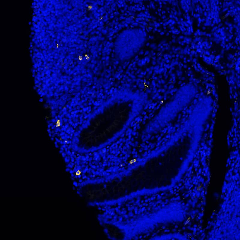
DR555RB) and counterstained with DAPI (blue; Lunaphore Catalog #
DR100). Specific staining was localized to the membrane. Protocol available in
COMET™ Panel Builder." class="big_thumb" />
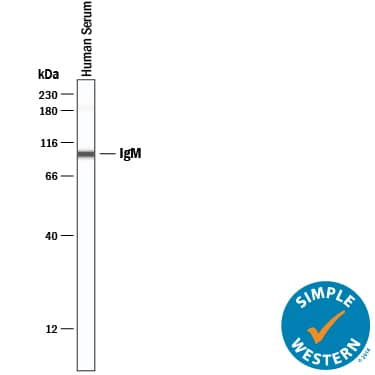
HAF008). A specific band was detected for IgM at approximately 65 kDa (as indicated). This experiment was conducted under reducing conditions and using
Immunoblot Buffer Group 1." class="big_lightbox">
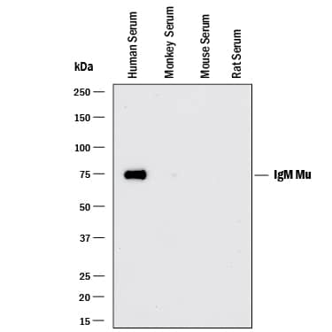
HAF008). A specific band was detected for IgM at approximately 65 kDa (as indicated). This experiment was conducted under reducing conditions and using
Immunoblot Buffer Group 1." class="big_thumb" />
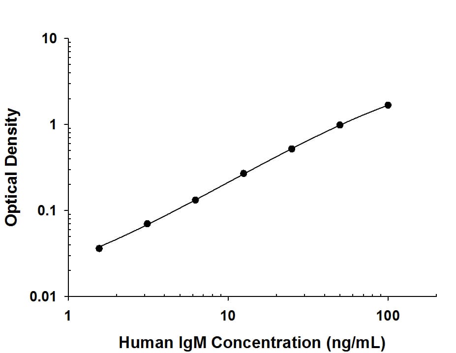
DY990). Rabbit Anti-Human IgM Monoclonal Antibody (
MAB94351) was biotinylated and incubated with the protein captured on the plate. Detection of the standard curve was achieved by incubating Streptavidin-HRP (
DY998) followed by Substrate Solution (
DY999) and stopping the enzymatic reaction with Stop Solution (
DY994)." class="big_lightbox">
DY990). Rabbit Anti-Human IgM Monoclonal Antibody (
MAB94351) was biotinylated and incubated with the protein captured on the plate. Detection of the standard curve was achieved by incubating Streptavidin-HRP (
DY998) followed by Substrate Solution (
DY999) and stopping the enzymatic reaction with Stop Solution (
DY994)." class="big_thumb" />
HAF008) or the Anti-Rabbit IgG VisUCyte™ HRP Polymer Antibody (Catalog #
VC003). Before incubation with the primary antibody, tissue was subjected to heat-induced epitope retrieval using VisUCyte Antigen Retrieval Reagent-Basic (Catalog #
VCTS021). Tissue was stained using DAB (brown) and counterstained with hematoxylin (blue). Specific staining was localized to the membrane of B cells. View our protocol for
Chromogenic IHC Staining of Paraffin-embedded Tissue Sections." class="big_lightbox">

HAF008) or the Anti-Rabbit IgG VisUCyte™ HRP Polymer Antibody (Catalog #
VC003). Before incubation with the primary antibody, tissue was subjected to heat-induced epitope retrieval using VisUCyte Antigen Retrieval Reagent-Basic (Catalog #
VCTS021). Tissue was stained using DAB (brown) and counterstained with hematoxylin (blue). Specific staining was localized to the membrane of B cells. View our protocol for
Chromogenic IHC Staining of Paraffin-embedded Tissue Sections." class="big_thumb" />
 DR555RB) and counterstained with DAPI (blue; Lunaphore Catalog # DR100). Specific staining was localized to the membrane. Protocol available in COMET™ Panel Builder." alt="IgM was detected in immersion fixed paraffin-embedded sections of human Non-Hodgkins Lymphoma using Rabbit Anti-Human IgM, Monoclonal Antibody (Catalog #MAB9435) at 0.2ug/mL at 37° Celsius for 4 minutes. Before incubation with the primary antibody, tissue underwent an all-in-one dewaxing and antigen retrieval preprocessing using PreTreatment Module (PT Module) and Dewax and HIER Buffer H (pH 9; Epredia Catalog # TA-999-DHBH). Tissue was stained using the Alexa Fluor™ Plus 555 Goat anti-Rabbit IgG Secondary Antibody at 1:100 at 37 ° Celsius for 2 minutes. (Yellow; Lunaphore Catalog # DR555RB) and counterstained with DAPI (blue; Lunaphore Catalog # DR100). Specific staining was localized to the membrane. Protocol available in COMET™ Panel Builder." class="big_thumb" />


![Western Blot: Goat anti-Monkey IgM Heavy Chain Secondary Antibody [NBP2-59719] - Western Blot of Goat anti-Monkey IgM Heavy Chain Secondary antibody. Lane 1: Monkey IgM. Lane 2: Monkey IgG.Lane 3: Monkey IgG (A and M Free).Lane 4: Monkey IgG F(c). Load: 50 ng per lane.Primary antibody: Goat anti-Monkey IgM Heavy Chain Secondary antibody at 1:1,000 for 1 hour at RT.Secondary antibody: Peroxidase conjugated Dk-a-Gt IgG at 1:40,000 for 30 min at RT.Block for 30 min at RT.Predicted/Observed size: 70 kDa, 70 kDa for Monkey IgM (mu chain).](http://images.novusbio.com/fullsize/Goat anti-Monkey IgM Heavy Chain Secondary Antibody-Western Blot-NBP2-59719-img0005.jpg "Western Blot: Goat anti-Monkey IgM Heavy Chain Secondary Antibody [NBP2-59719] - Western Blot of Goat anti-Monkey IgM Heavy Chain Secondary antibody. Lane 1: Monkey IgM. Lane 2: Monkey IgG.Lane 3: Monkey IgG (A and M Free).Lane 4: Monkey IgG F(c). Load: 50 ng per lane.Primary antibody: Goat anti-Monkey IgM Heavy Chain Secondary antibody at 1:1,000 for 1 hour at RT.Secondary antibody: Peroxidase conjugated Dk-a-Gt IgG at 1:40,000 for 30 min at RT.Block for 30 min at RT.Predicted/Observed size: 70 kDa, 70 kDa for Monkey IgM (mu chain).")
![Western Blot: Goat anti-Monkey IgM Heavy Chain Secondary Antibody [NBP2-59719] - Lane 1: Monkey IgM. Lane 2: None. Load: 50 ng per lane. Primary antibody: None. Secondary antibody: Fluorescein goat secondary antibody at 1:1,000 for 60 min at RT. Block: incubated with blocking buffer for 30 min t RT. Predicted/Observed size: 72 kDa, 72 kDa for Monkey IgM. Other band(s): None. Image using the Fluorescein form of this antibody.](http://images.novusbio.com/fullsize/Goat-anti-Monkey-IgM-Heavy-Chain-Secondary-Antibody-Western-Blot-NBP2-59719-img0003.jpg)
![N/A: Goat anti-Monkey IgM Heavy Chain Secondary Antibody [NBP2-59719]](http://images.novusbio.com/fullsize/Goat-anti-Monkey-IgM-Heavy-Chain-Secondary-Antibody-N-A-NBP2-59719-img0004.jpg)
![Flow Cytometry: IgM Antibody (II/41) [NBP1-43303] - Analysis of PE/Cy7 conjugate of NBP1-43303. Staining of C57BL/6 bone marrow cells with Anti-Human/Mouse CD45R (B220) FITC and 0.06 micrograms of Rat IgG2a ? Isotype Control PE-Cy7 (left) or 0.06 micrograms of Anti-Mouse IgM PE-Cy7 (right). Cells in the ly](http://images.novusbio.com/fullsize/Mouse-IgM-Antibody-II-41-Flow-Cytometry-NBP1-43303-img0008.jpg "Flow Cytometry: IgM Antibody (II/41) [NBP1-43303] - Analysis of PE/Cy7 conjugate of NBP1-43303. Staining of C57BL/6 bone marrow cells with Anti-Human/Mouse CD45R (B220) FITC and 0.06 micrograms of Rat IgG2a ? Isotype Control PE-Cy7 (left) or 0.06 micrograms of Anti-Mouse IgM PE-Cy7 (right). Cells in the ly")
![Flow Cytometry: IgM Antibody (II/41) [NBP1-43303] - Staining of C57BL/6 splenocytes with 0.5 ug of Rat IgG2a kappa Isotype Control Purified (open histogram) or 0.5 ug of Anti-Mouse IgM Purified (filled histogram) followed by Anti-Rat IgG Biotin and Streptavidin PE. Total viable cells were used for analysis.](http://images.novusbio.com/fullsize/Mouse-IgM-Antibody-II-41-Flow-Cytometry-NBP1-43303-img0001.jpg)
![Flow Cytometry: IgM Antibody (II/41) [NBP1-43303] - Analysis of Allophycocyanin conjugate of NBP1-43303. Staining of C57Bl/6 bone marrow cells with Anti-Human/Mouse CD45R (B220) FITC and 0.125 ug of Rat IgG2a K Isotype Control APC (left) or 0.125 ug of Anti-Mouse IgM APC (right). Cells in the small scatter](http://images.novusbio.com/fullsize/Mouse-IgM-Antibody-II-41-Flow-Cytometry-NBP1-43303-img0002.jpg)
![Flow Cytometry: IgM Antibody (II/41) [NBP1-43303] - Analysis of Allophycocyanin conjugate of NBP1-43303. Staining of C57Bl/6 bone marrow cells with Anti-Human/Mouse CD45R (B220) FITC and 0.125 ug of Rat IgG2a K Isotype Control APC (left) or 0.125 ug of Anti-Mouse IgM APC (right). Cells in the small scatter](http://images.novusbio.com/fullsize/Mouse-IgM-Antibody-II-41-Flow-Cytometry-NBP1-43303-img0003.jpg)
![Flow Cytometry: IgM Antibody (II/41) [NBP1-43303] - Analysis of Biotin conjugate of NBP1-43303. Staining of C57BL/6 bone marrow cells with Anti-Human/Mouse CD45R (B220) APC and 0.06 ug of Rat IgG2a kappa Isotype Control Biotin (left) or 0.06 ug of Anti-Mouse IgM Biotin (right) followed by Streptavidin PE .](http://images.novusbio.com/fullsize/Mouse-IgM-Antibody-II-41-Flow-Cytometry-NBP1-43303-img0004.jpg)
![Flow Cytometry: IgM Antibody (II/41) [NBP1-43303] - Analysis of FITC conjugate of NBP1-43303. Staining of C57BL/6 bone marrow cells with Anti-Human/Mouse CD45R (B220) APC and 0.125 ug of Rat IgG2a K Isotype Control FITC (left) or 0.125 ug of Anti-Mouse IgM FITC (right). Total viable cells were used for ana](http://images.novusbio.com/fullsize/Mouse-IgM-Antibody-II-41-Flow-Cytometry-NBP1-43303-img0005.jpg)
![Flow Cytometry: IgM Antibody (II/41) [NBP1-43303] - Analysis of PE conjugate of NBP1-43303. The II/41 antibody has been tested by flow cytometric mouse spleen cell suspension. This can be used at less than or equal to 1 ug per test. Cell number should be determined empirically but can range from 10^5to 10^](http://images.novusbio.com/fullsize/Mouse-IgM-Antibody-II-41-Flow-Cytometry-NBP1-43303-img0006.jpg)
![Flow Cytometry: IgM Antibody (II/41) [NBP1-43303] - Analysis of PE/Cy5 conjugate of NBP1-43303. Staining of C57Bl/6 bone marrow cells with Anti-Human/Mouse CD45R (B220) FITC and 0.06 ug of Rat IgG2a Isotype Control PE-Cy5 (left) or 0.06 ug of Anti-Mouse IgM PE-Cy5 (right). Total viable cells were used for](http://images.novusbio.com/fullsize/Mouse-IgM-Antibody-II-41-Flow-Cytometry-NBP1-43303-img0007.jpg)
 DR555RB) and counterstained with DAPI (blue; Lunaphore Catalog #
  DR555RB) and counterstained with DAPI (blue; Lunaphore Catalog #
  HAF008). A specific band was detected for IgM at approximately 65 kDa (as indicated). This experiment was conducted under reducing conditions and using
 DY990). Rabbit Anti-Human IgM Monoclonal Antibody (
 HAF008) or the Anti-Rabbit IgG VisUCyte™ HRP Polymer Antibody (Catalog #
 ![Western Blot: Goat anti-Chicken IgM Heavy Chain Secondary Antibody [NBP2-60690] - Lane 1: Chicken IgG whole molecule. Lane 2: Chicken IgG F(c) Fragment. Lane 3: Chicken IgG F(ab) Fragment. Lane 4: Chicken IgM Whole Molecule. All samples were reduced. Load: 50 ng per lane. Block: incubated with blocking buffer for 30 min at RT. Primary Antibody: Anti-Chicken IgM (mu chain) (GOAT) Antibody Peroxidase Conjugated 1:30,000 for 60 min at RT. Predicted/Observed Size: 25 and 72 kDa for Chicken IgG, 25 kDa for F(c) and Fab, 75 kDa for IgM. Chicken F(c) migrates slightly higher. Image using the Biotin form of this antibody.](http://images.novusbio.com/fullsize/Goat-anti-Chicken-IgM-Heavy-Chain-Secondary-Antibody-Western-Blot-NBP2-60690-img0001.jpg "Western Blot: Goat anti-Chicken IgM Heavy Chain Secondary Antibody [NBP2-60690] - Lane 1: Chicken IgG whole molecule. Lane 2: Chicken IgG F(c) Fragment. Lane 3: Chicken IgG F(ab) Fragment. Lane 4: Chicken IgM Whole Molecule. All samples were reduced. Load: 50 ng per lane. Block: incubated with blocking buffer for 30 min at RT. Primary Antibody: Anti-Chicken IgM (mu chain) (GOAT) Antibody Peroxidase Conjugated 1:30,000 for 60 min at RT. Predicted/Observed Size: 25 and 72 kDa for Chicken IgG, 25 kDa for F(c) and Fab, 75 kDa for IgM. Chicken F(c) migrates slightly higher. Image using the Biotin form of this antibody.")
![SDS-Page: Goat anti-Chicken IgM Heavy Chain Secondary Antibody [NBP2-60690] - Lane 1: Goat anti-Chicken IgM Secondary Antibody - Non-Reduced. Lane 2: Goat anti-Chicken IgM Secondary Antibody - Reduced. Load: 1.0 ug per lane. Predicted/Observed size: 50 kDa, 25 kDa for Reduced - Goat IgG. Other band(s): none.](http://images.novusbio.com/fullsize/Goat-anti-Chicken-IgM-Heavy-Chain-Secondary-Antibody-SDS-Page-NBP2-60690-img0002.jpg)
![Immunocytochemistry/Immunofluorescence: Goat anti-Human IgM Heavy Chain Secondary Antibody [NBP2-60670] - Immunocytochemistry/Immunofluorescence: Goat anti-Human IgM Secondary Antibody [DyLight 488] [NBP1-72860]](http://images.novusbio.com/fullsize/Goat-anti-Human-IgM-Heavy-Chain-Secondary-Antibody-Immunocytochemistry-Immunofluorescence-NBP2-60670-img0002.jpg "Immunocytochemistry/Immunofluorescence: Goat anti-Human IgM Heavy Chain Secondary Antibody [NBP2-60670] - Immunocytochemistry/Immunofluorescence: Goat anti-Human IgM Secondary Antibody [DyLight 488] [NBP1-72860]")
![Fluorophore-linked immunosorbent assay: Goat anti-Human IgM Heavy Chain Secondary Antibody [NBP2-60670] - Image using the DyLight 405 format of this antibody.](http://images.novusbio.com/fullsize/Goat-anti-Human-IgM-Heavy-Chain-Secondary-Antibody-Fluorophore-linked-immunosorbent-assay-NBP2-60670-img0003.jpg)
![Fluorophore-linked immunosorbent assay: Goat anti-Human IgM Heavy Chain Secondary Antibody [NBP2-60670] - Image using the DyLight 488 format of this antibody.](http://images.novusbio.com/fullsize/Goat-anti-Human-IgM-Heavy-Chain-Secondary-Antibody-Fluorophore-linked-immunosorbent-assay-NBP2-60670-img0004.jpg)
![ELISA: Goat anti-Human IgM Heavy Chain Secondary Antibody [NBP2-60670] - Image using the DyLight 680 format of this antibody.](http://images.novusbio.com/fullsize/Goat-anti-Human-IgM-Heavy-Chain-Secondary-Antibody-ELISA-NBP2-60670-img0005.jpg)
![Fluorophore-linked immunosorbent assay: Goat anti-Human IgM Heavy Chain Secondary Antibody [NBP2-60670] - Image using the DyLight 800 format of this antibody.](http://images.novusbio.com/fullsize/Goat-anti-Human-IgM-Heavy-Chain-Secondary-Antibody-Fluorophore-linked-immunosorbent-assay-NBP2-60670-img0008.jpg)
![ELISA: Goat anti-Human IgM Heavy Chain Secondary Antibody [NBP2-60670] - ELISA results of purified Goat anti-Human IgM Heavy Chain Secondary antibody tested against purified human IgM [RED line], human IgG F(ab')2 {PURPLE line], human IgG Fab [BLUE line], human IgG F(c) [GREEN line]. Each well was coated in duplicate with 1.0 ug of IgM, IgG F(ab')2, IgG Fab, IgG F(c). The starting dilution of antibody was 5ug/ml and the X-axis represents the Log10 of a 3-fold dilution. This titration is a 4-parameter curve fit where the IC50 is defined as the titer of the antibody. Assay performed using Blocking buffer, Donkey-anti-Goat IgG HRP conjugate 1:12, 000, and TMB substrate.](http://images.novusbio.com/fullsize/Goat-anti-Human-IgM-Heavy-Chain-Secondary-Antibody-ELISA-NBP2-60670-img0009.jpg)
![SDS-Page: Monkey IgM Isotype Control [NBP1-97060] - Lane 1: Monkey IgM, Non-Reduced. Lane 2: Monkey IgM, Reduced. Load: 1.0 ug per lane.](http://images.novusbio.com/fullsize/Monkey-IgM-Isotype-Control-SDS-Page-NBP1-97060-img0001.jpg "SDS-Page: Monkey IgM Isotype Control [NBP1-97060] - Lane 1: Monkey IgM, Non-Reduced. Lane 2: Monkey IgM, Reduced. Load: 1.0 ug per lane.")


