HAF016). A specific band was detected for RelA/NF kappa B p65 at approximately 70 kDa (as indicated). This experiment was conducted under reducing conditions and using
Immunoblot Buffer Group 1." class="big_lightbox">
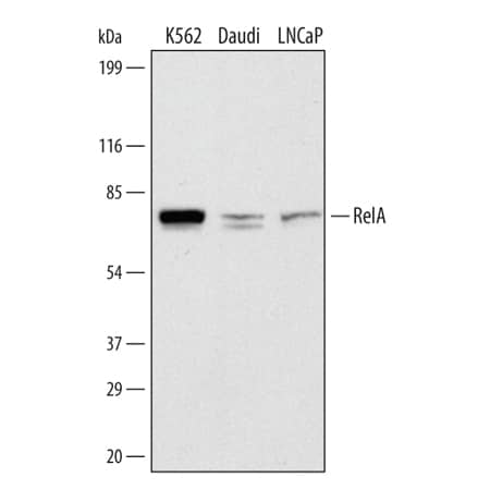
HAF016). A specific band was detected for RelA/NF kappa B p65 at approximately 70 kDa (as indicated). This experiment was conducted under reducing conditions and using
Immunoblot Buffer Group 1." alt="Western blot shows lysates of K562 human chronic myelogenous leukemia cell line, Daudi human Burkitt's lymphoma cell line, and LNCaP human prostate cancer cell line. PVDF membrane was probed with 1 µg/mL of Sheep Anti-Human/Mouse/Rat RelA/NF kappa B p65 Antigen Affinity-purified Polyclonal Antibody (Catalog # AF5078) followed by HRP-conjugated Anti-Sheep IgG Secondary Antibody (Catalog #
HAF016). A specific band was detected for RelA/NF kappa B p65 at approximately 70 kDa (as indicated). This experiment was conducted under reducing conditions and using
Immunoblot Buffer Group 1." class="big_thumb" />
HAF016). A specific band was detected for RelA/NF kappa B p65 at approximately 70 kDa (as indicated). This experiment was conducted under reducing conditions and using
Immunoblot Buffer Group 1." class="big_lightbox">
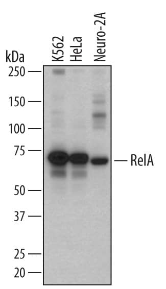
HAF016). A specific band was detected for RelA/NF kappa B p65 at approximately 70 kDa (as indicated). This experiment was conducted under reducing conditions and using
Immunoblot Buffer Group 1." class="big_thumb" />
HAF016). A specific band was detected for RelA/NF kappa B p65 at approximately 65 kDa (as indicated). This experiment was conducted under reducing conditions and using
Immunoblot Buffer Group 1." class="big_lightbox">
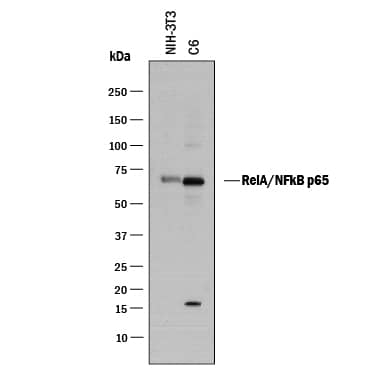
HAF016). A specific band was detected for RelA/NF kappa B p65 at approximately 65 kDa (as indicated). This experiment was conducted under reducing conditions and using
Immunoblot Buffer Group 1." class="big_thumb" />
5-001-A) for 15 minutes in an ultrasonic bath, followed by Biotinylated Anti-Sheep IgG Secondary Antibody (Catalog # [[catalogNumber:BAF016). Immunocomplexes were captured using 50 µL of MagCellect Streptavidin Ferrofluid (Catalog # [[catalogNumber:MAG999) and DNA was purified using chelating resin solution. The
p21promoter was detected by standard PCR." class="big_lightbox">
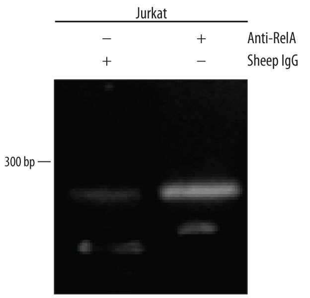
5-001-A) for 15 minutes in an ultrasonic bath, followed by Biotinylated Anti-Sheep IgG Secondary Antibody (Catalog # [[catalogNumber:BAF016). Immunocomplexes were captured using 50 µL of MagCellect Streptavidin Ferrofluid (Catalog # [[catalogNumber:MAG999) and DNA was purified using chelating resin solution. The
p21promoter was detected by standard PCR." class="big_thumb" />
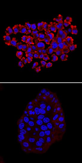
210-TA) for 10 minutes using Sheep Anti-Human/Mouse/Rat RelA/NF kappa B p65 Antigen Affinity-purified Polyclonal Antibody (Catalog # AF5078) at 10 µg/mL for 3 hours at room temperature. Cells were stained using the NorthernLights™ 557-conjugated Anti-Sheep IgG Secondary Antibody (red; Catalog #
NL010) and counterstained with DAPI (blue). Specific staining was localized to cytoplasm in untreated cells and nuclei in treated cells. View our protocol for
Fluorescent ICC Staining of Cells on Coverslips." class="big_lightbox">
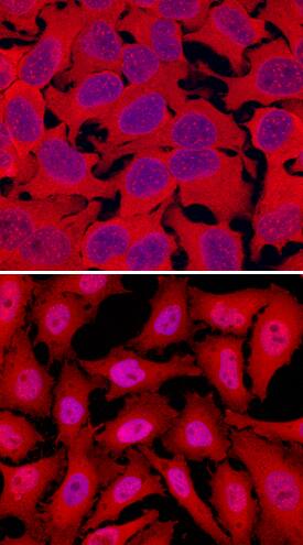
210-TA) for 10 minutes using Sheep Anti-Human/Mouse/Rat RelA/NF kappa B p65 Antigen Affinity-purified Polyclonal Antibody (Catalog # AF5078) at 10 µg/mL for 3 hours at room temperature. Cells were stained using the NorthernLights™ 557-conjugated Anti-Sheep IgG Secondary Antibody (red; Catalog #
NL010) and counterstained with DAPI (blue). Specific staining was localized to cytoplasm in untreated cells and nuclei in treated cells. View our protocol for
Fluorescent ICC Staining of Cells on Coverslips." class="big_thumb" />
HAF016). This experiment was conducted under reducing conditions and using the 12-230 kDa separation system." class="big_lightbox">
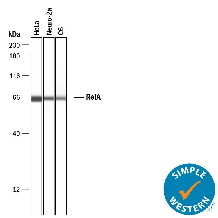
HAF016). This experiment was conducted under reducing conditions and using the 12-230 kDa separation system." class="big_thumb" />
HAF016). A specific band was detected for RelA/NF kappa B p65 at approximately 65 kDa (as indicated) in the parental HeLa cell line, but is not detectable in knockout HeLa cell line. GAPDH (Catalog #
AF5718) is shown as a loading control. This experiment was conducted under reducing conditions and using
Immunoblot Buffer Group 1." class="big_lightbox">
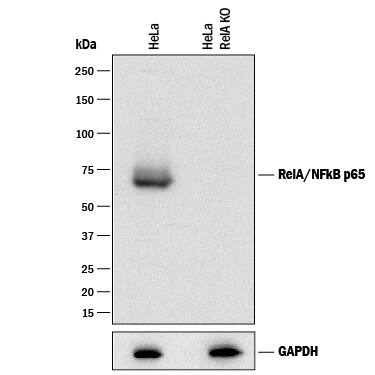
HAF016). A specific band was detected for RelA/NF kappa B p65 at approximately 65 kDa (as indicated) in the parental HeLa cell line, but is not detectable in knockout HeLa cell line. GAPDH (Catalog #
AF5718) is shown as a loading control. This experiment was conducted under reducing conditions and using
Immunoblot Buffer Group 1." class="big_thumb" />
 HAF018). A specific band was detected for RelA/NF kappa B p65 at approximately 65 kDa (as indicated). This experiment was conducted under reducing conditions and using Western Blot Buffer Group 1." alt="Western Blot shows lysates of HeLa human cervical epithelial carcinoma cell line and K562 human chronic myelogenous leukemia cell line. PVDF membrane was probed with 2 µg/ml of Mouse Anti-Human/Mouse/Rat RelA/NF kappa B p65 Monoclonal Antibody (Catalog # MAB5078) followed by HRP-conjugated Anti-Mouse IgG Secondary Antibody (Catalog # HAF018). A specific band was detected for RelA/NF kappa B p65 at approximately 65 kDa (as indicated). This experiment was conducted under reducing conditions and using Western Blot Buffer Group 1." class="big_thumb" />
 HAF016). A specific band was detected for RelA/NF kappa B p65 at approximately 70 kDa (as indicated). This experiment was conducted under reducing conditions and using Immunoblot Buffer Group 1." alt="Western blot shows lysates of K562 human chronic myelogenous leukemia cell line, Daudi human Burkitt's lymphoma cell line, and LNCaP human prostate cancer cell line. PVDF membrane was probed with 1 µg/mL of Sheep Anti-Human/Mouse/Rat RelA/NF kappa B p65 Antigen Affinity-purified Polyclonal Antibody (Catalog # AF5078) followed by HRP-conjugated Anti-Sheep IgG Secondary Antibody (Catalog # HAF016). A specific band was detected for RelA/NF kappa B p65 at approximately 70 kDa (as indicated). This experiment was conducted under reducing conditions and using Immunoblot Buffer Group 1." class="big_thumb" />
 1336) and 20 ng/mL Recombinant Human TNF-a (Catalog # 210-TA) for 10 minutes. PVDF membrane was probed with 2 µg/mL of Mouse Anti-Human Phospho-RelA/NF kappa B p65 (S536) Monoclonal Antibody (Catalog # MAB7226) followed by HRP-conjugated Anti-Mouse IgG Secondary Antibody (Catalog # HAF018). A specific band was detected for Phospho-RelA/ NF kappa B p65 (S536) at approximately 65 kDa (as indicated). This experiment was conducted under reducing conditions and using Immunoblot Buffer Group 2." alt="Western blot shows lysates of HT-29 human colon adenocarcinoma cell line and HeLa human cervical epithelial carcinoma cell line untreated (-) or treated (+) with 100 nM Calyculin A (Catalog # 1336) and 20 ng/mL Recombinant Human TNF-a (Catalog # 210-TA) for 10 minutes. PVDF membrane was probed with 2 µg/mL of Mouse Anti-Human Phospho-RelA/NF kappa B p65 (S536) Monoclonal Antibody (Catalog # MAB7226) followed by HRP-conjugated Anti-Mouse IgG Secondary Antibody (Catalog # HAF018). A specific band was detected for Phospho-RelA/ NF kappa B p65 (S536) at approximately 65 kDa (as indicated). This experiment was conducted under reducing conditions and using Immunoblot Buffer Group 2." class="big_thumb" />
 MAB006, open histogram), followed by Phycoerythrin-conjugated Anti-Rat IgG Secondary Antibody (Catalog # F0105B). To facilitate intracellular staining, cells were fixed with paraformaldehyde and permeabilized with methanol." alt="Daudi human Burkitt's lymphoma cell line was stained with Human RelA/NF kappa B p65 Monoclonal Antibody (Catalog # MAB50781, filled histogram) or isotype control antibody (Catalog # MAB006, open histogram), followed by Phycoerythrin-conjugated Anti-Rat IgG Secondary Antibody (Catalog # F0105B). To facilitate intracellular staining, cells were fixed with paraformaldehyde and permeabilized with methanol." class="big_thumb" />
 210-TA) for 10 minutes. PVDF membrane was probed with 1 µg/mL of Mouse Anti-Human Phospho-RelA/NF kappa B p65 (S529) Monoclonal Antibody (Catalog # MAB7624) followed by HRP-conjugated Anti-Mouse IgG Secondary Antibody (Catalog # HAF018). A specific band was detected for Phospho-RelA/NF kappa B p65 (S529) at approximately 65 kDa (as indicated). This experiment was conducted under reducing conditions and using Immunoblot Buffer Group 1." alt="Western blot shows lysates of HeLa human cervical epithelial carcinoma cell line untreated (-) or treated (+) with 20 ng/mL Recombinant Human TNF-a (Catalog # 210-TA) for 10 minutes. PVDF membrane was probed with 1 µg/mL of Mouse Anti-Human Phospho-RelA/NF kappa B p65 (S529) Monoclonal Antibody (Catalog # MAB7624) followed by HRP-conjugated Anti-Mouse IgG Secondary Antibody (Catalog # HAF018). A specific band was detected for Phospho-RelA/NF kappa B p65 (S529) at approximately 65 kDa (as indicated). This experiment was conducted under reducing conditions and using Immunoblot Buffer Group 1." class="big_thumb" />
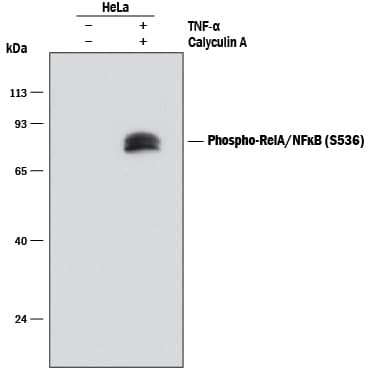 1336) and 20 ng/mL Recombinant Human TNF-a (Catalog # 210-TA) for 10 minutes. PVDF membrane was probed with 0.1 µg/mL of Rabbit Anti-Human Phospho-RelA/NF kappa B p65 (S536) Monoclonal Antibody (Catalog # MAB72261) followed by HRP-conjugated Anti-Rabbit IgG Secondary Antibody (Catalog # HAF008). A specific band was detected for Phospho-RelA/NF kappa B p65 (S536) at approximately 65 kDa (as indicated). This experiment was conducted under reducing conditions and using Immunoblot Buffer Group 1." alt="Western blot shows lysates of HeLa human cervical epithelial carcinoma cell line untreated (-) or treated (+) with 100 nM Calyculin A (Catalog # 1336) and 20 ng/mL Recombinant Human TNF-a (Catalog # 210-TA) for 10 minutes. PVDF membrane was probed with 0.1 µg/mL of Rabbit Anti-Human Phospho-RelA/NF kappa B p65 (S536) Monoclonal Antibody (Catalog # MAB72261) followed by HRP-conjugated Anti-Rabbit IgG Secondary Antibody (Catalog # HAF008). A specific band was detected for Phospho-RelA/NF kappa B p65 (S536) at approximately 65 kDa (as indicated). This experiment was conducted under reducing conditions and using Immunoblot Buffer Group 1." class="big_thumb" />


![Western Blot: RelA/NFkB p65 Antibody [NB100-2176] - Human glomerular endothelial cell lysates, time-response treatment with 10ng/ml TNF-alpha. This image was submitted via customer Review.](http://images.novusbio.com/fullsize/RelA-NFkB-p65-Antibody-Western-Blot-NB100-2176-img0006.jpg "<b>Biological Strategies Validation. </b>Western Blot: RelA/NFkB p65 Antibody [NB100-2176] - Human glomerular endothelial cell lysates, time-response treatment with 10ng/ml TNF-alpha. This image was submitted via customer Review.")
![<b>Biological Strategies Validation. </b>Immunohistochemistry-Paraffin: RelA/NFkB p65 Antibody [NB100-2176] - Staining of formalin-fixed, paraffin-embedded human DLBCL showing nuclear expression of NFkB p65 in the tumor cells.](http://images.novusbio.com/fullsize/RelA-NFkB-p65-Antibody-Immunohistochemistry-Paraffin-NB100-2176-img0004.jpg)
![Western Blot: RelA/NFkB p65 Antibody [NB100-2176] - Analysis of NFkB p65 expression in A431 (A) and HeLa (B) whole cell lysates.](http://images.novusbio.com/fullsize/RelA-NFkB-p65-Antibody-Western-Blot-NB100-2176-img0005.jpg)
![Immunohistochemistry-Paraffin: Rabbit Polyclonal RelA/NFkB p65 Antibody [NB100-2176] - RelA/NFkB p65 IHC (brown) in the mice liver tissue; bars, 200 μm. Image from a verified customer review.](http://images.novusbio.com/fullsize/antibody/nb100-2176_rabbit-polyclonal-rela-nfkb-p65-antibody-immunohistochemistry-paraffin-1111202410307..jpg)
![Immunocytochemistry/Immunofluorescence: Rabbit Polyclonal RelA/NFkB p65 Antibody [NB100-2176] - Mice hepatocytes stained for RelA/NFkB p65. Image from a verified customer review.](http://images.novusbio.com/fullsize/antibody/nb100-2176_rabbit-polyclonal-rela-nfkb-p65-antibody-immunocytochemistry-immunofluorescence-2511202410544..png)
![Immunocytochemistry/Immunofluorescence: RelA/NFkB p65 [p Ser536] Antibody [NB100-82088] - Methanol-fixed MEF cells](http://images.novusbio.com/fullsize/RelA-NFkB-p65-[p-Ser536]-Antibody-Immunocytochemistry-Immunofluorescence-NB100-82088-img0009.jpg "Immunocytochemistry/Immunofluorescence: RelA/NFkB p65 [p Ser536] Antibody [NB100-82088] - Methanol-fixed MEF cells")
![Immunohistochemistry-Paraffin: RelA/NFkB p65 [p Ser536] Antibody [NB100-82088] - Paraffin-embedded human breast carcinoma tissue.](http://images.novusbio.com/fullsize/RelA-NFkB-p65-[p-Ser536]-Antibody-Immunohistochemistry-Paraffin-NB100-82088-img0007.jpg)
![Immunohistochemistry-Paraffin: RelA/NFkB p65 [p Ser536] Antibody [NB100-82088] - Paraffin-embedded human Lung carcinoma tissue](http://images.novusbio.com/fullsize/RelA-NFkB-p65-[p-Ser536]-Antibody-Immunohistochemistry-Paraffin-NB100-82088-img0008.jpg)
![Western Blot: RelA/NFkB p65 [p Ser536] Antibody [NB100-82088] - Western Blot analysis of various cells using NB100-82088 diluted at 1:2000](http://images.novusbio.com/fullsize/nb100-82088_rabbit-polyclonal-rela-nfkb-p65-p-ser536-antibody-23420241545352.jpg)
![Immunocytochemistry/ Immunofluorescence: RelA/NFkB p65 [p Ser536] Antibody [NB100-82088] - Immunofluorescence staining of methanol-fixed Hela cells using NB100-82088.](http://images.novusbio.com/fullsize/nb100-82088_rabbit-polyclonal-rela-nfkb-p65-p-ser536-antibody-103202523464512.jpg)
![Immunohistochemistry-Paraffin: RelA/NFkB p65 [p Ser536] Antibody [NB100-82088] - Analysis of paraffin-embedded human breast carcinoma tissue using NB100-82088 incubated with blocking peptide (right)](http://images.novusbio.com/fullsize/nb100-82088_rabbit-polyclonal-rela-nfkb-p65-p-ser536-antibody-103202523464555.jpg)


![Immunohistochemistry-Paraffin: RelA/NFkB p65 [p Ser276] Antibody [NB100-82086] - Immunohistochemical analysis of paraffin- embedded human lung carcinoma tissue using NB100-82086](http://images.novusbio.com/fullsize/RelA-NFkB-p65-[p-Ser276]-Antibody-Immunohistochemistry-Paraffin-NB100-82086-img0009.jpg "Immunohistochemistry-Paraffin: RelA/NFkB p65 [p Ser276] Antibody [NB100-82086] - Immunohistochemical analysis of paraffin- embedded human lung carcinoma tissue using NB100-82086")
![Immunocytochemistry/Immunofluorescence: RelA/NFkB p65 [p Ser276] Antibody [NB100-82086] - Staining of methanol-fixed Hela cells.](http://images.novusbio.com/fullsize/RelA-NFkB-p65-[p-Ser276]-Antibody-Immunocytochemistry-Immunofluorescence-NB100-82086-img0014.jpg)
![Immunohistochemistry-Paraffin: RelA/NFkB p65 [p Ser276] Antibody [NB100-82086] - Human breast carcinoma tissue.](http://images.novusbio.com/fullsize/RelA-NFkB-p65-[p-Ser276]-Antibody-Immunohistochemistry-Paraffin-NB100-82086-img0010.jpg)
![Western Blot: RelA/NFkB p65 [p Ser276] Antibody [NB100-82086] - Western Blot analysis of various cells using NB100-82086 diluted at 1:1000](http://images.novusbio.com/fullsize/nb100-82086_rabbit-polyclonal-rela-nfkb-p65-p-ser276-antibody-23420241545358.jpg)
![Immunohistochemistry-Paraffin: RelA/NFkB p65 [p Ser276] Antibody [NB100-82086] - Analysis of paraffin- embedded human breast carcinoma tissue using NB100-82086 incubated with blocking peptide (right)](http://images.novusbio.com/fullsize/nb100-82086_rabbit-polyclonal-rela-nfkb-p65-p-ser276-antibody-103202523464543.jpg)
![Immunocytochemistry/Immunofluorescence: RelA/NFkB p65 [p Ser276] Antibody [NB100-82086] - Immunofluorescence staining of methanol-fixed MEF cells untreated or treated with TNF using NB100-82086](http://images.novusbio.com/fullsize/nb100-82086_rabbit-polyclonal-rela-nfkb-p65-p-ser276-antibody-11320251555922.jpg)
![Western Blot: RelA/NFkB p65 [p Ser276] Antibody [NB100-82086] - Lane 1: HEK293. Lane 2: Lentivirus mediated NFkB overexpression. We image submitted by a verified customer review.](http://images.novusbio.com/fullsize/antibody/nb100-82086_rabbit-polyclonal-rela-nfkb-p65-p-ser276-antibody-western-blot-1132025204015..jpg)
 HAF018). A specific band was detected for RelA/NF kappa B p65 at approximately 65 kDa (as indicated). This experiment was conducted under reducing conditions and using
  HAF018). A specific band was detected for RelA/NF kappa B p65 at approximately 65 kDa (as indicated). This experiment was conducted under reducing conditions and using
  NL007) and counterstained with DAPI (blue). View our protocol for
  MAB0041, open histogram), followed by Phycoerythrin-conjugated Anti-Mouse IgG Secondary Antibody (Catalog #
  HAF007). A specific band was detected for RelA/NF kappa B p65 at approximately 65 kDa (as indicated) in the parental HeLa cell line, but is not detectable in knockout HeLa cell line. GAPDH (Catalog #


![Western Blot: RelA/NFkB p65 [p Ser529, p Ser536] Inhibitor Peptide Set [NBP2-29321] - Inhibits TNF-induced NF-kB activation: KBM-5 cells were incubated with various concentrations of peptides for 1 hr and treated with 0.1nM TNF for 30min. Nuclear extracts were prepared and NF-kB activation was analyzed by EMSA (Please refer toTakada et al, 2004 for further details). PTD-p65-P6 suppresses TNF-induced NF-kB activation by 25% at 100 uM and completely at 150 uM, where as the control peptide, PTD or p65-P6 without PTD did not have any effect.](http://images.novusbio.com/fullsize/RelA-NFkB-p65-[p-Ser529--p-Ser536]-Inhibitor-Peptide-Set-Western-Blot-NBP2-29321-img0015.jpg "<b>Biological Strategies Validation. </b>Western Blot: RelA/NFkB p65 [p Ser529, p Ser536] Inhibitor Peptide Set [NBP2-29321] - Inhibits TNF-induced NF-kB activation: KBM-5 cells were incubated with various concentrations of peptides for 1 hr and treated with 0.1nM TNF for 30min. Nuclear extracts were prepared and NF-kB activation was analyzed by EMSA (Please refer toTakada et al, 2004 for further details). PTD-p65-P6 suppresses TNF-induced NF-kB activation by 25% at 100 uM and completely at 150 uM, where as the control peptide, PTD or p65-P6 without PTD did not have any effect.")
![<b>Biological Strategies Validation. </b>Western Blot: RelA/NFkB p65 [p Ser529, p Ser536] Inhibitor Peptide Set [NBP2-29321] - Effect of various p65-inhibitor peptides on binding of purified recombinant p65 protein to the DNA in vitro. Recombinant p65 protein (100 ng/sample) was incubated for 30 minutes with 50uM of various peptides in 0.025 ml and then assayed for DNA binding activity by EMSA (Please refer to Takada et al, 2004 (3) for further details).](http://images.novusbio.com/fullsize/RelA-NFkB-p65-[p-Ser529--p-Ser536]-Inhibitor-Peptide-Set-Western-Blot-NBP2-29321-img0013.jpg)
![Functional (Inhibition): RelA/NFkB p65 [p Ser529, p Ser536] Inhibitor Peptide Set [NBP2-29321] - TLR5/NF-kB/SEAPorter HEK 293 (NBP2-26277) cells were plated in 96-well plates at 5 x 10^4 cells/well for 16 h. Cells were preincubated with different concentrations (0, 1, 5, 10, 25 and 50 uM) of Inhibitory Peptide (NBP2-29321) and Control Peptide (NBP2-29334) for 1 h. Cells were then stimulated with 1 ng/ml Flagellin (NBP2-25289) for 24 h. Secreted alkaline phosphatase (SEAP) was analyzed using SEAPorter Assay Kit (NBP2-25285). *p < 0.05 versus control peptide at the corresponding concentrations (Mann-Whitney U test).](http://images.novusbio.com/fullsize/RelA-NFkB-p65-p-Ser529-p-Ser536-Inhibitor-Peptide-Set-Functional-Inhibition-NBP2-29321-img0012.jpg)
 HAF016). A specific band was detected for RelA/NF kappa B p65 at approximately 70 kDa (as indicated). This experiment was conducted under reducing conditions and using
  HAF016). A specific band was detected for RelA/NF kappa B p65 at approximately 65 kDa (as indicated). This experiment was conducted under reducing conditions and using
  5-001-A) for 15 minutes in an ultrasonic bath, followed by Biotinylated Anti-Sheep IgG Secondary Antibody (Catalog # [[catalogNumber:BAF016). Immunocomplexes were captured using 50 µL of MagCellect Streptavidin Ferrofluid (Catalog # [[catalogNumber:MAG999) and DNA was purified using chelating resin solution. Thep21promoter was detected by standard PCR." class="big_thumb" />
 210-TA) for 10 minutes using Sheep Anti-Human/Mouse/Rat RelA/NF kappa B p65 Antigen Affinity-purified Polyclonal Antibody (Catalog # AF5078) at 10 µg/mL for 3 hours at room temperature. Cells were stained using the NorthernLights™ 557-conjugated Anti-Sheep IgG Secondary Antibody (red; Catalog #
  CTS019) and counterstained with hematoxylin (blue). Specific staining was localized to cytoplasm in cancer cells. View our protocol for
  HAF016). This experiment was conducted under reducing conditions and using the 12-230 kDa separation system." class="big_thumb" />
 HAF016). A specific band was detected for RelA/NF kappa B p65 at approximately 65 kDa (as indicated) in the parental HeLa cell line, but is not detectable in knockout HeLa cell line. GAPDH (Catalog #
  1336) and Recombinant Human TNF-a (Catalog #
 ![Western Blot: RelA/NFkB p65 [p Ser529] Antibody [NBP1-77808] - TNF Induces phosphorylation of p65 in KBM-5 cells. Nuclear protein lysates prepared after 0, 5, 10, 15, 30 and 60 minutes of 0.1 nM TNF treatment of KBM-5 cells shows inducible phosphorylation using phospho specific polyclonal anti-human pS529 p65. Anti-beta-actin staining confirms loading of equivalent amounts of protein. HRP conjugated Gt-anti-Rabbit IgG used to develop blot using a chemiluminescent detection method. Other detection methods will yield similar results.](http://images.novusbio.com/fullsize/RelA-NFkB p65 [p Ser529] Antibody-Western Blot-NBP1-77808-img0005.jpg "<b>Biological Strategies Validation. </b>Western Blot: RelA/NFkB p65 [p Ser529] Antibody [NBP1-77808] - TNF Induces phosphorylation of p65 in KBM-5 cells. Nuclear protein lysates prepared after 0, 5, 10, 15, 30 and 60 minutes of 0.1 nM TNF treatment of KBM-5 cells shows inducible phosphorylation using phospho specific polyclonal anti-human pS529 p65. Anti-beta-actin staining confirms loading of equivalent amounts of protein. HRP conjugated Gt-anti-Rabbit IgG used to develop blot using a chemiluminescent detection method. Other detection methods will yield similar results.")
![<b>Biological Strategies Validation. </b>Western Blot: RelA/NFkB p65 [p Ser529] Antibody [NBP1-77808] - pS529 shows phospho p65 staining in carcinoma cells. western blot of total protein lysates from various human head and neck tumors shows phospho p65 staining in tumor cell lines using phospho specific polyclonal anti-human pS529 p65. Lanes 1-6 contain protein lysates from human squamal carcinoma cell lines. Lane 7 is a protein lysate from a primary culture of human keratinocytes. Lane 8 contains protein lysate from ATCC SCC9 cells (also a head and neck squamal carcinoma). Lane 9 contains lysate from EGF-induced human derived A431 cells. Lane 10 (not shown) contains a molecular weight standard. Concurrent staining with anti-beta microtubulin (not shown) was used to confirm equal protein loading in all lanes. HRP conjugated Gt-anti-Rabbit IgG was used to develop the blot using a chemiluminescent detection method.](http://images.novusbio.com/fullsize/RelA-NFkB-p65-[p-Ser529]-Antibody-Western-Blot-NBP1-77808-img0003.jpg)
![<b>Biological Strategies Validation. </b>Western Blot: RelA/NFkB p65 [p Ser529] Antibody [NBP1-77808] - TNF Induces phosphorylation of p65 in KBM-5 cells. Cytoplasmic and nuclear protein lysates prepared after 0, 5, 10, 15, 30 and 60 minutes of 0.1 nM TNF treatment of KBM-5 cells shows inducible phosphorylation using phospho specific polyclonal anti-human pS276 p65. Pan reactive anti p65 (NBP1-77808) was used a control to show the presence of total p65 in both the cytoplasmic and nuclear extracts. Phosphorylation of p65 occurs after approximately 10 min of TNF exposure. Migration of phosphorylated p65 into the nucleus occurs within a similar time frame. HRP conjugated Gt-anti-Rabbit IgG was used to develop the western blot using a chemi-luminescent detection method.](http://images.novusbio.com/fullsize/RelA-NFkB-p65-[p-Ser529]-Antibody-Western-Blot-NBP1-77808-img0004.jpg)
![Western Blot: RelA/NFkB p65 [p Ser276] Antibody [NBP1-77827] - Probed against Normal (Lane 1) and TNFalpha (Lane 2) Stimulated HeLa whole cell lysates. A band was observed between 55 and 72 kD corresponding to the expected MW of NFkB p65. The observed higher MW bands have not been characterized.](http://images.novusbio.com/fullsize/RelA-NFkB-p65-[p-Ser276]-Antibody-Western-Blot-NBP1-77827-img0001.jpg "Western Blot: RelA/NFkB p65 [p Ser276] Antibody [NBP1-77827] - Probed against Normal (Lane 1) and TNFalpha (Lane 2) Stimulated HeLa whole cell lysates. A band was observed between 55 and 72 kD corresponding to the expected MW of NFkB p65. The observed higher MW bands have not been characterized.")
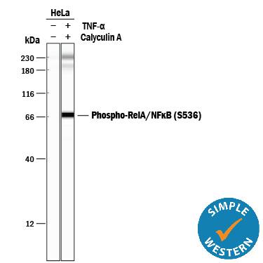 1336) and 20 ng/mL Recombinant Human TNF-a (Catalog #
  1336) and Recombinant Human TNF-a (Catalog #


