DR647MS) and counterstained with DAPI (blue; Lunaphore Catalog #
DR100). Specific staining was localized to the membrane. Protocol available in
COMET™ Panel Builder." class="big_lightbox">
DR647MS) and counterstained with DAPI (blue; Lunaphore Catalog #
DR100). Specific staining was localized to the membrane. Protocol available in
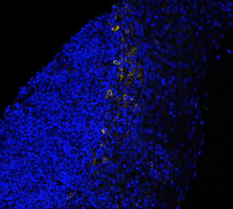
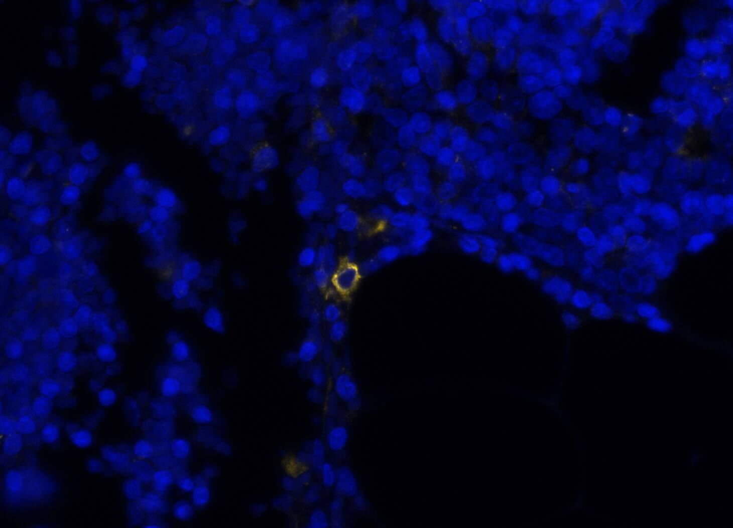
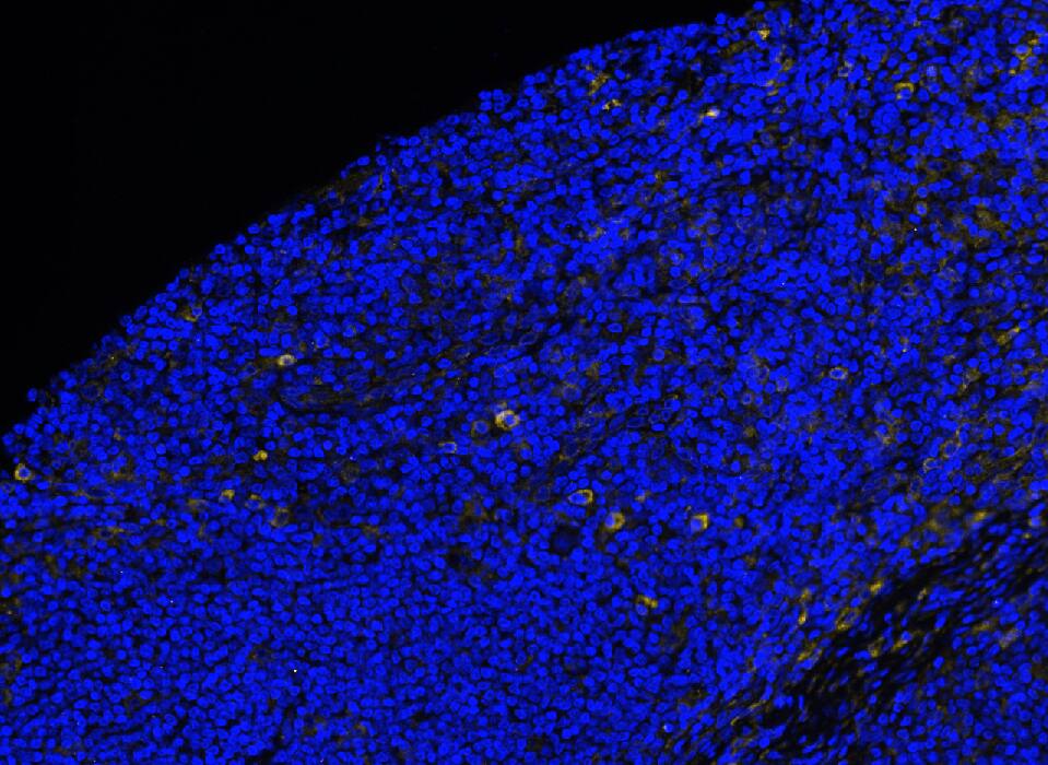
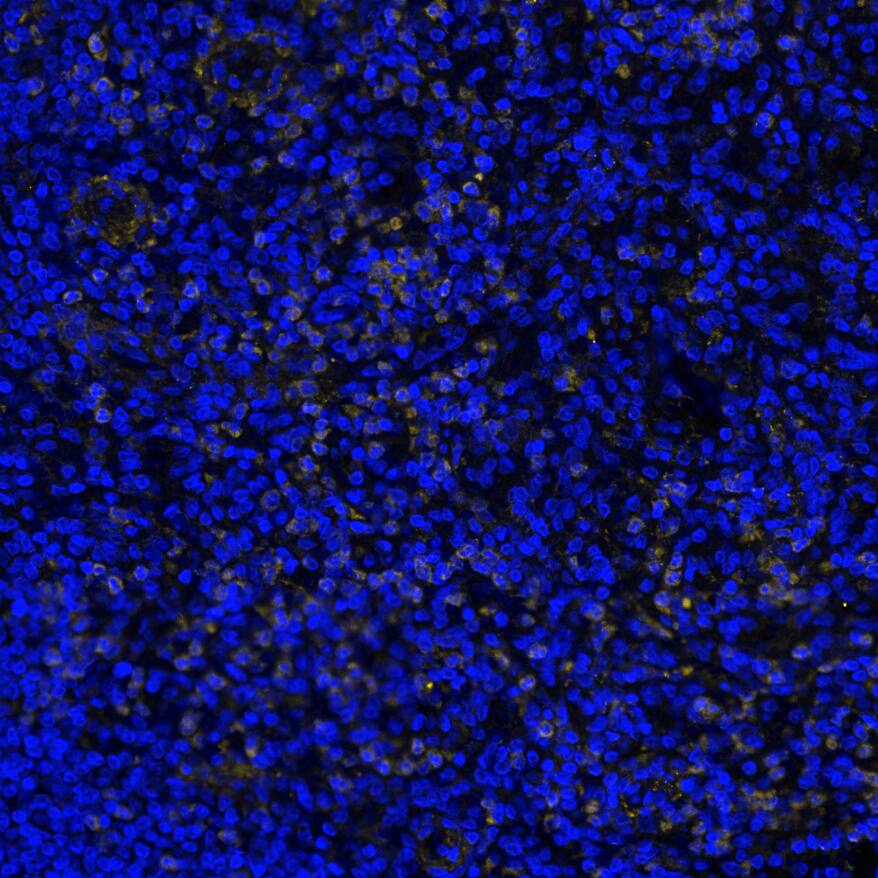
COMET™ Panel Builder." alt="CD33 was detected in immersion fixed paraffin-embedded sections of human tonsil using Mouse Anti-Human CD33, Monoclonal Antibody (Catalog # MAB11371) at 15ug/mL at 37 ° Celsius for 4 minutes. Before incubation with the primary antibody, tissue underwent an all-in-one dewaxing and antigen retrieval preprocessing using PreTreatment Module (PT Module) and Dewax and HIER Buffer H (pH 9; Epredia Catalog # TA-999-DHBH). Tissue was stained using the Alexa Fluor™ 647 Goat anti-Mouse IgG Secondary Antibody at 1:200 at 37 ° Celsius for 2 minutes. (Yellow; Lunaphore Catalog #
DR647MS) and counterstained with DAPI (blue; Lunaphore Catalog #
DR100). Specific staining was localized to the membrane. Protocol available in
COMET™ Panel Builder." class="big_thumb" />
DR647MS) and counterstained with DAPI (blue; Lunaphore Catalog #
DR100). Specific staining was localized to the membrane. Protocol available in
COMET™ Panel Builder." class="big_lightbox">
DR647MS) and counterstained with DAPI (blue; Lunaphore Catalog #
DR100). Specific staining was localized to the membrane. Protocol available in
COMET™ Panel Builder." class="big_thumb" />
DR647MS) and counterstained with DAPI (blue; Lunaphore Catalog #
DR100). Specific staining was localized to the membrane. Protocol available in
COMET™ Panel Builder." class="big_lightbox">
DR647MS) and counterstained with DAPI (blue; Lunaphore Catalog #
DR100). Specific staining was localized to the membrane. Protocol available in
COMET™ Panel Builder." class="big_thumb" />
DR647MS) and counterstained with DAPI (blue; Lunaphore Catalog #
DR100). Specific staining was localized to the membrane. Protocol available in
COMET™ Panel Builder." class="big_lightbox">
DR647MS) and counterstained with DAPI (blue; Lunaphore Catalog #
DR100). Specific staining was localized to the membrane. Protocol available in
COMET™ Panel Builder." class="big_thumb" />
VC001). Before incubation with the primary antibody, tissue was subjected to heat-induced epitope retrieval using Antigen Retrieval Reagent-Basic (Catalog #
CTS013). Tissue was stained using DAB (brown) and counterstained with hematoxylin (blue). Specific staining was localized to lymphocytes. View our protocol for
IHC Staining with VisUCyte HRP Polymer Detection Reagents." class="big_lightbox">
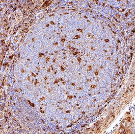
VC001). Before incubation with the primary antibody, tissue was subjected to heat-induced epitope retrieval using Antigen Retrieval Reagent-Basic (Catalog #
CTS013). Tissue was stained using DAB (brown) and counterstained with hematoxylin (blue). Specific staining was localized to lymphocytes. View our protocol for
IHC Staining with VisUCyte HRP Polymer Detection Reagents." class="big_thumb" />
HAF018). A specific band was detected for Siglec-3/CD33 at approximately 55 kDa (as indicated). This experiment was conducted under reducing conditions and using
Immunoblot Buffer Group 1." class="big_lightbox">
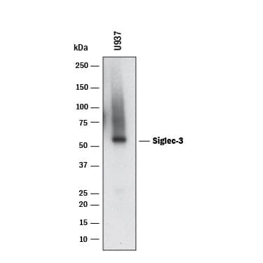
HAF018). A specific band was detected for Siglec-3/CD33 at approximately 55 kDa (as indicated). This experiment was conducted under reducing conditions and using
Immunoblot Buffer Group 1." class="big_thumb" />
 DR647MS) and counterstained with DAPI (blue; Lunaphore Catalog # DR100). Specific staining was localized to the membrane. Protocol available in COMET™ Panel Builder." alt="CD33 was detected in immersion fixed paraffin-embedded sections of human tonsil using Mouse Anti-Human CD33, Monoclonal Antibody (Catalog # MAB11371) at 15ug/mL at 37 ° Celsius for 4 minutes. Before incubation with the primary antibody, tissue underwent an all-in-one dewaxing and antigen retrieval preprocessing using PreTreatment Module (PT Module) and Dewax and HIER Buffer H (pH 9; Epredia Catalog # TA-999-DHBH). Tissue was stained using the Alexa Fluor™ 647 Goat anti-Mouse IgG Secondary Antibody at 1:200 at 37 ° Celsius for 2 minutes. (Yellow; Lunaphore Catalog # DR647MS) and counterstained with DAPI (blue; Lunaphore Catalog # DR100). Specific staining was localized to the membrane. Protocol available in COMET™ Panel Builder." class="big_thumb" />
 MAB002, open histogram) followed by anti-Mouse IgG PE-conjugated secondary antibody (Catalog # F0102B). View our protocol for Staining Membrane-associated Proteins." alt="Human peripheral blood granulocytes were stained with Mouse Anti-Human Siglec-3/CD33 Monoclonal Antibody (Catalog # MAB1137, filled histogram) or isotype control antibody (Catalog # MAB002, open histogram) followed by anti-Mouse IgG PE-conjugated secondary antibody (Catalog # F0102B). View our protocol for Staining Membrane-associated Proteins." class="big_thumb" />
 F0135). View our protocol for Staining Membrane-associated Proteins." alt="U937 Human histiocytic lymphoma cell line was stained with Human Anti-Human Siglec-3/CD33 (Research Grade Gemtuzumab Biosimilar) Monoclonal Antibody (Catalog # MAB9635, filled histogram) or irrelevant antibody (open histogram) followed by APC-conjugated Anti-Human IgG Secondary Antibody (Catalog # F0135). View our protocol for Staining Membrane-associated Proteins." class="big_thumb" />
 IC050A).View our protocol for Staining Membrane-associated Proteins." alt="HEK293 human embryonic kidney cell line transfected with either (A) human Siglec-3/CD33 or (B) irrelevant protein and eGFP was stained with Llama Anti-Human Siglec-3/CD33 Llamabody VHH His-tag Monoclonal Antibody (Catalog # LMAB10752) followed by Allophycocyanin-conjugated Anti-His Antibody (IC050A).View our protocol for Staining Membrane-associated Proteins." class="big_thumb" />
 MAB1137) is immobilized at 0.5 µg/mL, 100 µL/well, Biotinylated Recombinant Human Siglec-3/CD33 His-tag Avi-tag (Catalog # AVI1137) binds with an ED50 of 60-360 pg/mL." alt="When Human Siglec-3/CD33 (6C5/2) Antibody (Catalog # MAB1137) is immobilized at 0.5 µg/mL, 100 µL/well, Biotinylated Recombinant Human Siglec-3/CD33 His-tag Avi-tag (Catalog # AVI1137) binds with an ED50 of 60-360 pg/mL." class="big_thumb" />
 2 μg/lane of Biotinylated Recombinant Human Siglec-3/CD33 His-tag Avi-tag (Catalog # AVI1137) was resolved with SDS-PAGE under reducing (R) and non-reducing (NR) conditions and visualized by Coomassie® Blue staining, showing bands at 45-52 kDa.
 MAB1050) followed by Allophycocyanin-conjugated Anti-Rabbit IgG Secondary Antibody (Catalog # F0111). View our protocol for Staining Membrane-associated Proteins." alt="HEK293 human embryonic kidney cell line transfected with human Siglec-3/CD33 and eGFP was stained with (A) Rabbit Anti-Human Siglec-3/CD33 Monoclonal Antibody (Catalog # MAB11373) or (B) Rabbit IgG control antibody (Catalog # MAB1050) followed by Allophycocyanin-conjugated Anti-Rabbit IgG Secondary Antibody (Catalog # F0111). View our protocol for Staining Membrane-associated Proteins." class="big_thumb" />
 IC050A).View our protocol for Staining Membrane-associated Proteins." alt="HEK293 human embryonic kidney cell line transfected with either (A) human Siglec-3/CD33 or (B) irrelevant protein and eGFP was stained with Llama Anti-Human Siglec-3/CD33 Llamabody VHH His-tag Monoclonal Antibody (Catalog # LMAB10740) followed by Allophycocyanin-conjugated Anti-His Antibody (IC050A).View our protocol for Staining Membrane-associated Proteins." class="big_thumb" />


![Western Blot: Siglec-3/CD33 Antibody (44M12D3) [NBP2-22377] - Analysis of CD33 in A) a partial recombinant protein and B) HCT-116 lysate using CD33 antibody at 2 ug/ml. goat anti-mouse Ig HRP secondary antibody and PicoTect ECL substrate solution were used for this test.](http://images.novusbio.com/fullsize/Siglec-3-CD33-Antibody-44M12D3-Western-Blot-NBP2-22377-img0005.jpg "Western Blot: Siglec-3/CD33 Antibody (44M12D3) [NBP2-22377] - Analysis of CD33 in A) a partial recombinant protein and B) HCT-116 lysate using CD33 antibody at 2 ug/ml. goat anti-mouse Ig HRP secondary antibody and PicoTect ECL substrate solution were used for this test.")
![Immunohistochemistry-Paraffin: Siglec-3/CD33 Antibody (44M12D3) [NBP2-22377] - Human lung tissue stained with CD33 antibody (3 ug/ml), peroxidase-conjugate and DAB chromogen. Note cytoplasmic and membrane staining.](http://images.novusbio.com/fullsize/Siglec-3-CD33-Antibody-44M12D3-Immunohistochemistry-Paraffin-NBP2-22377-img0006.jpg)
 DR647MS) and counterstained with DAPI (blue; Lunaphore Catalog #
  DR647MS) and counterstained with DAPI (blue; Lunaphore Catalog #
  DR647MS) and counterstained with DAPI (blue; Lunaphore Catalog #
  VC001). Before incubation with the primary antibody, tissue was subjected to heat-induced epitope retrieval using Antigen Retrieval Reagent-Basic (Catalog #
  HAF018). A specific band was detected for Siglec-3/CD33 at approximately 55 kDa (as indicated). This experiment was conducted under reducing conditions and using
  NL007) and counterstained with DAPI (blue). Specific staining was localized to cell surfaces. View our protocol for
 ![Western Blot: Siglec-3/CD33 Antibody [NBP2-29619] - Analysis of CD33 in the partial recombinant protein.](http://images.novusbio.com/fullsize/CD33-Antibody-Western-Blot-NBP2-29619-img0001.jpg "Western Blot: Siglec-3/CD33 Antibody [NBP2-29619] - Analysis of CD33 in the partial recombinant protein.")
![Immunohistochemistry-Paraffin: Siglec-3/CD33 Antibody [NBP2-29619] - Analysis of CD33 protein in a section of human small intestinal malignant stromal tumor using 5 ug/mL concentration of CD33 antibody. The cancer cells developed the expected membrane-cytoplasmic staining pattern.](http://images.novusbio.com/fullsize/CD33-Antibody-Immunohistochemistry-Paraffin-NBP2-29619-img0002.jpg)
![Western Blot: Siglec-3/CD33 Antibody (2C6B7) [NBP2-37388] - Analysis using CD33 mAb against HEK293 (1) and CD33 (AA: 15-237)-hIgGFc transfected HEK293 (2) cell lysate.](http://images.novusbio.com/fullsize/Siglec-3-CD33-Antibody-2C6B7-Western-Blot-NBP2-37388-img0003.jpg "Western Blot: Siglec-3/CD33 Antibody (2C6B7) [NBP2-37388] - Analysis using CD33 mAb against HEK293 (1) and CD33 (AA: 15-237)-hIgGFc transfected HEK293 (2) cell lysate.")
![Immunohistochemistry: Siglec-3/CD33 Antibody (2C6B7) [NBP2-37388] - Analysis of paraffin-embedded endometrium tissues using CD33 mouse mAb with DAB staining.](http://images.novusbio.com/fullsize/Siglec-3-CD33-Antibody-2C6B7-Immunohistochemistry-NBP2-37388-img0007.jpg)
![Flow Cytometry: Siglec-3/CD33 Antibody (2C6B7) [NBP2-37388] - Analysis of HepG2 cells using CD33 mouse mAb (green) and negative control (purple).](http://images.novusbio.com/fullsize/Siglec-3-CD33-Antibody-2C6B7-Flow-Cytometry-NBP2-37388-img0004.jpg)
![Western Blot: Siglec-3/CD33 Antibody (2C6B7) [NBP2-37388] - Analysis using CD33 mAb against human CD33 recombinant protein. (Expected MW is 49.2 kDa)](http://images.novusbio.com/fullsize/Siglec-3-CD33-Antibody-2C6B7-Western-Blot-NBP2-37388-img0002.jpg)
![Immunohistochemistry: Siglec-3/CD33 Antibody (2C6B7) [NBP2-37388] - Analysis of paraffin-embedded esophageal cancer tissues using CD33 mouse mAb with DAB staining.](http://images.novusbio.com/fullsize/Siglec-3-CD33-Antibody-2C6B7-Immunohistochemistry-NBP2-37388-img0005.jpg)
![ELISA: Siglec-3/CD33 Antibody (2C6B7) [NBP2-37388] - Red: Control Antigen (100ng); Purple: Antigen (10ng); Green: Antigen (50ng); Blue: Antigen (100ng);](http://images.novusbio.com/fullsize/Siglec-3-CD33-Antibody-2C6B7-ELISA-NBP2-37388-img0001.jpg)

 Fluorescent ICC Staining of Cells on Coverslips." class="big_thumb" />
 MAB1050, open histogram) followed by APC-conjugated Anti-Rabbit IgG Secondary Antibody (Catalog #
  Fluorescent ICC Staining of Cells on Coverslips." class="big_thumb" />


