HAF018). A specific band was detected for STAT3 at approximately 86 kDa (as indicated). This experiment was conducted under reducing conditions and using
Western Blot Buffer Group 1." class="big_lightbox">
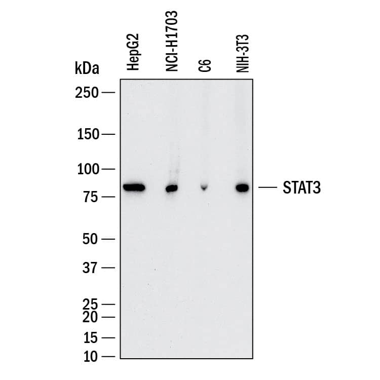
HAF018). A specific band was detected for STAT3 at approximately 86 kDa (as indicated). This experiment was conducted under reducing conditions and using
Western Blot Buffer Group 1." alt="Western Blot shows lysates of HepG2 human hepatocellular carcinoma cell line, NCI‑H1703 human non-small cell lung carcinoma cell line, C6 rat glioma cell line and NIH‑3T3 mouse embryonic fibroblast cell line. PVDF membrane was probed with 1 µg/ml of Mouse Anti-Human/Mouse/Rat STAT3 Monoclonal Antibody (Catalog # MAB1799) followed by HRP-conjugated Anti-Mouse IgG Secondary Antibody (Catalog #
HAF018). A specific band was detected for STAT3 at approximately 86 kDa (as indicated). This experiment was conducted under reducing conditions and using
Western Blot Buffer Group 1." class="big_thumb" />
MAB0041, open histogram), followed by Allophycocyanin-conjugated Anti-Mouse IgG F(ab')
2Secondary Antibody (Catalog #
F0101B). To facilitate intracellular staining, cells were fixed with paraformaldehyde and permeabilized with methanol." class="big_lightbox">

MAB0041, open histogram), followed by Allophycocyanin-conjugated Anti-Mouse IgG F(ab')
2Secondary Antibody (Catalog #
F0101B). To facilitate intracellular staining, cells were fixed with paraformaldehyde and permeabilized with methanol." class="big_thumb" />
MAB0041, open histogram) followed by anti-Mouse IgG PE-conjugated secondary antibody (Catalog #
F0102B). To facilitate intracellular staining, cells were fixed with Flow Cytometry Fixation Buffer (Catalog #
FC004) and permeabilized with Flow Cytometry Permeabilization/Wash Buffer I (Catalog #
FC005). View our protocol for
Staining Intracellular Molecules." class="big_lightbox">
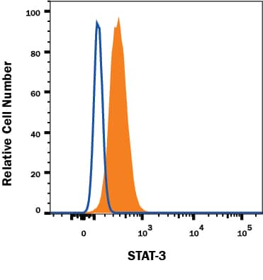
MAB0041, open histogram) followed by anti-Mouse IgG PE-conjugated secondary antibody (Catalog #
F0102B). To facilitate intracellular staining, cells were fixed with Flow Cytometry Fixation Buffer (Catalog #
FC004) and permeabilized with Flow Cytometry Permeabilization/Wash Buffer I (Catalog #
FC005). View our protocol for
Staining Intracellular Molecules." class="big_thumb" />
HAF018). A specific band was detected for STAT3 at approximately 80 kDa (as indicated) in the parental HeLa cell line, but is not detectable in the STAT3 knockout HeLa cell line. GAPDH (Catalog #
AF5718) is shown as a loading control. This experiment was conducted under reducing conditions and using
Immunoblot Buffer Group 1." class="big_lightbox">
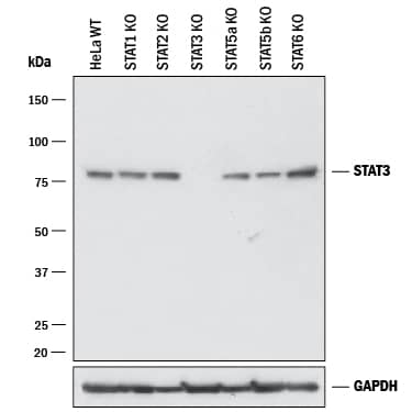
HAF018). A specific band was detected for STAT3 at approximately 80 kDa (as indicated) in the parental HeLa cell line, but is not detectable in the STAT3 knockout HeLa cell line. GAPDH (Catalog #
AF5718) is shown as a loading control. This experiment was conducted under reducing conditions and using
Immunoblot Buffer Group 1." class="big_thumb" />
MAB0041, open histogram) followed by anti-Mouse IgG PE-conjugated secondary antibody (Catalog #
F0102B). No staining in the STAT3 knockout HeLa cell line was observed. To facilitate intracellular staining, cells were fixed with Flow Cytometry Fixation Buffer (Catalog #
FC004) and permeabilized with Flow Cytometry Permeabilization/Wash Buffer I (Catalog #
FC005). View our protocol for
Staining Intracellular Molecules." class="big_lightbox">
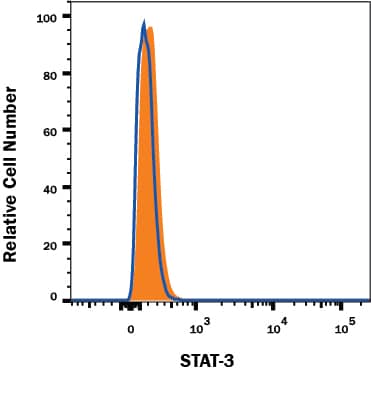
MAB0041, open histogram) followed by anti-Mouse IgG PE-conjugated secondary antibody (Catalog #
F0102B). No staining in the STAT3 knockout HeLa cell line was observed. To facilitate intracellular staining, cells were fixed with Flow Cytometry Fixation Buffer (Catalog #
FC004) and permeabilized with Flow Cytometry Permeabilization/Wash Buffer I (Catalog #
FC005). View our protocol for
Staining Intracellular Molecules." class="big_thumb" />
 HAF018). A specific band was detected for STAT3 at approximately 86 kDa (as indicated). This experiment was conducted under reducing conditions and using Western Blot Buffer Group 1." alt="Western Blot shows lysates of HepG2 human hepatocellular carcinoma cell line, NCI‑H1703 human non-small cell lung carcinoma cell line, C6 rat glioma cell line and NIH‑3T3 mouse embryonic fibroblast cell line. PVDF membrane was probed with 1 µg/ml of Mouse Anti-Human/Mouse/Rat STAT3 Monoclonal Antibody (Catalog # MAB1799) followed by HRP-conjugated Anti-Mouse IgG Secondary Antibody (Catalog # HAF018). A specific band was detected for STAT3 at approximately 86 kDa (as indicated). This experiment was conducted under reducing conditions and using Western Blot Buffer Group 1." class="big_thumb" />
 11100-1) for 20 minutes or 50 µg/mL Recombinant Human IL-22 (Catalog # 782-IL) for 15 minutes. PVDF membrane was probed with 0.5 µg/mL of Rabbit Anti-Human Phospho-STAT3 (Y705) Antigen Affinity-purified Polyclonal Antibody (Catalog # AF4607) followed by HRP-conjugated Anti-Rabbit IgG Secondary Antibody (Catalog # HAF008). A specific band was detected for STAT3 at approximately 95 kDa (as indicated). This experiment was conducted under reducing conditions and using Immunoblot Buffer Group 1." alt="Western blot shows lysates of Daudi human Burkitt's lymphoma cell line and HepG2 human hepatocellular carcinoma cell line untreated (-) or treated (+) with 500 U/mL Recombinant Human IFN-aA (Catalog # 11100-1) for 20 minutes or 50 µg/mL Recombinant Human IL-22 (Catalog # 782-IL) for 15 minutes. PVDF membrane was probed with 0.5 µg/mL of Rabbit Anti-Human Phospho-STAT3 (Y705) Antigen Affinity-purified Polyclonal Antibody (Catalog # AF4607) followed by HRP-conjugated Anti-Rabbit IgG Secondary Antibody (Catalog # HAF008). A specific band was detected for STAT3 at approximately 95 kDa (as indicated). This experiment was conducted under reducing conditions and using Immunoblot Buffer Group 1." class="big_thumb" />
 206-IL) for 15 minutes. PVDF membrane was probed with 1 µg/mL of Mouse Anti-Human Phospho-STAT3 (S727) Monoclonal Antibody (Catalog # MAB4934) followed by HRP-conjugated Anti-Mouse IgG Secondary Antibody (Catalog # HAF018). A specific band was detected for Phospho-STAT3 (S727) at approximately 95 kDa (as indicated). This experiment was conducted under reducing conditions and using Immunoblot Buffer Group 1." alt="Western blot shows lysates of HepG2 human hepatocellular carcinoma cell line untreated (-) or treated (+) with 100 ng/mL Recombinant Human IL-6 (Catalog # 206-IL) for 15 minutes. PVDF membrane was probed with 1 µg/mL of Mouse Anti-Human Phospho-STAT3 (S727) Monoclonal Antibody (Catalog # MAB4934) followed by HRP-conjugated Anti-Mouse IgG Secondary Antibody (Catalog # HAF018). A specific band was detected for Phospho-STAT3 (S727) at approximately 95 kDa (as indicated). This experiment was conducted under reducing conditions and using Immunoblot Buffer Group 1." class="big_thumb" />
 11100-1) for 20 minutes or 50 µg/mL Recombinant Human IL-6 (Catalog # 206-IL) for 15 minutes. PVDF membrane was probed with 2 µg/mL of Rat Anti-Human Phospho-STAT3 (Y705) Monoclonal Antibody (Catalog # MAB4607) followed by HRP-conjugated Anti-Rat IgG Secondary Antibody (Catalog # HAF005). A specific band was detected for Phospho-STAT3 (Y705) at approximately 95 kDa (as indicated). This experiment was conducted under reducing conditions and using Immunoblot Buffer Group 1." alt="Western blot shows lysates of Daudi human Burkitt's lymphoma cell line and HepG2 human hepatocellular carcinoma cell line untreated (-) or treated (+) with 500 U/mL Recombinant Human IFN-aA (Catalog # 11100-1) for 20 minutes or 50 µg/mL Recombinant Human IL-6 (Catalog # 206-IL) for 15 minutes. PVDF membrane was probed with 2 µg/mL of Rat Anti-Human Phospho-STAT3 (Y705) Monoclonal Antibody (Catalog # MAB4607) followed by HRP-conjugated Anti-Rat IgG Secondary Antibody (Catalog # HAF005). A specific band was detected for Phospho-STAT3 (Y705) at approximately 95 kDa (as indicated). This experiment was conducted under reducing conditions and using Immunoblot Buffer Group 1." class="big_thumb" />


 NL007) and counterstained with DAPI (blue). Specific staining was localized to cytoplasm and nuclei. View our protocol for
  MAB0041, open histogram), followed by Allophycocyanin-conjugated Anti-Mouse IgG F(ab')2Secondary Antibody (Catalog #
  MAB0041, open histogram) followed by anti-Mouse IgG PE-conjugated secondary antibody (Catalog #
  NL007) and counterstained with DAPI (blue). Specific staining was localized to cytoplasm and nuclei. View our protocol for
  HAF018). A specific band was detected for STAT3 at approximately 80 kDa (as indicated) in the parental HeLa cell line, but is not detectable in the STAT3 knockout HeLa cell line. GAPDH (Catalog #
  MAB0041, open histogram) followed by anti-Mouse IgG PE-conjugated secondary antibody (Catalog #


![Western Blot: STAT3 Antibody (9D8) [NBP2-22471] - Human breast cancer cell MDA-MB-231 was treated with carboplatin for 72 hours and the expression of p-Stat3 at Y705 and total Stat3 were detected by western blot. From verified customer review.](http://images.novusbio.com/fullsize/STAT3-Antibody-9D8-Western-Blot-NBP2-22471-img0023.jpg "<b>Biological Strategies Validation. </b>Western Blot: STAT3 Antibody (9D8) [NBP2-22471] - Human breast cancer cell MDA-MB-231 was treated with carboplatin for 72 hours and the expression of p-Stat3 at Y705 and total Stat3 were detected by western blot. From verified customer review.")
![<b>Biological Strategies Validation. </b>Immunocytochemistry/Immunofluorescence: STAT3 Antibody (9D8) [NBP2-22471] - Analysis of STAT3 using anti-STAT3 (9D8) monoclonal antibody (shown in green) in HeLa cells. Formalin fixed cells were permeabilized with 0.1% Triton X-100 in TBS for 10 minutes at room temperature. Cells were then blocked with 1% Blocker BSA for 15 minutes at room temperature. Cells were probed with a mouse monoclonal antibody recognizing STAT3, at a dilution of 1:100 for at least 1 hour at room temperature. Cells were then washed with PBS and incubated with DyLight 488 goat-anti-mouse secondary antibody at a dilution of 1:400 for 30 minutes at room temperature. Nuclei (blue) were stained with Hoechst 33342 dye.](http://images.novusbio.com/fullsize/STAT3-Antibody-9D8-Immunocytochemistry-Immunofluorescence-NBP2-22471-img0016.jpg)
![Immunohistochemistry-Paraffin: STAT3 Antibody (9D8) [NBP2-22471] - Biopsies of normal and cancer tissues.](http://images.novusbio.com/fullsize/STAT3-Antibody-9D8-Immunohistochemistry-Paraffin-NBP2-22471-img0017.jpg)
![<b>Biological Strategies Validation. </b>Chromatin Immunoprecipitation: STAT3 Antibody (9D8) [NBP2-22471] - Analysis performed using cross-linked chromatin from rat hepatoma cells treated with insulin. IP performed using a multiplex microplate Matrix ChIP assay with STAT3 monoclonal antibody. Chromatin aliquots from cells were used per ChIP pull-down. Quantitative PCR data done in quadruplicate using 1ul of DNA in 2ul SYBR real-time PCR reactions containing primers to amplify exon-1 or exon-28 of LAMC1.Quantitation of immunoprecipitated chromatin is presented as signal relative to the total amount of input chromatin. Results represent the mean +/- SEM for three experiments. A schematic representation of the rat LAMC1 locus is shown; boxes represent exons (black boxes = translated regions, white boxes = untranslated regions), the zigzag line represents an intron, and the straight line represents upstream sequence. Regions amplified by LAMC1 primers are represented by black bars. Data courtesy of the Innovators Program.](http://images.novusbio.com/fullsize/STAT3-Antibody-9D8-Chromatin-Immunoprecipitation-ChIP-NBP2-22471-img0020.jpg)
![Western Blot: STAT3 Antibody (9D8) [NBP2-22471] - Analysis of 25ug HepG2 total lysate.](http://images.novusbio.com/fullsize/STAT3-Antibody-9D8-Western-Blot-NBP2-22471-img0010.jpg)
![Western Blot: STAT3 Antibody (9D8) [NBP2-22471] - Analysis of 25ug of various whole cell lysates.](http://images.novusbio.com/fullsize/STAT3-Antibody-9D8-Western-Blot-NBP2-22471-img0019.jpg)
![Western Blot: STAT3 Antibody (9D8) [NBP2-22471] - Analysis of 25ug of U2-OS lysate from STAT3 SMART pool siRNA transfected or non-targeting control transfected U2-OS cells onto a 4-20% Tris-HCl polyacrylamide gel.](http://images.novusbio.com/fullsize/STAT3-Antibody-9D8-Western-Blot-NBP2-22471-img0022.jpg)
![Immunoprecipitation: STAT3 Antibody (9D8) [NBP2-22471] - Analysis of STAT3 was performed on HepG2 cells. The antigen: antibody complex was formed by incubating 750ug whole cell lysate with 2ug of mouse monoclonal antibody recognizing STAT3 overnight on a rocking platform at 4C. The immune-complex was then captured on 50ul Protein A/G Plus Agarose. Captured immune-complexes were then washed extensively and proteins eluted with 5X Reducing Sample Loading Dye. Samples were then resolved on a 4-20% Tris-HCl polyacrylamide gel. Proteins were transferred to PVDF membrane and blocked with 5% Milk/TBS-0.1%Tween for at least 1 hour. Membranes were then probed with a mouse monoclonal antibody recognizing STAT3 at a dilution of 1:5000 overnight rotating at 4C. Membranes were washed in TBST and probed with Clean-blot IP detection reagent at a dilution of 1:2000 for at least one hour. Membranes were washed and chemiluminescent detection was performed using Super Signal West Dura.](http://images.novusbio.com/fullsize/STAT3-Antibody-9D8-Immunoprecipitation-NBP2-22471-img0021.jpg)

 AB-105-C, open histogram), followed by Phycoerythrin-conjugated Anti-Rabbit IgG Secondary Antibody (Catalog #
  NL004) and counterstained with DAPI (blue). View our protocol for


![Immunocytochemistry/Immunofluorescence: STAT3 Antibody [NB100-799] - Immunofluorescence analysis of paraformaldehyde fixed HeLa cells, permeabilized with 0.15% Triton. Primary incubation 1hr (10 ug/mL) followed by Alexa Fluor 488 secondary antibody (2 ug/mL), showing cytoplasmic and weak nuclear staining. Actin filaments were stained with phalloidin (red) and the nuclear stain is DAPI (blue). Negative control: Unimmunized goat IgG (10 ug/mL) followed by Alexa Fluor 488 secondary antibody (2 ug/mL).](http://images.novusbio.com/fullsize/STAT3-Antibody-Immunocytochemistry-Immunofluorescence-NB100-799-img0005.jpg "Immunocytochemistry/Immunofluorescence: STAT3 Antibody [NB100-799] - Immunofluorescence analysis of paraformaldehyde fixed HeLa cells, permeabilized with 0.15% Triton. Primary incubation 1hr (10 ug/mL) followed by Alexa Fluor 488 secondary antibody (2 ug/mL), showing cytoplasmic and weak nuclear staining. Actin filaments were stained with phalloidin (red) and the nuclear stain is DAPI (blue). Negative control: Unimmunized goat IgG (10 ug/mL) followed by Alexa Fluor 488 secondary antibody (2 ug/mL).")
![Immunohistochemistry-Paraffin: STAT3 Antibody [NB100-799] - Immunofluorescence analysis of paraformaldehyde fixed A431 cells, permeabilized with 0.15% Triton. Primary incubation 1hr (10 ug/mL) followed by Alexa Fluor 488 secondary antibody (2 ug/mL), showing nuclear and cytoplasmic staining. Actin filaments were stained with phalloidin (red) and the nuclear stain is DAPI (blue). Negative control: Unimmunized goat IgG (10 ug/mL) followed by Alexa Fluor 488 secondary antibody (2 ug/mL).](http://images.novusbio.com/fullsize/STAT3-Antibody-Immunohistochemistry-Paraffin-NB100-799-img0006.jpg)
![Flow Cytometry: STAT3 Antibody [NB100-799] - Flow cytometric analysis of paraformaldehyde fixed A431 cells (blue line), permeabilized with 0.5% Triton. Primary incubation 1hr (10 ug/mL) followed by Alexa Fluor 488 secondary antibody (1 ug/mL). IgG control: Unimmunized goat IgG (black line) followed by Alexa Fluor 488 secondary antibody.](http://images.novusbio.com/fullsize/STAT3-Antibody-Flow-Cytometry-NB100-799-img0007.jpg)
![Immunohistochemistry-Paraffin: STAT3 Antibody [NB100-799] - Staining of Mouse Thymus. Antibody at 4 ug/mL. Microwaved antigen retrieval with citrate buffer pH 6, HRP-staining.](http://images.novusbio.com/fullsize/STAT3-Antibody-Immunohistochemistry-Paraffin-NB100-799-img0003.jpg)
![Immunohistochemistry-Paraffin: STAT3 Antibody [NB100-799] - Negative Control showing staining of Mouse Thymus, with no primary antibody.](http://images.novusbio.com/fullsize/STAT3-Antibody-Immunohistochemistry-Paraffin-NB100-799-img0004.jpg)
![Western Blot: STAT3 [p Tyr705] Antibody [NBP2-24463] - Human breast cancer MCF7 cells were treated with control (Con) or Adenosine (Ado) and the expression of phospho-Stat3 was detected. Image from verified customer review.](http://images.novusbio.com/fullsize/STAT3-[p-Tyr705]-Antibody-Western-Blot-NBP2-24463-img0003.jpg "<b>Biological Strategies Validation. </b>Western Blot: STAT3 [p Tyr705] Antibody [NBP2-24463] - Human breast cancer MCF7 cells were treated with control (Con) or Adenosine (Ado) and the expression of phospho-Stat3 was detected. Image from verified customer review.")
![<b>Biological Strategies Validation. </b>Western Blot: STAT3 [p Tyr705] Antibody [NBP2-24463] - Analysis of STAT3 (Tyr705) in recombinant fusion protein containing 1) a phosphorylated tyrosine at position 705 and 2) an unphosphorylated tyrosine at position 705, using STAT3 phospho antibody at 0.1 ug/mL. Goat anti-rabbit Ig HRP secondary antibody and PicoTect ECL substrate solution were used for this test.](http://images.novusbio.com/fullsize/STAT3-[p-Tyr705]-Antibody-Western-Blot-NBP2-24463-img0001.jpg)
![<b>Biological Strategies Validation. </b>Western Blot: STAT3 [p Tyr705] Antibody [NBP2-24463] - Human breast cancer cells MDA-MB-231 were treated with carboplatin for 72 hours and the expression of p-Stat3 at Y705 and total Stat3 were detected. Image from verified customer review.](http://images.novusbio.com/fullsize/STAT3-[p-Tyr705]-Antibody-Western-Blot-NBP2-24463-img0002.jpg)
 F0101B). To facilitate intracellular staining, cells were fixed with paraformaldehyde and permeabilized with methanol. View our protocol for


![Western Blot: STAT3 [p Tyr705] Antibody [NB100-82213] - Analysis of extracts from Hela cells untreated or treated with IFN a using STAT3](http://images.novusbio.com/fullsize/STAT3-[p-Tyr705]-Antibody-Western-Blot-NB100-82213-img0001.jpg "<b>Biological Strategies Validation. </b>Western Blot: STAT3 [p Tyr705] Antibody [NB100-82213] - Analysis of extracts from Hela cells untreated or treated with IFN a using STAT3")
![<b>Biological Strategies Validation. </b>Simple Western: STAT3 [p Tyr705] Antibody [NB100-82213] - STAT3-P assay on mouse megakaryocyte lysates. Simple Western image submitted by a verified customer review.](http://images.novusbio.com/fullsize/STAT3-[p-Tyr705]-Antibody-Simple-Western-NB100-82213-img0008.jpg)
![Immunohistochemistry-Paraffin: STAT3 [p Tyr705] Antibody [NB100-82213] - Immunohistochemical analysis of paraffin-embedded Human-lung-cancer tissue.](http://images.novusbio.com/fullsize/nb100-82213_rabbit-polyclonal-stat3-p-tyr705-antibody-23420241446223.jpg)
![Immunohistochemistry-Paraffin: STAT3 [p Tyr705] Antibody [NB100-82213] - Immunohistochemical analysis of paraffin-embedded Rat-lung tissue.](http://images.novusbio.com/fullsize/nb100-82213_rabbit-polyclonal-stat3-p-tyr705-antibody-23420241446229.jpg)
![Immunohistochemistry: STAT3 [p Tyr705] Antibody [NB100-82213] - Immunofluorescence analysis of mouse-spleen tissue.](http://images.novusbio.com/fullsize/nb100-82213_rabbit-polyclonal-stat3-p-tyr705-antibody-103202523464550.png)
![Western Blot: STAT3 [p Ser727] Antibody (SY24-09) [NBP2-67433] - Analysis of STAT3 [p Ser727] on NIH-3T3 cell lysates. Lane 1: NIH-3T3 cells, whole cell lysates, 10 ug/lane. Lane 2: NIH-3T3 cells were starved 6h, whole cell lysates, 10 ug/lane. Lane 3/4: NIH-3T3 cells were starved 6h, then treated with 200 ng/ml EGF for 10 min, whole cell lysates, 10 ug/lane. Lane 5: NIH-3T3 cells were starved 6h, then treated with 200 ng/ml EGF for 10 min, and then treated with 2.8ug/ul lambda-PP for 30 minutes, whole cell lysates, 10 ug/lane. The primary antibody Anti-STAT3 [p Ser727] (1/500) , Anti-STAT3 antibody ( 1/500) and Anti-GAPDH antibody (1/10,000)was used in 5% BSA at room temperature for 2 hours. Goat Anti-Rabbit IgG H&L (HRP) Secondary Antibody at 1:200,000 dilution was used for 1 hour at room temperature. Predicted band size: 88 kDa Observed band size: 88 kDa](http://images.novusbio.com/fullsize/STAT3-p-Ser727-Antibody-SY24-09-Western-Blot-NBP2-67433-img0010.jpg "<b>Biological Strategies Validation. </b>Western Blot: STAT3 [p Ser727] Antibody (SY24-09) [NBP2-67433] - Analysis of STAT3 [p Ser727] on NIH-3T3 cell lysates. Lane 1: NIH-3T3 cells, whole cell lysates, 10 ug/lane. Lane 2: NIH-3T3 cells were starved 6h, whole cell lysates, 10 ug/lane. Lane 3/4: NIH-3T3 cells were starved 6h, then treated with 200 ng/ml EGF for 10 min, whole cell lysates, 10 ug/lane. Lane 5: NIH-3T3 cells were starved 6h, then treated with 200 ng/ml EGF for 10 min, and then treated with 2.8ug/ul lambda-PP for 30 minutes, whole cell lysates, 10 ug/lane. The primary antibody Anti-STAT3 [p Ser727] (1/500) , Anti-STAT3 antibody ( 1/500) and Anti-GAPDH antibody (1/10,000)was used in 5% BSA at room temperature for 2 hours. Goat Anti-Rabbit IgG H&L (HRP) Secondary Antibody at 1:200,000 dilution was used for 1 hour at room temperature. Predicted band size: 88 kDa Observed band size: 88 kDa")
![<b>Biological Strategies Validation. </b>Immunocytochemistry/Immunofluorescence: STAT3 [p Ser727] Antibody (SY24-09) [NBP2-67433] - Staining Phospho-STAT3(S727) in NIH/3T3 cells (green). The nuclear counter stain is DAPI (blue). Cells were fixed in paraformaldehyde, permeabilised with 0.25% Triton X100/PBS.](http://images.novusbio.com/fullsize/STAT3-p-Ser727-Antibody-SY24-09-Immunocytochemistry-Immunofluorescence-NBP2-67433-img0003.jpg)
![Immunohistochemistry-Paraffin: STAT3 [p Ser727] Antibody (SY24-09) [NBP2-67433] - Analysis of paraffin-embedded human kidney tissue with Rabbit anti-STAT3 [p Ser727] antibody washed with ddH2O and PBS, and then probed with the primary antibody at 1:500 dilution for 1 hour at room temperature. The detection was performed using an HRP conjugated compact polymer system. DAB was used as the chromogen. Tissues were counterstained with hematoxylin and mounted with DPX.](http://images.novusbio.com/fullsize/STAT3-p-Ser727-Antibody-SY24-09-Immunohistochemistry-Paraffin-NBP2-67433-img0014.jpg)
![Western Blot: STAT3 [p Ser727] Antibody (SY24-09) [NBP2-67433] - Analysis of Phospho-STAT3(S727) on different lysates. Proteins were transferred to a PVDF membrane and blocked with 5% BSA in PBS for 1 hour at room temperature. The primary antibody (1/500) was used in 5% BSA at room temperature for 2 hours. Goat Anti-Rabbit IgG - HRP Secondary Antibody at 1:5,000 dilution was used for 1 hour at room temperature. Positive control: Lane 1: Hela cell lysate Lane 2: A431 cell lysate Lane 1: NIH/3T3 cell lysate Lane 2: Jurkat cell lysate](http://images.novusbio.com/fullsize/STAT3-p-Ser727-Antibody-SY24-09-Western-Blot-NBP2-67433-img0008.jpg)
![<b>Biological Strategies Validation. </b>Western Blot: STAT3 [p Ser727] Antibody (SY24-09) [NBP2-67433] - Human U251 glioma cell line, whole cell lysates.Cells were treated with SP600125, DB07268, JNK-IN-8 and SB203580. WB image submitted by a verified customer review.](http://images.novusbio.com/fullsize/STAT3-p-Ser727-Antibody-SY24-09-Western-Blot-NBP2-67433-img0009.jpg)
![Immunocytochemistry/Immunofluorescence: STAT3 [p Ser727] Antibody (SY24-09) [NBP2-67433] - Staining Phospho-STAT3(S727) in A431 cells (green). The nuclear counter stain is DAPI (blue). Cells were fixed in paraformaldehyde, permeabilised with 0.25% Triton X100/PBS.](http://images.novusbio.com/fullsize/STAT3-p-Ser727-Antibody-SY24-09-Immunocytochemistry-Immunofluorescence-NBP2-67433-img0001.jpg)
![Immunocytochemistry/Immunofluorescence: STAT3 [p Ser727] Antibody (SY24-09) [NBP2-67433] - Staining Phospho-STAT3(S727) in Hela cells (green). The nuclear counter stain is DAPI (blue). Cells were fixed in paraformaldehyde, permeabilised with 0.25% Triton X100/PBS.](http://images.novusbio.com/fullsize/STAT3-p-Ser727-Antibody-SY24-09-Immunocytochemistry-Immunofluorescence-NBP2-67433-img0002.jpg)
![Immunohistochemistry-Paraffin: STAT3 [p Ser727] Antibody (SY24-09) [NBP2-67433] - Analysis of paraffin-embedded human lung tissue using anti-Phospho-STAT3(S727) antibody. Counter stained with hematoxylin.](http://images.novusbio.com/fullsize/STAT3-p-Ser727-Antibody-SY24-09-Immunohistochemistry-Paraffin-NBP2-67433-img0004.jpg)
![Immunohistochemistry-Paraffin: STAT3 [p Ser727] Antibody (SY24-09) [NBP2-67433] - Analysis of paraffin-embedded rat brain tissue using anti-Phospho-STAT3(S727) antibody. Counter stained with hematoxylin.](http://images.novusbio.com/fullsize/STAT3-p-Ser727-Antibody-SY24-09-Immunohistochemistry-Paraffin-NBP2-67433-img0005.jpg)
![Immunohistochemistry-Paraffin: STAT3 [p Ser727] Antibody (SY24-09) [NBP2-67433] - Analysis of paraffin-embedded rat kidney tissue using anti-Phospho-STAT3(S727) antibody. Counter stained with hematoxylin.](http://images.novusbio.com/fullsize/STAT3-p-Ser727-Antibody-SY24-09-Immunohistochemistry-Paraffin-NBP2-67433-img0006.jpg)
![Immunohistochemistry-Paraffin: STAT3 [p Ser727] Antibody (SY24-09) [NBP2-67433] - Analysis of paraffin-embedded rat liver tissue using anti-Phospho-STAT3(S727) antibody. Counter stained with hematoxylin.](http://images.novusbio.com/fullsize/STAT3-p-Ser727-Antibody-SY24-09-Immunohistochemistry-Paraffin-NBP2-67433-img0007.jpg)
![<b>Biological Strategies Validation. </b>Immunohistochemistry-Paraffin: STAT3 [p Ser727] Antibody (SY24-09) [NBP2-67433] - Analysis of paraffin-embedded rat kidney tissue using anti-Phospho-STAT3 (S727) antibody. The section was pre-treated using heat mediated antigen retrieval with Tris-EDTA buffer (pH 8.0-8.4) for 20 minutes.The tissues were blocked in 5% BSA for 30 minutes at room temperature, washed with ddH2O and PBS, and then probed with the primary antibody (1/50) for 30 minutes at room temperature. The detection was performed using an HRP conjugated compact polymer system. DAB was used as the chromogen. Tissues were counterstained with hematoxylin and mounted with DPX.](http://images.novusbio.com/fullsize/STAT3-p-Ser727-Antibody-SY24-09-Immunohistochemistry-Paraffin-NBP2-67433-img0011.jpg)
![Immunohistochemistry-Paraffin: STAT3 [p Ser727] Antibody (SY24-09) [NBP2-67433] - Analysis of paraffin-embedded rat hippocampus tissue with Rabbit anti-STAT3 [p Ser727] antibody washed with ddH2O and PBS, and then probed with the primary antibody at 1:500 dilution for 1 hour at room temperature. The detection was performed using an HRP conjugated compact polymer system. DAB was used as the chromogen. Tissues were counterstained with hematoxylin and mounted with DPX.](http://images.novusbio.com/fullsize/STAT3-p-Ser727-Antibody-SY24-09-Immunohistochemistry-Paraffin-NBP2-67433-img0012.jpg)
![Immunohistochemistry-Paraffin: STAT3 [p Ser727] Antibody (SY24-09) [NBP2-67433] - Analysis of paraffin-embedded rat brain tissue with Rabbit anti-STAT3 [p Ser727] antibody washed with ddH2O and PBS, and then probed with the primary antibody at 1:500 dilution for 1 hour at room temperature. The detection was performed using an HRP conjugated compact polymer system. DAB was used as the chromogen. Tissues were counterstained with hematoxylin and mounted with DPX.](http://images.novusbio.com/fullsize/STAT3-p-Ser727-Antibody-SY24-09-Immunohistochemistry-Paraffin-NBP2-67433-img0013.jpg)
![Western blot analysis of Hela cell lysate with NBP2-67433 at 1/1,000 dilution. Lysates/proteins at 15 µg/Lane. Exposure time: 30 seconds Blocking: 5% NFDM/TBST, 1 hour at room temperature Primary antibody: NBP2-67433, 1/1,000 in 5% NFDM/TBST, overnight at 4 ℃ Secondary antibody: Goat Anti-Rabbit IgG [HRP] Secondary Antibody, 1/50,000 in 5% NFDM/TBST, 1 hour at room temperature Predicted band size: 88.1 kDa Observed band size: 88 kDa](http://images.novusbio.com/fullsize/nbp2-67433_-western-blot-639204586668891819.jpg)
![Western blot analysis of Hela cell lysate with NBP2-67433 at 1/2,000 dilution. Lane 1: NIH/3T3 cell lysate Lane 2: C6 cell lysate Lysates/proteins at 20 µg/Lane. Exposure time: 30 seconds Blocking: 5% NFDM/TBST, 1 hour at room temperature Primary antibody: NBP2-67433, 1/2,000 in 5% NFDM/TBST, overnight at 4 ℃ Secondary antibody: Goat Anti-Rabbit IgG [HRP] Secondary Antibody, 1/50,000 in 5% NFDM/TBST, 1 hour at room temperature Predicted band size: 88.1 kDa Observed band size: 88 kDa](http://images.novusbio.com/fullsize/nbp2-67433_-western-blot-639204597490422236.jpg)
![Western blot analysis on different lysates with NBP2-67433 at 1/1,000 dilution. Lane 1: PC-12 cell lysate (20 µg/Lane) Lane 2: C6 cell lysate (20 µg/Lane) Lane 3: C6 treated with 200ng/mL TPA for 35 minutes cell lysate (20 µg/Lane) Lane 4: Rat cerebellum tissue lysate (40 µg/Lane) Predicted band size: 88 kDa Observed band size: 88 kDa Exposure time: 1 minute 50 seconds; 4-20% SDS-PAGE gel. Proteins were transferred to a PVDF membrane and blocked with 5% NFDM/TBST for 1 hour at room temperature. NBP2-67433 at 1/1,000 dilution was used in 5% NFDM/TBST at 4℃ overnight. Goat Anti-Rabbit IgG [HRP] Secondary Antibody at 1/50,000 dilution was used for 1 hour at room temperature.](http://images.novusbio.com/fullsize/nbp2-67433_-western-blot-639204598086887785.jpg)
![Western Blot: STAT3 Antibody (SY24-08) [NBP2-67432] - Western blot analysis of STAT3 on different lysates. Proteins were transferred to a PVDF membrane and blocked with 5% BSA in PBS for 1 hour at room temperature. The primary antibody (1/500) was used in 5% BSA at room temperature for 2 hours. Goat Anti-Rab](http://images.novusbio.com/fullsize/STAT3-Antibody-SY24-08-Western-Blot-NBP2-67432-img0009.jpg "Western Blot: STAT3 Antibody (SY24-08) [NBP2-67432] - Western blot analysis of STAT3 on different lysates. Proteins were transferred to a PVDF membrane and blocked with 5% BSA in PBS for 1 hour at room temperature. The primary antibody (1/500) was used in 5% BSA at room temperature for 2 hours. Goat Anti-Rab")
![Immunocytochemistry/Immunofluorescence: STAT3 Antibody (SY24-08) [NBP2-67432] - Staining Stat3 in CRC cells (green). The nuclear counter stain is DAPI (blue). Cells were fixed in paraformaldehyde, permeabilised with 0.25% Triton X100/PBS.](http://images.novusbio.com/fullsize/STAT3-Antibody-SY24-08-Immunocytochemistry-Immunofluorescence-NBP2-67432-img0006.jpg)
![<b>Biological Strategies Validation. </b>Immunohistochemistry-Paraffin: STAT3 Antibody (SY24-08) [NBP2-67432] - Immunohistochemical analysis of paraffin-embedded rat pancreas tissue using anti-STAT3 antibody. The section was pre-treated using heat mediated antigen retrieval with Tris-EDTA buffer (pH 8.0-8.4) for 20 minutes. The tissues were blocked in 5% BSA for 30 minutes at room temperature, washed with ddH2O and PBS, and then probed with the primary antibody (ET1607-38, 1/50) for 30 minutes at room temperature. The detection was performed using an HRP conjugated compact polymer system. DAB was used as the chromogen. Tissues were counterstained with hematoxylin and mounted with DPX.](http://images.novusbio.com/fullsize/STAT3-Antibody-SY24-08-Immunohistochemistry-Paraffin-NBP2-67432-img0008.jpg)
![Western Blot: STAT3 Antibody (SY24-08) [NBP2-67432] - Analysis of Stat3 on different lysates using anti-Stat3 antibody at 1/1,000 dilution. Positive control: Lane 1: Hela Lane 2: SHG-44 Lane 3: SW480](http://images.novusbio.com/fullsize/STAT3-Antibody-SY24-08-Western-Blot-NBP2-67432-img0005.jpg)
![Immunohistochemistry-Paraffin: STAT3 Antibody (SY24-08) [NBP2-67432] - Analysis of paraffin-embedded human colon cancer tissue using anti-Stat3 antibody. Counter stained with hematoxylin.](http://images.novusbio.com/fullsize/STAT3-Antibody-SY24-08-Immunohistochemistry-Paraffin-NBP2-67432-img0001.jpg)
![Immunohistochemistry-Paraffin: STAT3 Antibody (SY24-08) [NBP2-67432] - Analysis of paraffin-embedded human pancreas tissue using anti-Stat3 antibody. Counter stained with hematoxylin.](http://images.novusbio.com/fullsize/STAT3-Antibody-SY24-08-Immunohistochemistry-Paraffin-NBP2-67432-img0002.jpg)
![Immunohistochemistry-Paraffin: STAT3 Antibody (SY24-08) [NBP2-67432] - Analysis of paraffin-embedded mouse brain tissue using anti-Stat3 antibody. Counter stained with hematoxylin.](http://images.novusbio.com/fullsize/STAT3-Antibody-SY24-08-Immunohistochemistry-Paraffin-NBP2-67432-img0003.jpg)
![Immunohistochemistry-Paraffin: STAT3 Antibody (SY24-08) [NBP2-67432] - Analysis of paraffin-embedded mouse pancreas tissue using anti-Stat3 antibody. Counter stained with hematoxylin.](http://images.novusbio.com/fullsize/STAT3-Antibody-SY24-08-Immunohistochemistry-Paraffin-NBP2-67432-img0004.jpg)
![<b>Biological Strategies Validation. </b>Immunohistochemistry-Paraffin: STAT3 Antibody (SY24-08) [NBP2-67432] - Immunohistochemical analysis of paraffin-embedded rat brain tissue using anti-STAT3 antibody. The section was pre-treated using heat mediated antigen retrieval with Tris-EDTA buffer (pH 8.0-8.4) for 20 minutes. The tissues were blocked in 5% BSA for 30 minutes at room temperature, washed with ddH2O and PBS, and then probed with the primary antibody (ET1607-38, 1/200) for 30 minutes at room temperature. The detection was performed using an HRP conjugated compact polymer system. DAB was used as the chromogen. Tissues were counterstained with hematoxylin and mounted with DPX.](http://images.novusbio.com/fullsize/STAT3-Antibody-SY24-08-Immunohistochemistry-Paraffin-NBP2-67432-img0007.jpg)
![Western blot analysis on different lysates with NBP2-67432 at 1/2,000 dilution. Lane 1: HeLa cell lysate Lane 2: A431 cell lysate Lane 3: PANC-1 cell lysate Lane 4: NIH/3T3 cell lysate Lane 5: RAW264.7 cell lysate Lane 6: PC-12 cell lysate Lane 7: C6 cell lysate Lysates/proteins at 20 µg/Lane. Predicted band size: 88 kDa Observed band size: 88 kDa Exposure time: 2 minutes 24 seconds; 4-20% SDS-PAGE gel. Proteins were transferred to a PVDF membrane and blocked with 5% NFDM/TBST for 1 hour at room temperature. NBP2-67432 at 1/2,000 dilution was used in 5% NFDM/TBST at 4℃ overnight. Goat Anti-Rabbit IgG [HRP] Secondary Antibody at 1/50,000 dilution was used for 1 hour at room temperature.](http://images.novusbio.com/fullsize/nbp2-67432_-western-blot-639204597791042768.jpg)

![Western Blot: STAT3 Antibody (CL0492) [NBP2-52936] - Analysis in RT-4 cells transfected with control siRNA, target specific siRNA probe #1 and #2, using Anti-STAT3 antibody. Remaining relative intensity is presented. Loading control: Anti-PPIB.](http://images.novusbio.com/fullsize/STAT3-Antibody-CL0492-Western-Blot-NBP2-52936-img0006.jpg "<b>Genetic Strategies Validation. </b>Western Blot: STAT3 Antibody (CL0492) [NBP2-52936] - Analysis in RT-4 cells transfected with control siRNA, target specific siRNA probe #1 and #2, using Anti-STAT3 antibody. Remaining relative intensity is presented. Loading control: Anti-PPIB.")
![<b>Genetic Strategies Validation and Orthogonal Strategies Validation.</b>Immunohistochemistry-Paraffin: STAT3 Antibody (CL0492) [NBP2-52936] - Staining in human endometrium and pancreas tissues. Corresponding STAT3 RNA-seq data are presented for the same tissues.](http://images.novusbio.com/fullsize/STAT3-Antibody-CL0492-Immunohistochemistry-Paraffin-NBP2-52936-img0014.jpg)
![Western Blot: STAT3 Antibody (CL0492) [NBP2-52936] - Lane 1: Marker [kDa], Lane 2: Human cell line RT-4](http://images.novusbio.com/fullsize/STAT3-Antibody-CL0492-Western-Blot-NBP2-52936-img0005.jpg)
![Immunohistochemistry-Paraffin: STAT3 Antibody (CL0492) [NBP2-52936] - Staining of human endometrium shows strong nuclear and moderate cytoplasmic positivity in glandular cells.](http://images.novusbio.com/fullsize/STAT3-Antibody-CL0492-Immunohistochemistry-Paraffin-NBP2-52936-img0008.jpg)
![Immunohistochemistry-Paraffin: STAT3 Antibody (CL0492) [NBP2-52936] - Staining of human kidney shows strong nuclear and moderate cytoplasmic positivity in cells in tubules.](http://images.novusbio.com/fullsize/STAT3-Antibody-CL0492-Immunohistochemistry-Paraffin-NBP2-52936-img0009.jpg)
![Immunohistochemistry-Paraffin: STAT3 Antibody (CL0492) [NBP2-52936] - Staining of human rectum shows moderate to strong nuclear and cytoplasmic positivity in glandular cells.](http://images.novusbio.com/fullsize/STAT3-Antibody-CL0492-Immunohistochemistry-Paraffin-NBP2-52936-img0010.jpg)
![Immunohistochemistry-Paraffin: STAT3 Antibody (CL0492) [NBP2-52936] - Staining of human pancreas shows no positivity in exocrine glandular cells as expected.](http://images.novusbio.com/fullsize/STAT3-Antibody-CL0492-Immunohistochemistry-Paraffin-NBP2-52936-img0011.jpg)

![Western Blot: STAT3 Antibody (OTI21E7) [NBP1-48014] - Equivalent amounts of cell lysates (10 ug per lane) of wild-type Hela cells and STAT3-Knockout Hela cells were separated by SDS-PAGE and immunoblotted with anti-STAT3 monoclonal antibody. Then the blotted membrane was stripped and reprobed with anti-b-actin antibody as a loading control.(1:500)](http://images.novusbio.com/fullsize/STAT3-Antibody-OTI21E7-Western-Blot-NBP1-48014-img0013.jpg "<b>Genetic Strategies Validation. </b>Western Blot: STAT3 Antibody (OTI21E7) [NBP1-48014] - Equivalent amounts of cell lysates (10 ug per lane) of wild-type Hela cells and STAT3-Knockout Hela cells were separated by SDS-PAGE and immunoblotted with anti-STAT3 monoclonal antibody. Then the blotted membrane was stripped and reprobed with anti-b-actin antibody as a loading control.(1:500)")
![<b>Genetic Strategies Validation. </b>Western Blot: STAT3 Antibody (OTI21E7) [NBP1-48014] - Analysis of extracts (35ug) from different cell line by using anti-STAT3 monoclonal antibody. (1:500)](http://images.novusbio.com/fullsize/STAT3-Antibody-OTI21E7-Western-Blot-NBP1-48014-img0011.jpg)
![Immunohistochemistry-Paraffin: STAT3 Antibody (21E7) [NBP1-48014] Staining of paraffin-embedded thyroid using anti-STAT3 mouse monoclonal antibody.](http://images.novusbio.com/fullsize/STAT3-Antibody-21E7-Immunohistochemistry-Paraffin-NBP1-48014-img0009.jpg)
![Western Blot: STAT3 Antibody (21E7) [NBP1-48014] - HEK293T cells were transfected with the pCMV6-ENTRY control (Left lane) or pCMV6-ENTRY STAT3(Right lane) cDNA for 48 hrs and lysed. Equivalent amounts of cell lysates (5 ug per lane) were separated by SDS-PAGE and immunoblotted with anti-STAT3.](http://images.novusbio.com/fullsize/STAT3-Antibody-21E7-Western-Blot-NBP1-48014-img0001.jpg)
![Western Blot: STAT3 Antibody (OTI21E7) [NBP1-48014] - Analysis of extracts (35ug) from different cell line by using anti-STAT3 monoclonal antibody. (1:500)](http://images.novusbio.com/fullsize/STAT3-Antibody-OTI21E7-Western-Blot-NBP1-48014-img0010.jpg)
![Immunohistochemistry-Paraffin: STAT3 Antibody (21E7) [NBP1-48014] - Staining of paraffin-embedded breast using anti-STAT3 mouse monoclonal antibody.](http://images.novusbio.com/fullsize/STAT3-Antibody-21E7-Immunohistochemistry-Paraffin-NBP1-48014-img0002.jpg)
![Immunohistochemistry-Paraffin: STAT3 Antibody (21E7) [NBP1-48014] - Staining of paraffin-embedded Carcinoma of kidney using anti-STAT3 mouse monoclonal antibody.](http://images.novusbio.com/fullsize/STAT3-Antibody-21E7-Immunohistochemistry-Paraffin-NBP1-48014-img0003.jpg)
![Immunohistochemistry-Paraffin: STAT3 Antibody (21E7) [NBP1-48014] - Staining of paraffin-embedded Carcinoma of lung using anti-STAT3 mouse monoclonal antibody.](http://images.novusbio.com/fullsize/STAT3-Antibody-21E7-Immunohistochemistry-Paraffin-NBP1-48014-img0004.jpg)
![Immunohistochemistry-Paraffin: STAT3 Antibody (21E7) [NBP1-48014] - Staining of paraffin-embedded Carcinoma of pancreas using anti-STAT3 mouse monoclonal antibody.](http://images.novusbio.com/fullsize/STAT3-Antibody-21E7-Immunohistochemistry-Paraffin-NBP1-48014-img0005.jpg)
![Immunohistochemistry-Paraffin: STAT3 Antibody (21E7) [NBP1-48014] - Staining of paraffin-embedded colon using anti-STAT3 mouse monoclonal antibody.](http://images.novusbio.com/fullsize/STAT3-Antibody-21E7-Immunohistochemistry-Paraffin-NBP1-48014-img0006.jpg)
![Immunohistochemistry-Paraffin: STAT3 Antibody (21E7) [NBP1-48014] - Staining of paraffin-embedded kidney using anti-STAT3 mouse monoclonal antibody.](http://images.novusbio.com/fullsize/STAT3-Antibody-21E7-Immunohistochemistry-Paraffin-NBP1-48014-img0007.jpg)
![Immunohistochemistry-Paraffin: STAT3 Antibody (21E7) [NBP1-48014] - Staining of paraffin-embedded lung using anti-STAT3 mouse monoclonal antibody.](http://images.novusbio.com/fullsize/STAT3-Antibody-21E7-Immunohistochemistry-Paraffin-NBP1-48014-img0008.jpg)


