HAF017). A specific band was detected for SOX1 at approximately 39 kDa (as indicated). This experiment was conducted under reducing conditions and using
Immunoblot Buffer Group 1." class="big_lightbox">
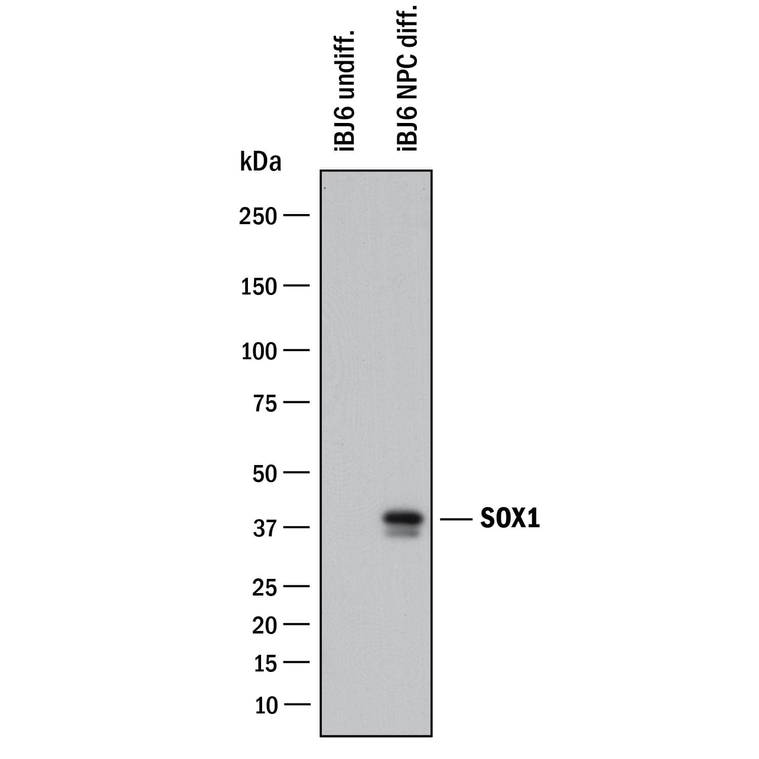
HAF017). A specific band was detected for SOX1 at approximately 39 kDa (as indicated). This experiment was conducted under reducing conditions and using
Immunoblot Buffer Group 1." alt="Western blot shows lysates of undifferentiated iBJ6 human iPS cells and iBJ6 human iPS cells differentiated into neuroprogenitor cells. PVDF membrane was probed with 1 µg/mL of Goat Anti-Human/Mouse/Rat SOX1 Antigen Affinity-purified Polyclonal Antibody (Catalog # AF3369) followed by HRP-conjugated Anti-Goat IgG Secondary Antibody (
HAF017). A specific band was detected for SOX1 at approximately 39 kDa (as indicated). This experiment was conducted under reducing conditions and using
Immunoblot Buffer Group 1." class="big_thumb" />
NL001) and counterstained with DAPI (blue). Nestin was also detected using Mouse Anti-Mouse/Rat Nestin Monoclonal Antibody (
MAB2736) and stained using the NorthernLights™ 493-conjugated Anti-Mouse IgG Secondary Antibody (green; Catalog #
NL009). Specific staining of SOX1 was localized to nuclei. View our protocol for
Fluorescent ICC Staining of Stem Cells on Coverslips." class="big_lightbox">
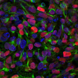
NL001) and counterstained with DAPI (blue). Nestin was also detected using Mouse Anti-Mouse/Rat Nestin Monoclonal Antibody (
MAB2736) and stained using the NorthernLights™ 493-conjugated Anti-Mouse IgG Secondary Antibody (green; Catalog #
NL009). Specific staining of SOX1 was localized to nuclei. View our protocol for
Fluorescent ICC Staining of Stem Cells on Coverslips." class="big_thumb" />
NSC001) using Goat Anti-Human/Mouse/Rat SOX1 Antigen Affinity-purified Polyclonal Antibody (Catalog # AF3369) at 10 µg/mL for 3 hours at room temperature. Cells were stained using the NorthernLights™ 557-conjugated Anti-Goat IgG Secondary Antibody (red, upper panel;
NL001) and counterstained with DAPI (blue, lower panel). Specific staining was localized to nuclei. View our protocol for
Fluorescent ICC Staining of Cells on Coverslips." class="big_lightbox">
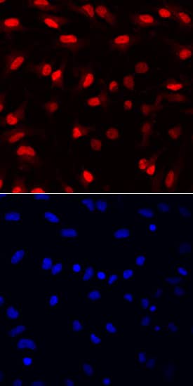
NSC001) using Goat Anti-Human/Mouse/Rat SOX1 Antigen Affinity-purified Polyclonal Antibody (Catalog # AF3369) at 10 µg/mL for 3 hours at room temperature. Cells were stained using the NorthernLights™ 557-conjugated Anti-Goat IgG Secondary Antibody (red, upper panel;
NL001) and counterstained with DAPI (blue, lower panel). Specific staining was localized to nuclei. View our protocol for
Fluorescent ICC Staining of Cells on Coverslips." class="big_thumb" />
NSC002) using Goat Anti-Human/Mouse/Rat SOX1 Antigen Affinity-purified Polyclonal Antibody (Catalog # AF3369) at 10 µg/mL for 3 hours at room temperature. Cells were stained using the NorthernLights™ 557-conjugated Anti-Goat IgG Secondary Antibody (red, upper panel;
NL001) and counterstained with DAPI (blue, lower panel). Specific staining was localized to nuclei. View our protocol for
Fluorescent ICC Staining of Cells on Coverslips." class="big_lightbox">
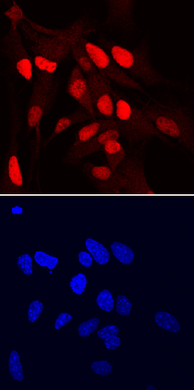
NSC002) using Goat Anti-Human/Mouse/Rat SOX1 Antigen Affinity-purified Polyclonal Antibody (Catalog # AF3369) at 10 µg/mL for 3 hours at room temperature. Cells were stained using the NorthernLights™ 557-conjugated Anti-Goat IgG Secondary Antibody (red, upper panel;
NL001) and counterstained with DAPI (blue, lower panel). Specific staining was localized to nuclei. View our protocol for
Fluorescent ICC Staining of Cells on Coverslips." class="big_thumb" />
VC001). Before incubation with the primary antibody, tissue was subjected to heat-induced epitope retrieval using Antigen Retrieval Reagent-Basic (
CTS013). Tissue was stained using DAB (brown) and counterstained with hematoxylin (blue). Specific staining was localized to nuclei in neuron. Staining was performed using our protocol for IHC Staining with VisUCyte HRP Polymer Detection Reagents." class="big_lightbox">
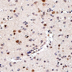
VC001). Before incubation with the primary antibody, tissue was subjected to heat-induced epitope retrieval using Antigen Retrieval Reagent-Basic (
CTS013). Tissue was stained using DAB (brown) and counterstained with hematoxylin (blue). Specific staining was localized to nuclei in neuron. Staining was performed using our protocol for IHC Staining with VisUCyte HRP Polymer Detection Reagents." class="big_thumb" />
HAF109). This experiment was conducted under reducing conditions and using the 12-230 kDa separation system." class="big_lightbox">
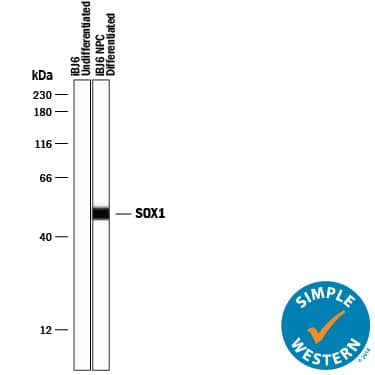
HAF109). This experiment was conducted under reducing conditions and using the 12-230 kDa separation system." class="big_thumb" />
 HAF017). A specific band was detected for SOX1 at approximately 39 kDa (as indicated). This experiment was conducted under reducing conditions and using Immunoblot Buffer Group 1." alt="Western blot shows lysates of undifferentiated iBJ6 human iPS cells and iBJ6 human iPS cells differentiated into neuroprogenitor cells. PVDF membrane was probed with 1 µg/mL of Goat Anti-Human/Mouse/Rat SOX1 Antigen Affinity-purified Polyclonal Antibody (Catalog # AF3369) followed by HRP-conjugated Anti-Goat IgG Secondary Antibody (HAF017). A specific band was detected for SOX1 at approximately 39 kDa (as indicated). This experiment was conducted under reducing conditions and using Immunoblot Buffer Group 1." class="big_thumb" />


 NL001) and counterstained with DAPI (blue). Nestin was also detected using Mouse Anti-Mouse/Rat Nestin Monoclonal Antibody (
 NL003) and counterstained with DAPI (blue, lower panel). View our protocol for
  NSC001) using Goat Anti-Human/Mouse/Rat SOX1 Antigen Affinity-purified Polyclonal Antibody (Catalog # AF3369) at 10 µg/mL for 3 hours at room temperature. Cells were stained using the NorthernLights™ 557-conjugated Anti-Goat IgG Secondary Antibody (red, upper panel;
  NSC002) using Goat Anti-Human/Mouse/Rat SOX1 Antigen Affinity-purified Polyclonal Antibody (Catalog # AF3369) at 10 µg/mL for 3 hours at room temperature. Cells were stained using the NorthernLights™ 557-conjugated Anti-Goat IgG Secondary Antibody (red, upper panel;
  VC001). Before incubation with the primary antibody, tissue was subjected to heat-induced epitope retrieval using Antigen Retrieval Reagent-Basic (
 HAF109). This experiment was conducted under reducing conditions and using the 12-230 kDa separation system." class="big_thumb" />


![Western Blot: SOX1 Antibody [NBP2-24486] - Analysis of extracts from mouse brain using SOX1 antibody (NBP2-24486, 1:100). Image from verified customer review.](http://images.novusbio.com/fullsize/SOX1-Antibody-Western-Blot-NBP2-24486-img0005.jpg "Western Blot: SOX1 Antibody [NBP2-24486] - Analysis of extracts from mouse brain using SOX1 antibody (NBP2-24486, 1:100). Image from verified customer review.")
![Western Blot: SOX1 Antibody [NBP2-24486] - Analysis of SOX1 in human brain lysate in the 1) absence and 2) presence of immunizing peptide. Goat anti-rabbit Ig HRP secondary antibody and PicoTect ECL substrate solution were used for this test.](http://images.novusbio.com/fullsize/SOX1-Antibody-Western-Blot-NBP2-24486-img0003.jpg)
![Western Blot: SOX1 Antibody [NBP2-24486] - Analysis in D3 cell lysate in the 1) absence and 2) presence of immunizing peptide using NBP2-24486 at 1 ug/mL. Goat anti-rabbit Ig HRP secondary antibody (Novus, 20301) and PicoTect ECL substrate solution (Novus, 10087K) were used.](http://images.novusbio.com/fullsize/SOX1-Antibody-Western-Blot-NBP2-24486-img0004.jpg)
![Immunocytochemistry/Immunofluorescence: SOX1 Antibody (JJ20-40) [NBP2-67864] - Staining SOX1 in SHG-44 cells (green). The nuclear counter stain is DAPI (blue). Cells were fixed in paraformaldehyde, permeabilised with 0.25% Triton X100/PBS.](http://images.novusbio.com/fullsize/SOX1-Antibody-JJ20-40-Immunocytochemistry-Immunofluorescence-NBP2-67864-img0001.jpg "Immunocytochemistry/Immunofluorescence: SOX1 Antibody (JJ20-40) [NBP2-67864] - Staining SOX1 in SHG-44 cells (green). The nuclear counter stain is DAPI (blue). Cells were fixed in paraformaldehyde, permeabilised with 0.25% Triton X100/PBS.")
![<b>Biological Strategies Validation. </b>Immunohistochemistry-Paraffin: SOX1 Antibody (JJ20-40) [NBP2-67864] - Immunohistochemical analysis of paraffin-embedded rat brain tissue using anti-SOX1 antibody. The section was pre-treated using heat mediated antigen retrieval with sodium citrate buffer (pH 6.0) for 20 minutes. The tissues were blocked in 5% BSA for 30 minutes at room temperature, washed with ddH2O and PBS, and then probed with the primary antibody (1/50) for 30 minutes at room temperature. The detection was performed using an HRP conjugated compact polymer system. DAB was used as the chromogen. Tissues were counterstained with hematoxylin and mounted with DPX.](http://images.novusbio.com/fullsize/SOX1-Antibody-JJ20-40-Immunohistochemistry-Paraffin-NBP2-67864-img0004.jpg)
![<b>Biological Strategies Validation. </b>Immunohistochemistry-Paraffin: SOX1 Antibody (JJ20-40) [NBP2-67864] - Immunohistochemical analysis of paraffin-embedded mouse brain tissue using anti-SOX1 antibody. The section was pre-treated using heat mediated antigen retrieval with sodium citrate buffer (pH 6.0) for 20 minutes. The tissues were blocked in 5% BSA for 30 minutes at room temperature, washed with ddH2O and PBS, and then probed with the primary antibody (1/50) for 30 minutes at room temperature. The detection was performed using an HRP conjugated compact polymer system. DAB was used as the chromogen. Tissues were counterstained with hematoxylin and mounted with DPX.](http://images.novusbio.com/fullsize/SOX1-Antibody-JJ20-40-Immunohistochemistry-Paraffin-NBP2-67864-img0002.jpg)
![<b>Biological Strategies Validation. </b>Immunohistochemistry-Paraffin: SOX1 Antibody (JJ20-40) [NBP2-67864] - Human brain tissue with Rabbit anti-SOX1 antibody at 1/200 dilution.The section was pre-treated using heat mediated antigen retrieval with sodium citrate buffer (pH 6.0) for 2 minutes. The tissues were blocked in 1% BSA for 20 minutes at room temperature, washed with ddH2O and PBS, and then probed with the primary antibody at 1/200 dilution for 1 hour at room temperature. The detection was performed using an HRP conjugated compact polymer system. DAB was used as the chromogen. Tissues were counterstained with hematoxylin and mounted with DPX.](http://images.novusbio.com/fullsize/nbp2-67864_rabbit-monoclonal-sox1-antibody-jj20-40-immunohistochemistry-paraffin-197202492310..jpg)
![Western blot analysis of E14.0 mouse embryonic brain tissue lysates with NBP2-67864 at 1/1,000 dilution. Lysates/proteins at 30 µg/Lane. Predicted band size: 39 kDa Observed band size: 39 kDa Exposure time: 3 minutes; 4-20% SDS-PAGE gel. Proteins were transferred to a PVDF membrane and blocked with 5% NFDM/TBST for 1 hour at room temperature. NBP2-67864 at 1/1,000 dilution was used in 5% NFDM/TBST at 4℃ overnight. Goat Anti-Rabbit IgG [HRP] Secondary Antibody at 1/50,000 dilution was used for 1 hour at room temperature.](http://images.novusbio.com/fullsize/nbp2-67864_-western-blot-639204586671208730.jpg)
![Western blot analysis on different lysates with NBP2-67864 at 1/2,000 dilution. Lane 1: NCCIT cell lysate Lane 2: Mouse brain tissue lysate Lane 3: Rat brain tissue lysate Lysates/proteins at 20 µg/Lane. Predicted band size: 39 kDa Observed band size: 39 kDa Exposure time: 3 minutes; 4-20% SDS-PAGE gel. Proteins were transferred to a PVDF membrane and blocked with 5% NFDM/TBST for 1 hour at room temperature. NBP2-67864 at 1/2,000 dilution was used in 5% NFDM/TBST at 4℃ overnight. Goat Anti-Rabbit IgG [HRP] Secondary Antibody at 1/50,000 dilution was used for 1 hour at room temperature.](http://images.novusbio.com/fullsize/nbp2-67864_-western-blot-639204597490695559.jpg)
![Western Blot: SOX1 Antibody [NBP2-24466] - Western blot analysis of SOX1 in human brain lysate in the 1) absence and 2) presence of immunizing peptide and 3) mouse brain lysate using NBP2-24466 at 0.5 ug/ml, 0.5 ug/ml and 2 ug/ml respectively.](http://images.novusbio.com/fullsize/SOX1-Antibody-Western-Blot-NBP2-24466-img0002.jpg "Western Blot: SOX1 Antibody [NBP2-24466] - Western blot analysis of SOX1 in human brain lysate in the 1) absence and 2) presence of immunizing peptide and 3) mouse brain lysate using NBP2-24466 at 0.5 ug/ml, 0.5 ug/ml and 2 ug/ml respectively.")
![Western Blot: SOX1 Antibody [NBP3-16153] - Western blot analysis of extracts of various cell lines, using SOX1 antibody (NBP3-16153) at 1:1000 dilution. Secondary antibody: HRP Goat Anti-Rabbit IgG (H+L) at 1:10000 dilution. Lysates/proteins: 25ug per lane. Blocking buffer: 3% nonfat dry milk in TBST. Detection: ECL Enhanced Kit. Exposure time: 180s.](http://images.novusbio.com/fullsize/SOX1-Antibody-Western-Blot-NBP3-16153-img0002.jpg "Western Blot: SOX1 Antibody [NBP3-16153] - Western blot analysis of extracts of various cell lines, using SOX1 antibody (NBP3-16153) at 1:1000 dilution. Secondary antibody: HRP Goat Anti-Rabbit IgG (H+L) at 1:10000 dilution. Lysates/proteins: 25ug per lane. Blocking buffer: 3% nonfat dry milk in TBST. Detection: ECL Enhanced Kit. Exposure time: 180s.")
![Immunohistochemistry-Paraffin: SOX1 Antibody [NBP3-16153] - Immunohistochemistry of paraffin-embedded mouse embryos using SOX1 Rabbit pAb (NBP3-16153) at dilution of 1:25 (40x lens).Perform high pressure antigen retrieval with 10 mM citrate buffer pH 6.0 before commencing with IHC staining protocol.](http://images.novusbio.com/fullsize/SOX1-Antibody-Immunohistochemistry-Paraffin-NBP3-16153-img0001.jpg)
![Western Blot: SOX1 Antibody (0H4C1) [NBP3-33424] - Western blot analysis of lysates from C6 cells, using SOX1 Rabbit mAb at 1:2000 dilution.<br/>Secondary antibody: HRP-conjugated Goat anti-Rabbit IgG (H+L) at 1:10000 dilution.<br/>Lysates/proteins: 25ug per lane.<br/>Blocking buffer: 3% nonfat dry milk in TBST.<br/>Detection: ECL Basic Kit.<br/>Exposure time: 180s.](http://images.novusbio.com/fullsize/nbp3-33424_rabbit-sox1-mab-0h4c1-91220241519517.jpg "Western Blot: SOX1 Antibody (0H4C1) [NBP3-33424] - Western blot analysis of lysates from C6 cells, using SOX1 Rabbit mAb at 1:2000 dilution.<br/>Secondary antibody: HRP-conjugated Goat anti-Rabbit IgG (H+L) at 1:10000 dilution.<br/>Lysates/proteins: 25ug per lane.<br/>Blocking buffer: 3% nonfat dry milk in TBST.<br/>Detection: ECL Basic Kit.<br/>Exposure time: 180s.")
![Western Blot: SOX1 Antibody (0H4C1) [NBP3-33424] - Western blot analysis of various lysates, using SOX1 Rabbit mAb at 1:2000 dilution.<br/>Secondary antibody: HRP-conjugated Goat anti-Rabbit IgG (H+L) at 1:10000 dilution.<br/>Lysates/proteins: 25ug per lane.<br/>Blocking buffer: 3% nonfat dry milk in TBST.<br/>Detection: ECL Basic Kit.<br/>Exposure time: 10s.](http://images.novusbio.com/fullsize/nbp3-33424_rabbit-sox1-mab-0h4c1-912202415121327.jpg)
![Western Blot: SOX1 Antibody (9L8Z4) [NBP3-16619] - Western blot analysis of extracts of A-549 cells, using SOX1 Rabbit mAb (NBP3-16619) at 1:1000 dilution. Secondary antibody: HRP Goat Anti-Rabbit IgG (H+L) at 1:10000 dilution. Lysates/proteins: 25ug per lane. Blocking buffer: 3% nonfat dry milk in TBST. Detection: ECL Enhanced Kit. Exposure time: 180s.](http://images.novusbio.com/fullsize/SOX1-Antibody-9L8Z4-Western-Blot-NBP3-16619-img0001.jpg "Western Blot: SOX1 Antibody (9L8Z4) [NBP3-16619] - Western blot analysis of extracts of A-549 cells, using SOX1 Rabbit mAb (NBP3-16619) at 1:1000 dilution. Secondary antibody: HRP Goat Anti-Rabbit IgG (H+L) at 1:10000 dilution. Lysates/proteins: 25ug per lane. Blocking buffer: 3% nonfat dry milk in TBST. Detection: ECL Enhanced Kit. Exposure time: 180s.")
![Western Blot: SOX1 Antibody [NBP3-10486] - Western blot analysis using NBP3-10486 on Human Jurkat as a positive control. Antibody Titration: 0.2-1 ug/ml](http://images.novusbio.com/fullsize/SOX1-Antibody-Western-Blot-NBP3-10486-img0001.jpg "Western Blot: SOX1 Antibody [NBP3-10486] - Western blot analysis using NBP3-10486 on Human Jurkat as a positive control. Antibody Titration: 0.2-1 ug/ml")


