HAF109). For additional reference, recombinant human Smad2 and Smad3 were included. Specific bands for Smad2 were detected at approximately 64 and 58 kDa (as indicated). This experiment was conducted under reducing conditions and using
Immunoblot Buffer Group 2." class="big_lightbox">
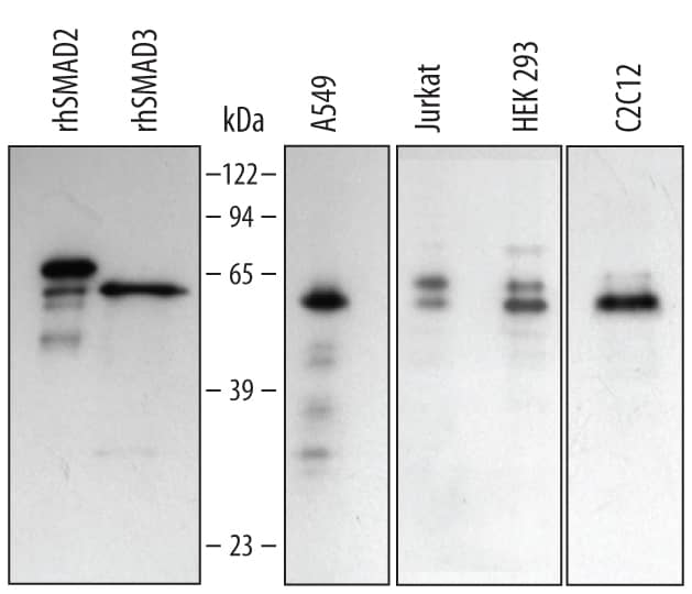
HAF109). For additional reference, recombinant human Smad2 and Smad3 were included. Specific bands for Smad2 were detected at approximately 64 and 58 kDa (as indicated). This experiment was conducted under reducing conditions and using
Immunoblot Buffer Group 2." alt="Western blot shows lysates of A549 human lung carcinoma cell line, Jurkat human acute T cell leukemia cell line, HEK293 human embryonic kidney cell line, and C2C12 mouse myoblast cell line. PVDF membrane was probed with 0.5 µg/mL Goat Anti-Human/Mouse Smad2/3 Antigen Affinity-purified Polyclonal Antibody (Catalog # AF3797) followed by HRP-conjugated Anti-Goat IgG Secondary Antibody (Catalog #
HAF109). For additional reference, recombinant human Smad2 and Smad3 were included. Specific bands for Smad2 were detected at approximately 64 and 58 kDa (as indicated). This experiment was conducted under reducing conditions and using
Immunoblot Buffer Group 2." class="big_thumb" />
AB-108-C) for 15 minutes in an ultrasonic bath, followed by Biotinylated Anti-Goat IgG Secondary Antibody (Catalog #
BAF109). Immunocomplexes were captured using 50 µL of MagCellect Streptavidin Ferrofluid (Catalog #
MAG999) and DNA was purified using chelating resin solution. The
c-mycpromoter was detected by standard PCR." class="big_lightbox">
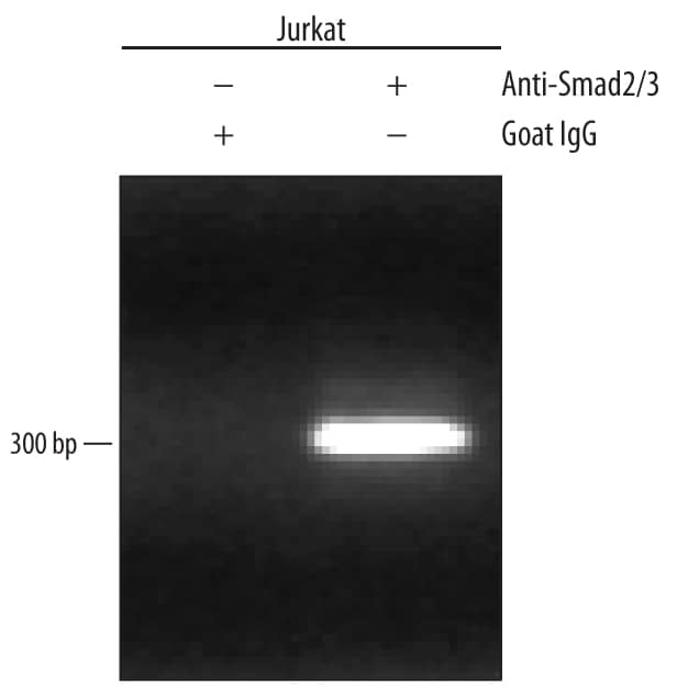
AB-108-C) for 15 minutes in an ultrasonic bath, followed by Biotinylated Anti-Goat IgG Secondary Antibody (Catalog #
BAF109). Immunocomplexes were captured using 50 µL of MagCellect Streptavidin Ferrofluid (Catalog #
MAG999) and DNA was purified using chelating resin solution. The
c-mycpromoter was detected by standard PCR." class="big_thumb" />
HAF109). This experiment was conducted under reducing conditions and using the 12-230 kDa separation system." class="big_lightbox">
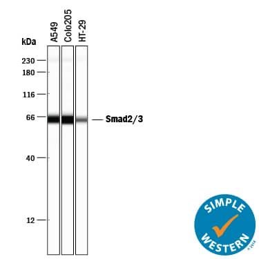
HAF109). This experiment was conducted under reducing conditions and using the 12-230 kDa separation system." class="big_thumb" />
VC004). Before incubation with the primary antibody, tissue was subjected to heat-induced epitope retrieval using Antigen Retrieval Reagent-Basic (
CTS013). Tissue was stained using DAB (brown) and counterstained with hematoxylin (blue). Specific staining was localized to cytoplasm in neurons. Staining was performed using our protocol for IHC Staining with VisUCyte HRP Polymer Detection Reagents." class="big_lightbox">
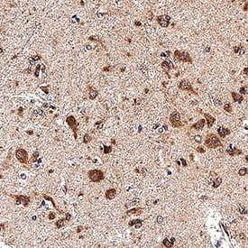
VC004). Before incubation with the primary antibody, tissue was subjected to heat-induced epitope retrieval using Antigen Retrieval Reagent-Basic (
CTS013). Tissue was stained using DAB (brown) and counterstained with hematoxylin (blue). Specific staining was localized to cytoplasm in neurons. Staining was performed using our protocol for IHC Staining with VisUCyte HRP Polymer Detection Reagents." class="big_thumb" />
VC004). Before incubation with the primary antibody, tissue was subjected to heat-induced epitope retrieval using Antigen Retrieval Reagent-Basic (
CTS013). Tissue was stained using DAB (brown) and counterstained with hematoxylin (blue). Specific staining was localized to cytoplasm in Purkinje neurons. Staining was performed using our protocol for IHC Staining with VisUCyte HRP Polymer Detection Reagents." class="big_lightbox">
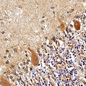
VC004). Before incubation with the primary antibody, tissue was subjected to heat-induced epitope retrieval using Antigen Retrieval Reagent-Basic (
CTS013). Tissue was stained using DAB (brown) and counterstained with hematoxylin (blue). Specific staining was localized to cytoplasm in Purkinje neurons. Staining was performed using our protocol for IHC Staining with VisUCyte HRP Polymer Detection Reagents." class="big_thumb" />
 HAF109). For additional reference, recombinant human Smad2 and Smad3 were included. Specific bands for Smad2 were detected at approximately 64 and 58 kDa (as indicated). This experiment was conducted under reducing conditions and using Immunoblot Buffer Group 2." alt="Western blot shows lysates of A549 human lung carcinoma cell line, Jurkat human acute T cell leukemia cell line, HEK293 human embryonic kidney cell line, and C2C12 mouse myoblast cell line. PVDF membrane was probed with 0.5 µg/mL Goat Anti-Human/Mouse Smad2/3 Antigen Affinity-purified Polyclonal Antibody (Catalog # AF3797) followed by HRP-conjugated Anti-Goat IgG Secondary Antibody (Catalog # HAF109). For additional reference, recombinant human Smad2 and Smad3 were included. Specific bands for Smad2 were detected at approximately 64 and 58 kDa (as indicated). This experiment was conducted under reducing conditions and using Immunoblot Buffer Group 2." class="big_thumb" />


 AB-108-C) for 15 minutes in an ultrasonic bath, followed by Biotinylated Anti-Goat IgG Secondary Antibody (Catalog #
  NL001) and counterstained with DAPI (blue). Specific staining was localized to cytoplasm and, in EMT-induced cells, nuclei. View our protocol for
  HAF109). This experiment was conducted under reducing conditions and using the 12-230 kDa separation system." class="big_thumb" />
 VC004). Before incubation with the primary antibody, tissue was subjected to heat-induced epitope retrieval using Antigen Retrieval Reagent-Basic (
 VC004). Before incubation with the primary antibody, tissue was subjected to heat-induced epitope retrieval using Antigen Retrieval Reagent-Basic (

![Western Blot: Smad2/3 Antibody [NBP3-15589] - Western blot analysis of extracts of various cell lines, using Smad2/3 antibody (NBP3-15589) at 1:1000 dilution. Secondary antibody: HRP Goat Anti-Rabbit IgG (H+L) at 1:10000 dilution. Lysates/proteins: 25ug per lane. Blocking buffer: 3% nonfat dry milk in TBST. Detection: ECL Enhanced Kit. Exposure time: 3min.](http://images.novusbio.com/fullsize/Smad2-3-Antibody-Western-Blot-NBP3-15589-img0001.jpg "Western Blot: Smad2/3 Antibody [NBP3-15589] - Western blot analysis of extracts of various cell lines, using Smad2/3 antibody (NBP3-15589) at 1:1000 dilution. Secondary antibody: HRP Goat Anti-Rabbit IgG (H+L) at 1:10000 dilution. Lysates/proteins: 25ug per lane. Blocking buffer: 3% nonfat dry milk in TBST. Detection: ECL Enhanced Kit. Exposure time: 3min.")
![Immunocytochemistry/ Immunofluorescence: Smad2/3 Antibody - Azide and BSA Free [Smad2/3] - Immunofluorescence analysis of Hep G2 cells using Smad2/3 Rabbit pAb at a dilution of 1:100 (40x lens). Secondary antibody: Cy3-conjugated Goat anti-Rabbit IgG (H+L) at 1:500 dilution. Blue: DAPI for nuclear staining.](http://images.novusbio.com/fullsize/nbp3-15589_rabbit-smad2-3-pab-52202517361286.jpg)
![Immunohistochemistry: Smad2/3 Antibody - Azide and BSA Free [Smad2/3] - Immunohistochemistry analysis of paraffin-embedded Rat intestine tissue using Smad2/3 Rabbit pAb at a dilution of 1:100 (40x lens). High pressure antigen retrieval performed with 0.01M Citrate Buffer(pH 6.0) prior to IHC staining.](http://images.novusbio.com/fullsize/nbp3-15589_rabbit-smad2-3-pab-52202517311036.jpg)
![Immunohistochemistry: Smad2/3 Antibody - Azide and BSA Free [Smad2/3] - Immunohistochemistry analysis of paraffin-embedded Mouse kidney tissue using Smad2/3 Rabbit pAb at a dilution of 1:100 (40x lens). High pressure antigen retrieval performed with 0.01M Citrate Buffer(pH 6.0) prior to IHC staining.](http://images.novusbio.com/fullsize/nbp3-15589_rabbit-smad2-3-pab-52202517303110.jpg)
![Immunohistochemistry: Smad2/3 Antibody - Azide and BSA Free [Smad2/3] - Immunohistochemistry analysis of paraffin-embedded Human kidney tissue using Smad2/3 Rabbit pAb at a dilution of 1:100 (40x lens). High pressure antigen retrieval performed with 0.01M Citrate Buffer(pH 6.0) prior to IHC staining.](http://images.novusbio.com/fullsize/nbp3-15589_rabbit-smad2-3-pab-52202517352770.jpg)


