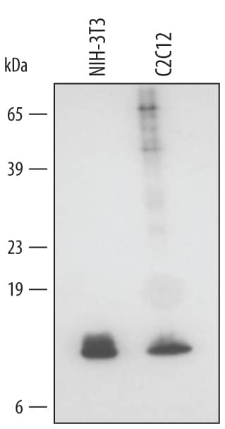
 HAF109). A specific band was detected for Glutaredoxin 1 at approximately 12 kDa (as indicated). This experiment was conducted using Immunoblot Buffer Group 2." alt="Western blot shows lysates of NIH-3T3 mouse embryonic fibroblast cell line and C2C12 mouse myoblast cell line. PVDF membrane was probed with 0.5 µg/mL of Mouse Glutaredoxin 1 Antigen Affinity-purified Polyclonal Antibody (Catalog # AF3119) followed by HRP-conjugated Anti-Goat IgG Secondary Antibody (Catalog # HAF109). A specific band was detected for Glutaredoxin 1 at approximately 12 kDa (as indicated). This experiment was conducted using Immunoblot Buffer Group 2." class="big_thumb" />
| Formulation | Catalog # | Availability | Price | |
|---|---|---|---|---|
| Carrier Free | AF3119-SP | |||
 HAF109). A specific band was detected for Glutaredoxin 1 at approximately 12 kDa (as indicated). This experiment was conducted under reducing conditions and using Immunoblot Buffer Group 2." alt="Western blot shows lysates of HepG2 human hepatocellular carcinoma cell line, Jurkat human acute T cell leukemia cell line, NIH-3T3 mouse embryonic fibroblast cell line, and C2C12 mouse myoblast cell line. PVDF membrane was probed with 0.5 µg/mL of Goat Anti-Human/Mouse Glutaredoxin 1 Antigen Affinity-purified Polyclonal Antibody (Catalog # AF3399) followed by HRP-conjugated Anti-Goat IgG Secondary Antibody (Catalog # HAF109). A specific band was detected for Glutaredoxin 1 at approximately 12 kDa (as indicated). This experiment was conducted under reducing conditions and using Immunoblot Buffer Group 2." class="big_thumb" />
| Formulation | Catalog # | Availability | Price | |
|---|---|---|---|---|
| Carrier Free | AF3399-SP | |||
| Formulation | Catalog # | Availability | Price | |
|---|---|---|---|---|
| BSA Free | NBP2-55346-25ul | |||


 HAF109). This experiment was conducted under reducing conditions and using the 12-230 kDa separation system." class="big_thumb" />
![Western Blot: Glutaredoxin 1/GLRX1 Antibody [NBP2-55346] - Analysis in human cell line RT-4, human cell line U-251 MG, human plasma, human liver tissue and human tonsil tissue.](http://images.novusbio.com/fullsize/Glutaredoxin-1-GLRX1-Antibody-Western-Blot-NBP2-55346-img0002.jpg "Western Blot: Glutaredoxin 1/GLRX1 Antibody [NBP2-55346] - Analysis in human cell line RT-4, human cell line U-251 MG, human plasma, human liver tissue and human tonsil tissue.")
![Immunohistochemistry-Paraffin: Glutaredoxin 1/GLRX1 Antibody [NBP2-55346] - Staining of human kidney shows strong cytoplasmic and nuclear positivity in cells in tubules.](http://images.novusbio.com/fullsize/Glutaredoxin-1-GLRX1-Antibody-Immunohistochemistry-Paraffin-NBP2-55346-img0001.jpg)


![Western Blot: Glutaredoxin 1/GLRX1 Antibody [NBP1-56684] - Human Brain lysate, concentration 0.2-1 ug/ml.](http://images.novusbio.com/fullsize/Glutaredoxin-1-GLRX1-Antibody-Western-Blot-NBP1-56684-img0003.jpg "Western Blot: Glutaredoxin 1/GLRX1 Antibody [NBP1-56684] - Human Brain lysate, concentration 0.2-1 ug/ml.")


