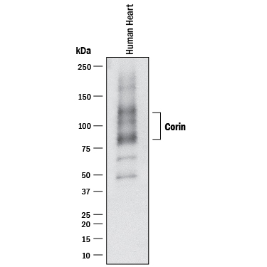
HAF017). A specific band was detected for Corin at approximately 85 and 130 kDa (as indicated). This experiment was conducted under reducing conditions and using
Immunoblot Buffer Group 1." class="big_lightbox">
HAF017). A specific band was detected for Corin at approximately 85 and 130 kDa (as indicated). This experiment was conducted under reducing conditions and using
Immunoblot Buffer Group 1." alt="Western blot shows lysates of human heart tissue. PVDF membrane was probed with 1 µg/mL of Goat Anti-Human Corin Antigen Affinity-purified Polyclonal Antibody (Catalog # af2209) followed by HRP-conjugated Anti-Goat IgG Secondary Antibody (Catalog #
HAF017). A specific band was detected for Corin at approximately 85 and 130 kDa (as indicated). This experiment was conducted under reducing conditions and using
Immunoblot Buffer Group 1." class="big_thumb" />
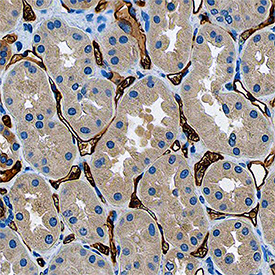
VC004). Before incubation with the primary antibody, tissue was subjected to heat-induced epitope retrieval using Antigen Retrieval Reagent-Basic (Catalog #
CTS013). Tissue was stained using DAB (brown) and counterstained with hematoxylin (blue). Specific staining was localized to endothelial cells in canaliculi. View our protocol for
IHC Staining with VisUCyte HRP Polymer Detection Reagents." class="big_lightbox">
VC004). Before incubation with the primary antibody, tissue was subjected to heat-induced epitope retrieval using Antigen Retrieval Reagent-Basic (Catalog #
CTS013). Tissue was stained using DAB (brown) and counterstained with hematoxylin (blue). Specific staining was localized to endothelial cells in canaliculi. View our protocol for
IHC Staining with VisUCyte HRP Polymer Detection Reagents." class="big_thumb" />
VC004). Before incubation with the primary antibody, tissue was subjected to heat-induced epitope retrieval using Antigen Retrieval Reagent-Basic (Catalog #
CTS013). Tissue was stained using DAB (brown) and counterstained with hematoxylin (blue). Specific staining was localized to cytoplasm in cardiomyocytes. View our protocol for
IHC Staining with VisUCyte HRP Polymer Detection Reagents." class="big_lightbox">
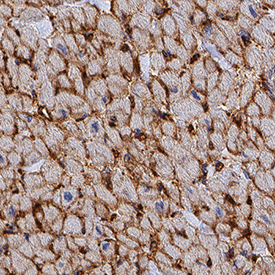
VC004). Before incubation with the primary antibody, tissue was subjected to heat-induced epitope retrieval using Antigen Retrieval Reagent-Basic (Catalog #
CTS013). Tissue was stained using DAB (brown) and counterstained with hematoxylin (blue). Specific staining was localized to cytoplasm in cardiomyocytes. View our protocol for
IHC Staining with VisUCyte HRP Polymer Detection Reagents." class="big_thumb" />
MAB2209) coated on a Clear Polystyrene Microplate (Catalog #
DY990). Goat Anti-Human Corin Antigen Affinity-purified Polyclonal Antibody (Catalog # AF2209) was biotinylated and incubated with the protein captured on the plate. Detection of the standard curve was achieved by incubating Streptavidin-HRP (Catalog #
DY998) followed by Substrate Solution (Catalog #
DY999) and stopping the enzymatic reaction with Stop Solution (Catalog #
DY994)." class="big_lightbox">
MAB2209) coated on a Clear Polystyrene Microplate (Catalog #
DY990). Goat Anti-Human Corin Antigen Affinity-purified Polyclonal Antibody (Catalog # AF2209) was biotinylated and incubated with the protein captured on the plate. Detection of the standard curve was achieved by incubating Streptavidin-HRP (Catalog #
DY998) followed by Substrate Solution (Catalog #
DY999) and stopping the enzymatic reaction with Stop Solution (Catalog #
DY994)." class="big_thumb" />
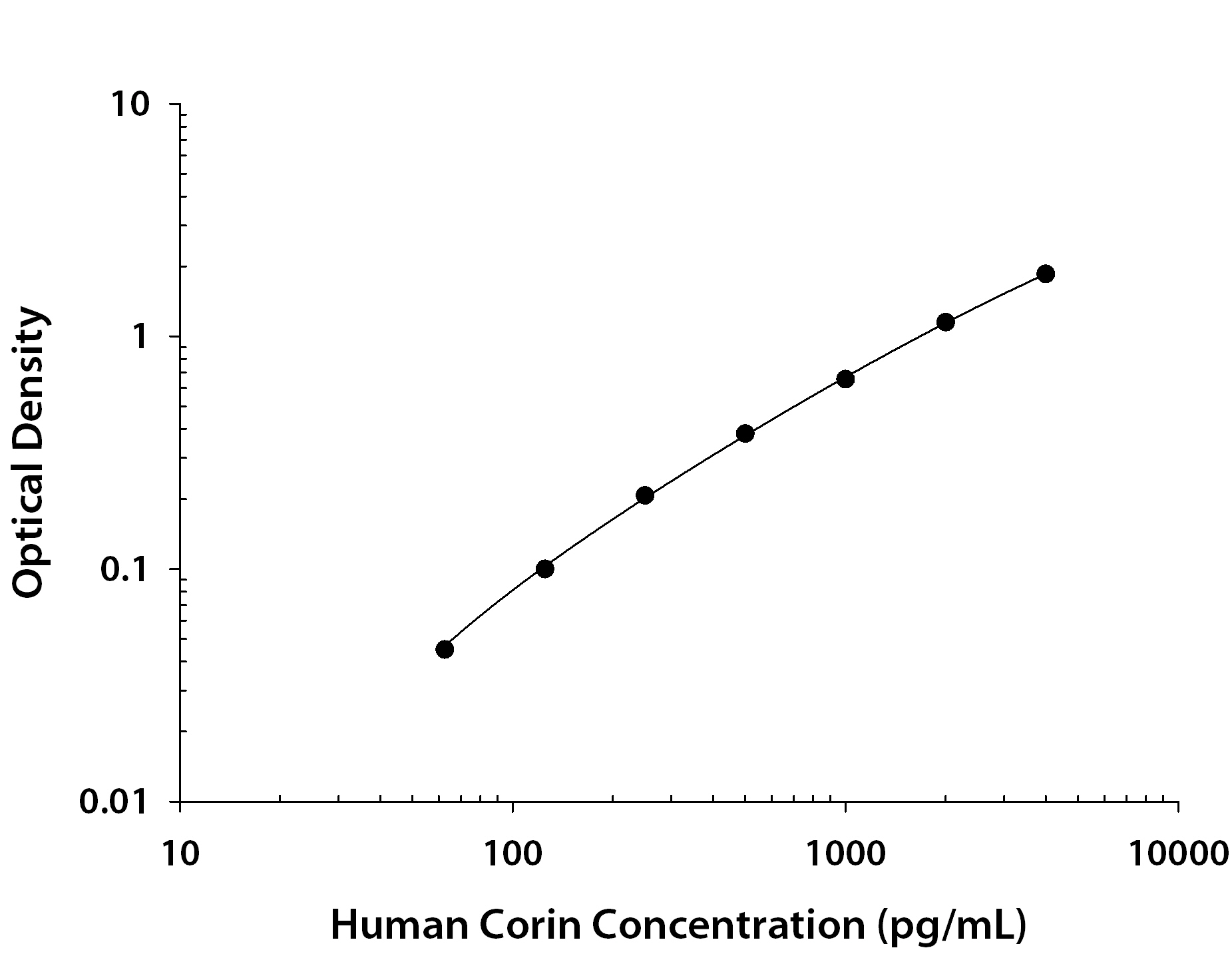
3560-ZN) was serially diluted and captured by Rat Anti-Human Corin Monoclonal Antibody (Catalog #
MAB2209) coated on a Clear Polystyrene Microplate (Catalog #
DY990). Goat Anti-Human Corin Antigen Affinity-purified Polyclonal Antibody (Catalog # AF2209) was biotinylated and incubated with the protein captured on the plate. Detection of the standard curve was achieved by incubating Streptavidin-HRP (Catalog #
DY998)" class="big_lightbox">

3560-ZN) was serially diluted and captured by Rat Anti-Human Corin Monoclonal Antibody (Catalog #
MAB2209) coated on a Clear Polystyrene Microplate (Catalog #
DY990). Goat Anti-Human Corin Antigen Affinity-purified Polyclonal Antibody (Catalog # AF2209) was biotinylated and incubated with the protein captured on the plate. Detection of the standard curve was achieved by incubating Streptavidin-HRP (Catalog #
DY998)" class="big_thumb" />
 HAF017). A specific band was detected for Corin at approximately 85 and 130 kDa (as indicated). This experiment was conducted under reducing conditions and using Immunoblot Buffer Group 1." alt="Western blot shows lysates of human heart tissue. PVDF membrane was probed with 1 µg/mL of Goat Anti-Human Corin Antigen Affinity-purified Polyclonal Antibody (Catalog # af2209) followed by HRP-conjugated Anti-Goat IgG Secondary Antibody (Catalog # HAF017). A specific band was detected for Corin at approximately 85 and 130 kDa (as indicated). This experiment was conducted under reducing conditions and using Immunoblot Buffer Group 1." class="big_thumb" />
 NL013) and counterstained with DAPI (blue). Specific staining was localized to plasma membranes. View our protocol for Fluorescent ICC Staining of Cells on Coverslips." alt="Corin was detected in immersion fixed HK2 human kidney proximal tubule cell line using Rat Anti-Human Corin Monoclonal Antibody (Catalog # MAB2209) at 25 µg/mL for 3 hours at room temperature. Cells were stained using the NorthernLights™ 557-conjugated Anti-Rat IgG Secondary Antibody (red; Catalog # NL013) and counterstained with DAPI (blue). Specific staining was localized to plasma membranes. View our protocol for Fluorescent ICC Staining of Cells on Coverslips." class="big_thumb" />


![N/A Corin [Biotin]](http://images.novusbio.com/fullsize/elisa/DATA_Corin_DY2209_ELISA_1787.jpg "N/A Corin [Biotin]")
 VC004). Before incubation with the primary antibody, tissue was subjected to heat-induced epitope retrieval using Antigen Retrieval Reagent-Basic (Catalog #
  VC004). Before incubation with the primary antibody, tissue was subjected to heat-induced epitope retrieval using Antigen Retrieval Reagent-Basic (Catalog #
  MAB2209) coated on a Clear Polystyrene Microplate (Catalog #
  3560-ZN) was serially diluted and captured by Rat Anti-Human Corin Monoclonal Antibody (Catalog #
 ![N/A Corin [HRP]](http://images.novusbio.com/fullsize/elisa/DATA_Corin_DCRN00_ELISA_635.jpg "N/A Corin [HRP]")
![N/A Corin [HRP]](http://images.novusbio.com/fullsize/elisa/Corin_DCRN00_ELISA_68.jpg)
 DY990). Goat Anti-Human Corin Antigen Affinity-purified Polyclonal Antibody (Catalog #
  3560-ZN) was serially diluted and captured by Rat Anti-Human Corin Monoclonal Antibody (Catalog # MAB2209) coated on a Clear Polystyrene Microplate (Catalog #
 ![Western Blot: CORIN Antibody [NBP1-59663] - HepG2 cell lysate, concentration 2.5 ug/ml.](http://images.novusbio.com/fullsize/CORIN-Antibody-Western-Blot-NBP1-59663-img0002.jpg "Western Blot: CORIN Antibody [NBP1-59663] - HepG2 cell lysate, concentration 2.5 ug/ml.")

![Sandwich ELISA: Corin Antibody (5B6) [H00010699-M01] - Detection limit for recombinant GST tagged CORIN is approximately 3ng/ml as a capture antibody.](http://images.novusbio.com/fullsize/Corin-Antibody-5B6-Sandwich-ELISA-H00010699-M01-img0002.jpg "Sandwich ELISA: Corin Antibody (5B6) [H00010699-M01] - Detection limit for recombinant GST tagged CORIN is approximately 3ng/ml as a capture antibody.")


